| Research Article | ||
Open Vet J. 2024; 14(10): 2634-2641 Open Veterinary Journal, (2024), Vol. 14(10): 2634-2641 Research Article Study of morphological and histological properties of the pancreas in crow (Linnaecus corvus) and Iraqi black partridge (Melanoperdix niger)Rabab AbdAlameer Naser1, Salah Hasan Almaliki2, Fatimah Swadi Zghair3 and Ali Ibrahim Ali Al-Ezzy4*1Department of Anatomy and Histology, College of Veterinary Medicine, University of Diyala, Baqubah, Iraq 2Department of Anatomy and Histology, College of Veterinary Medicine, University of Baghdad, Baghdad, Iraq 3Department of Anatomy and Histology, College of Veterinary Medicine, Al-Qadisiyah University, Al-Diwaniyah, Iraq 4Department of Pathology, College of Veterinary Medicine, University of Diyala, Baqubah, Iraq *Corresponding Author: Ali Ibrahim Ali Al-Ezzy. Department of Pathology, College of Veterinary Medicine, University of Diyala, Baqubah, Iraq. Email: alizziibrahim [at] gmail.com Submitted: 13/07/2024 Accepted: 19/09/2024 Published: 31/10/2024 © 2024 Open Veterinary Journal
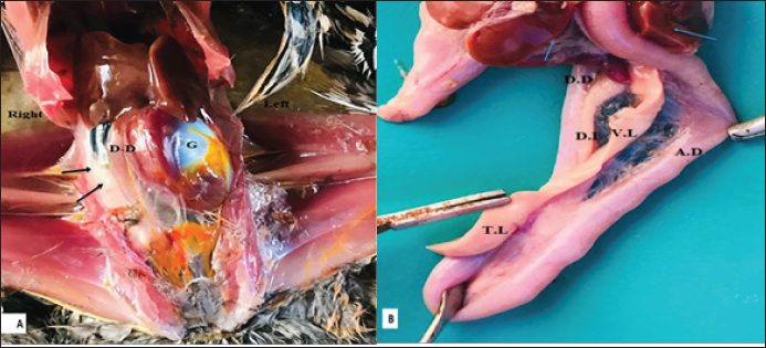
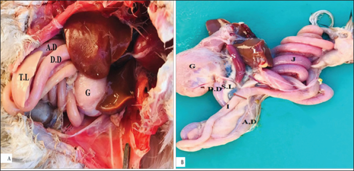
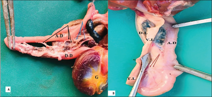
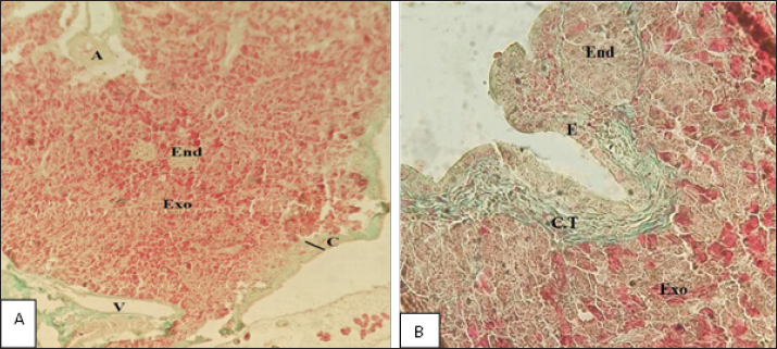
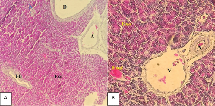
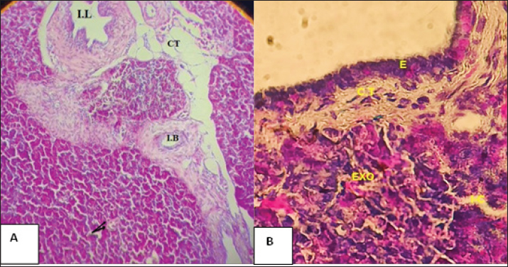
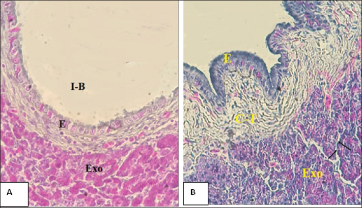
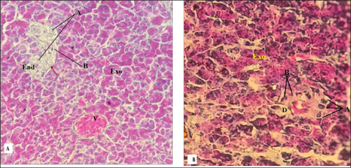
AbstractBackground: The structures of the pancreas in crow (Linnaecus corvus) and Iraqi black partridge (Melanoperdix niger) were the targets for histological and morphometric differences in both birds. Aim: To study the comparative histomorphology of the pancreas in two species black partridge and local crow. Methods: Five healthy black partridge and five local crows were used in the current study. Results: The anatomical study reveals that the pancreas in both species is situated within the coelomic compartment on the right side. It is composed of four lobes including splenic, ventral, third, and dorsal lobes. It showed three ducts of the pancreas located between two duodenal limbs. Histologically, the pancreas of both birds contained two portions, endocrine and exocrine zone. The portion that occupied a large area of the pancreas was the exocrine which consisted of acini made of pyramid cells varying in shape and size. In black partridge, the acini have centroacinar cells but no centroacinar in crow. The duct system starting from the intercalated duct, interlobular and terminated by the main duct was folded with line simple columnar epithelium. The islet Langerhans was oval in black partridge and had a distinctive border containing two types of cells (Alpha and Beta), while a Delta, in addition to Alpha Beta cells, was detected in the crow islet Langerhans which was a sphincter in shape. Conclusion: The pancreas of both bird black partridge (Melanoperdix niger) and crow (Linnaecus corvus) was a lobulated organ, that has a similar location in coelomic cavity. The pancreas in the crow was longer. In addition to the presence of some differences in histological structures between the two birds, a better understanding of the function of the pancreas in these species is needed. Keywords: Anatomy, Histology, Pancreas, Black partridge, Crow. IntroductionThe black partridge (Melanoperdix niger) is characterized by brown iris, grey legs, a thick bill, and has small body size. This bird is well-named wood partridge. It is spread in Southeast Asia, especially in Sumatra and Malaysia, and is found in the rainforests of those lowlands (Stępien-Pysniak et al., 2018). The crow (Linnaecus corvus) is distributed in many countries, and their head, tail, throat, and wings were blackish in color. It is an omnivorous feeder and opportunistic forager. The pancreas is considered an important accessory organ of the digestive system (Sturkie, 1986). It is a lobulated organ, situated in the mesentery adjacent to the duodenum, on the right side of the coelomic cavity (Saadatfar and Asadian 2009). Moreover, some previous research studies the pancreas microscopically and grossly, such as in geese (Al-Sharoot, 2016) and guinea fowl (Iniyah, 2018). This organ is subdivided microscopically into two portions, one portion is endocrine and the other one is exocrine portion (Gülmez et al., 2004; Jaafar, 2019). The exocrine in most avians includes a tubuloacinar unit connected with a number of the ductus (Faris, 2012). In pigeons; the endocrine portion is found to be islets Langerhans (Mutoh et al., 1997). The pancreatic juice and bile emptied into the ascending duodenal loop, these secretion is obtained by cannulating ducts, one or three ducts (Sturkie, 1986). Materials and MethodsAnatomy studyTen healthy adult birds: five black partridges (Melanoperdix niger) and five local crows, were obtained from bird hunters in Diyala province. The birds were sacrificed for this experiment by following the regulations of the animal ethics committee at the College of Veterinary Medicine, Diyala University. The mean weight of the black partridge (Melanoperdix niger) was 283.18 ± 0.03 and the crow (Linnaecus corvus) was 206.818 ± 0.22, respectively, then immediately sacrificed by a high dose of anesthesia, by injection of xylazine (alfasan R) of 10 mg/kg BW with Ketamin (VMDR)15 mg/kg BW (Bakhshi et al., 2019). The abdomens of all birds were dissected, then removed the pancreas carefully and washed with distilled water; after that examined grossly and measured the weight of the pancreas in each bird was by a sensitive balance. Histological studyThe specimens from each of the lobes of the pancreas were taken for histological study and were fixed in 10% formalin. All samples were processed routinely to obtain the histological sections that were stained with hematoxylin and eosin (H&E) for general description. Masson trichrome to detect the fibers and connective tissue (elastic, reticular, and collagen fibers), and Gomori staining method for studying the cells of islet of Langerhans (Bancroft and Gamble, 2008). Also, slides of the pancreas were examined by light microscopy (Genex microscope) and images were taken by the camera (Sony) with a resolution of (5) mega pixels. Statistical analysisMacroscopic and microscopic parameters were expressed as mean± Standard error (Hameed et al., 2024). T-test was used to clarify the difference between parameters (Hameed et al., 2020). The difference is considered significant when p value (0.05 ) (Hassan et al., 2020) and p value (0.001) as highly significant (Hameed and Al-Ezzy, 2024). Ethical approvalThe current study was approved by the scientific committee at the Department of Anatomy and Histology, College of Veterinary Medicine, University of Diyala, and following the regulations of the animal ethics committee at the College of Veterinary Medicine, Diyala University. ResultsAnatomical studyMeasurements of body weight and pancreas in black partridge and Crow are shown in Table 1. The macroscopic examination of the pancreas in the current study showed that in both birds the pancreas is situated in the cavity of coelomic at the right side, and comprises the lobes of the splenic, ventral, third, and dorsal (Fig. 1A, B). These lobes are housed in the duodenal between two duodenal limbs. The pancreas is fixed by a pancreatic duodenal ligament and blood supply (Fig. 2A). In black partridge (Melanoperdix niger), the pancreas appeared whitish to pinkish in color (Fig. 1B). Ventral lobe was shorter than dorsal one, while the splenic was most minor lobe, it extends forward opposite the spleen, and the third lobe most prominent lobe triangular in shape (Fig. 1B), whereas the pancreas in crow consists of four lobes, the dorsal lobe was a rod in shape while the ventral lobe is longer and the third lobe was protruding between the duodenal limb, but the spleen lobe is located parallel to the spleen (Fig. 2B). The pancreas of black partridge and the crow contains three pancreatic ducts empties and secreted in ascending lobe of the duodenum (Fig. 3A, B). Histological studyIn both birds, the pancreas is surrounded by thin capsules from connective tissue, consisting of the reticular and elastic fibers with little collagen, but in the crows, it was thicker than the black partridge (Table 2) which contains more reticular and elastic fibers, as reported in ducks. The septa contain blood lymphatic vessel nerve and excretory ducts (Fig. 4A, B) and excretory ducts (Fig. 4A, B). The mean thickness of capsules in black partridge and crow were (12.54 ±2.9 µm) and (15.25 ± 0.3 µm), respectively. Two portions were observed in the gland parenchyma which is represented by endocrine (islets Langerhans) and exocrine parts (Fig. 5A). In the black partridge, the exocrine part is composed of the tubuloacinar gland that occupied most parenchyma which consists of cells columnar (tall to the pyramid) that have dark round nuclei and observed the blood vessels scattered in the septa of the gland, (Fig. 5A). While the crow pancreas has a similar structure, but there are no centroacinar cells (Fig. 5B). While the excretory portion in black partridge made of the pyramid cells release the production into centroacini and finally release in intracalated duct (Fig. 6A). In both birds, the excretory portion has intercalated ducts lined by flat epithelium into interlobular ducts lined with simple cuboidal cells underlined by smooth muscle fibers and fibers, (Fig. 6B, 7A). The mucosa of the main pancreatic ducts in both birds was folded longitudinally and covered with epithelium of columnar type (Figure. 7, B). In both species, the endocrine portion occupied a small area in the pancreas parenchyma that is oval or circular in shape and varies in size (Fig. 5A). In the black partridge the islet is oval in shape and surrounded by a capsule composed of reticular fibers that give a distinct border (Fig. 8A). While in the crow bird, the islets Langerhans have no distinctive border (Fig. 8B). The islets Langerhans in black partridge is consisting of two types of cells alpha and beta (Fig. 8A) with a mean diameter (62.5 ± 3.7µm). The islet Langerhans in crow birds was circular in shape and contains cells of Alpha, Beta as well as the third type which is named Delta. Cells of alpha were polygonal in shape and had rounded nuclei that are occupied the peripheral area of the islet Langerhans while beta cells were round in shape and possessed oval nuclei that occupied the center of the islet. Whereas Delta cells are irregular cells and pale in color and does not have distinct (Fig. 8B), and the mean diameter (45.5 ± 0.89 µm) (Table 2). Table 1. Measurements of body weight and pancreas in black partridge and crow.
Fig. 1. Photograph shows the position of the pancreas in the coelomic cavity of the black partridge shows: duodenum (black arrow) descending & ascending duodenal limbs) (D–D & A–D), cranial flexure of the duodenum: lobes of the dorsal lobe (DL), ventral (VL), third lobe (T.L), liver: (blue arrow), third (TL), liver (blue arrow), gizzard (G).
Fig. 2. Photographs show the position and relationship of the pancreas in crow with another organ, duodenal loop, descending & ascending (D–D & A–D), gall bladder (brown arrow), liver (L), gizzard (G), spleen(S), dorsal loop (D. L). ventral loop (V.L), third loop (T.L).
Fig. 3. Photographs illustrate the pancreatic ducts pancreas in black partridge (A) and Crow (B) three pancreatic ducts (black arrow) and three bile ducts (blue arrow), duodenal loop, descending & ascending (D.D & A.D), gall bladder black partridge (g), liver (L), gizzard. And three ducts in the crow (black arrow) dorsal loop (D. L) ventral loop (V.L), and third loop (T.L). Table 2. Measurement microparameters of the pancreas in black partridge and crow.
Fig. 4. Photomicroscope section of the pancreas in black partridge (A) and crow (B) illustrate. The capsule (C), exocrine portion (Exo), an endocrine portion (End), mucosal fold), interlobular duct (I-B), vein (V), Masson’s Trichrome stain X100.
Fig. 5. Photomicroscope section of pancreas black partridge(A) and crow pancreas (B) illustrate., exocrine portion (Exo) and endocrine (Blue arrow), intralobular (I–B), duct (D), blood vessel artery (A), vein (V) (A: H & E Stain X100; B: Gomori stain X200).
Fig. 6. Photomicroscope section of pancreas illustrate. The duct system in black partridge (A) and in crow. (B), interlobular (I. L), intralobular duct ( I.B), intercalated duct (black arrow ):, Note lined by cuboidal epithelium, connect tissue (C.T) (A: H&E stain X100; B: Gomori stain A: X400). DiscussionAnatomical studyThe pancreas, in both birds of the study, is a lobulated organ located in the right side along the right side of the median plane in the coelomic cavity, it consists of four lobes as described in chicken and quail (Simsek and Alabay, 2008), in goose (Al-Sharoot 2016), in Mynah (Saadatfar and Asadian 2009), and in falcon (Jaafar 2019). In both birds, the pancreases were pale whitish and it is a ribbon-like organ situated between two duodenal limbs (Kadhim et al., 2010). While the shortest pancreas is found in bustards, (Bailey et al., 1997); in guinea fowl (Iniyah, 2018), and white-eared bulbul (Al-Khakani et al., 2019) which consist of two lobes due to the food of these birds being fruits and grains. The pancreas of both birds has three pancreatic ducts that empty the pancreatic juice in ascending duodenum (Liu et al., 1998). These results are discordant with those reported in guinea fowl (Zghair and Khaleel, 2019), in pigeon (Faris 2012) and in turkey (Naser and Khaleel, 2020).
Fig. 7. Photomicroscope section of the pancreas in black partridge (A) and in crow (B) illustrate. The duct system in the exocrine portion (Exo), intralobular duct (I.B). Note lined by cuboidal epithelium (IL), connective tissue. (A: Gomori stain X200; B: mucosa folded longitudinally in duct (I.B) Gomori stain X200).
Fig. 8. Photomicroscope section of the pancreas of black partridge (A) and crow (B), illustrate. The islets of Langerhans are composed of alpha cell (A), beta (B), and delta cells (D)present only in crow, collagen fiber surrounding islet Langerhans (E), center -acinar (C). (A: Gomori stain X400; B: Gomori stain X400). Histological studyIn both birds, the pancreas is surrounded by a thin capsule, but in crows was thicker than the black partridge as reported in ducks by Al-Shaeli (2010) and Mobini (2011), in geese (Beheiry et al., 2018), and in white-eared bulbul (Al-Khakani et al., 2019). These structures of the exocrine glands in both birds were clusters of acini-containing centro-acini cells connecting intercalated ducts in black partridge. These results are similar to those described in pigeon and adult homing pigeons (Faris, 2012) and in kestrel (Al-Haaik, 2019). There are no centroacinar cells in the crow pancreas but there was a central lumen of acini as described in Mynan (Gülmez et al., 2004). In the current study of both species there are no differences in the structure of the ducts of pancreases. The main ducts are folded and lead to interlobular than intralobular and terminated by an intercalated duct (smallest duct) in all ducts lined by cells initially form columnar to flattened cells. Similar results were reported in local broiler fowl (Kadhim, 2017), in duck (Liu et al., 1998; Wang et al., 2009), and in the red jungle (Kadhim et al., 2010). In goose, the main duct is lined by stratified columnar cells (Al-Sharoot, 2016), whereas in Golden Eagle the interlobular duct is lined by pseudostratified columnar epithelium (Al-Agele, 2012). In the present study, in both birds, the islets of Langerhans were single areas scattered in the exocrine part as described in ostrich (Tarakcy et al., 2007). In black partridge, the islets of Langerhans have distinct borders whereas in crow the islets had no distinct border. The black partridge is similar to some avian species such as pigeon pancreas (Faris, 2012); in both common gull and Guinea fowl (Hamodi et al., 2013), and in goose (Anser anser) (Gülmez et al., 2004; Al-Sharoot 2016). The islets Langerhans in black partridge possess two types of cells, the Alpha cells were longer than beta, similar to the results in Japanese quill (Simsek and Alabay, 2008); in golden eagles (Al-Agele 2012), in guinea fowl (Iniyah 2018) and in Kestrel that suggest these birds are eating one type of food. While the islets of crow were large in diameter (Table 2) and consisted of three types of cells (Alpha, Beta, and a few Delta). Alpha cells are situated peripherally but Beta is located in the central islets of Langerhans are much more complex in some species of birds due to different lifestyles metabolic demands, and diets have most likely influenced the great diversity in both structure and cell type content of islets in lower vertebrate species (MacGregor et al., 2006). Langerhans of the crow is similar to Columbia (AL-Hathry, 2006); in Japanese quail (Simsek and Alabay, 2008), in duck (Al-Shaeli, 2010), and in Common Moorhen and broiler fowl (Kadhim, 2017). These birds contain three cells that release glucagon, insulin, somatostatin, and avian pancreatic polypeptide beta cells responsible for insulin synthesis (Sturkie, 1986). ConclusionThe pancreas of both bird black partridge (Melanoperdix niger) and crow (Linnaecus corvus) is almost the same shape as the pancreas is responsible to secrete pancreatic juice in ascending duodenum due to different feeding habits in these birds. Histologically, the pancreas in both birds had a similar major structure but the differences in islets langerhans cells because each cell is responsible for releasing hormone type. It plays an important role in carbohydrate metabolism. AcknowledgmentThe authors would like to thank the technical staff of the anatomy department at the veterinary college in Diyala and Baghdad Universities for their valuable assistance in the current study. Conflict of interestThe authors declared no conflict of interest. FundingThe manuscript was funded by authors, with no funding agencies. Authors’ contributionsThe authors are equally contributing to the planning, practical work, data collection and analysis, writing of draft and final manuscript. Data availabilityAll data were available in the main text of the current manuscript. ReferencesAl-Agele, R.A.A. and Mohammed, F.S. 2012. Architecture morphology and histological investigations of pancreas in golden eagles (Aquila Chrysaetos). Al-Anbar J. Vet. Sci. 5, 149–155. Al-Haaik, A. 2019. A gross anatomical and histological study of pancreas in adult Kestrel (Falco tinnunculus). Iraqi J. Vet. Sci. 33(2), 175–180. AL-Hathry, S.A. 2006. Histological study of the Langerhans islets of Columbidae. J. Univ. Thi-qar. 2(2), 151–154. Al-Khakani, S.S.A., Zabiba, I., Al-zubaidi, K. and Al-alwany, E. 2019. Morphometrical and histochemical foundation of pancreas and ductal system in white-eared bulbul (Pycnonotus leucotis). Iraqi J. Vet. Sci. 33(1), 99–104. Al-Shaeli, S. 2010. Anatomical and histological study of pancreas in local breed ducks (Anas platyrhnchos, Mallard). Doctoral dissertation, M.Sc. Thesis, College of Veterinary Medicine, University of Baghdad, Baghdad, Iraq. Al-Sharoot, H.A. 2016. Anatomical, histological and histochemical architecture of pancreases in early hatched goose (Anser anser). Kufa J. V. Med. Sci. 7(1), 147–156. Bailey, T.A., Mensah-Brown, E.P., Samour, J.H., Naldo, J., Lawrence, P. and Garner, A. 1997. Comparative morphology of the alimentary tract and its glandular derivatives of captive bustards. J. Anat.191, 387–398. Bakhshi, S., Aghchelou, M.R. and Saadati, D. 2019. The clinical comparison of intraosseous and intravenous anesthesia (Thiopental-Na) in pigeon. Iran. J. Vet. Med. 13(4), 401–409. Bancroft, J.D. and Gamble, M. 2008. Theory and practice of histological techniques, Elsevier Health Sciences, Oxford, UK. Beheiry, R.R., Abdel-Raheem, W.A.A., Balah, A.M., Salem, H. F. and Karkit, M. W. 2018. Morphological, histological and ultrastructural studies on the exocrine pancreas of goose. Beni-Suef Univ. J. Basic Appl. Sci 7(3), 353–358. Faris, S.A. 2012. Anatomical and histological study of the pancreas of pigeon. J. Coll. Edu. Pure. Sci. 2(4), 64–72. Gülmez, N., Kocamiş, H., Aslan, Ş. and Nazli, M. 2004. Immunohistochemical distribution of cells containing insulin, glucagon and somatostatin in the goose (Anser anser) pancreas. Turkish J. Vet. Anim. Sci. 28(2), 403–407. Hameed, M.S. and Al-Ezzy, A.I.A. 2024. Evaluation of antioxidant, nephroprotective and immunomodulatory activity of vitamins C and E -sodium selenite in mice intoxicated with sodium nitrate. Adv. Anim. Vet. Sci. 12(6), 1018–1027. Hameed, M.S., Al-Zubaidi, R.M.H. and Al-Ezzy, A.I.A. 2024. Effectiveness of aqueous versus alcoholic extracts of Melia azedarach in Amelioration of lipid profile, liver enzymes and innate inflammatory indices for white New Zealand rabbits. Adv. Anim. Vet. Sci. 12(7), 1256–1265. Hameed, M.S., Ali Ibrahim Ali, A.E., Waleed, I.J. and Akram, A.H.A. K. 2020. Impact of stress factors on physiological level of interleukin 10 in healthy calves in Diyala province–Iraq. Int. J.Pharma. Res. 12(2), 3050–3056. Hamodi, H.M., Abed, A.A. and Taha, A.M. 2013. Comparative anatomical, histological and histochemical study of the pancreas in two species of birds. Res. Rev. Biosci. 8(1), 26–34. Hassan, A.A., Hameed, M.S. and Al-Ezzy, A.I.A. 2020. Effect of drinking water quality on physiological blood parameters and performance of laying hens in Diyala province, Iraq. Bioch. Cell Arch. 20(1), 2649–2654. Iniyah, K. 2018. Macro and microanatomy of pancreas in guinea fowl. J. Pharm. Phyto. 7(1S), 2895–2896. Jaafar, D.A. 2019. Histological study of pancreas gland in falcon (Falco peregrinus). J. Pharma. Sci. Res. 11(7), 2755–2758. Kadhim, D.M. 2017. A comparative morphological, histological and histochemical study for the liver, pancreas and gall bladder between local broiler fowl (gallus gallus domesticus) and common moorhen (gallinule chloropus). M.Sc., University of Basra, Basrah, Iraq. Kadhim, K.K., Zuki, A., Noordin, M., Babjee, S. and Zamri-Saad, M. 2010. Morphological study of pancreatic duct in red jungle fowl. Afr. J. Biotech. 9(42), 7209–7215. Liu, J., Evans, H., Larsen, P., Pan, D., Xu, S., Dong, H., Deng, X., Wan, B. and Gi, T. 1998. Gross anatomy of the pancreatic lobes and ducts in six breeds of domestic ducks and six species of wild ducks in china. Anat. Histo. Embryo. 27(6), 413–417. MacGregor, R., Williams, S., Tong, P., Kover, K., Moore, W. and Stehno-Bittel, L. 2006. Small rat islets are superior to large islets in in vitro function and in transplantation outcomes. Am. J. Phys.-Endocrin. Metabo. 290(5), E771–E779. Mobini, B. 2011. Histological studies on pancreas of goose (Anser Albifrons). Faculty of Veterinary Medicine, Urmia University, Urmia, Iran. Mutoh, K.I., Wakuri, L., Bo, H. and Taniguchi, K. 1997. Intercalated duct cells in the chicken pancreatic islet. Okajimas Folia Anat. Jap. 74(4), 147–153. Naser, R.A.A. and Khaleel, I.M. 2020. Morphometrical study of small and large intestine in adult bronze male turkeys (Meleagris gallopavo). Bioch. Cell. Arch. 20(2), 6335–6339. Saadatfar, Z. and Asadian, M. 2009. Anatomy of pancreas in mynah (Acridotheres tristis). J. Appl. Anim. Res. 36(2), 191–193. Simsek, N. and Alabay, B. 2008. Light and electron microscopic examinations of the pancreas in quails (Coturnix coturnix japonica). Revue de Med. Vet. 159(4), 198–206. Stępien-Pysniak, D., Hauschild, T., Nowaczek, A., Marek, A. and Dec, M. 2018. Wild Birds as a potential source of known and novel multilocus sequence types of antibiotic-resistant Enterococcus Faecalis. J. Wildl. Dis. 54(2), 219–228. Sturkie, P.D. 1986. Avian physiology. New York, NY: Springer–Verlag. Tarakcy, B.G., Yaman, M. and Bayrakdar, A. 2007. Immunohistochemical study on the endocrine cells in the pancreas of the ostrich (Struthio camelus). J. Anim. Vet. Adv. 6(5), 693–696. Wang, B.J., Liang, H.Y. and Cui, Z.J. 2009. Duck pancreatic acinar cell as a unique model for independent cholinergic stimulation–secretion coupling. Cell. Mol. Neurobio. 29(5), 747–756. Zghair, F.S. and Khaleel, I. 2019. Morphometrical study of small intestine in the adult guinea Fowl (Numidia meleagris). Indian J. Nat. Sci. 9(53), 16965–16974. | ||
| How to Cite this Article |
| Pubmed Style Naser RA, Almaliki SH, Zghair FS, Al-ezzy AIA. Study of morphological and histological properties of the pancreas in crow (Linnaecus corvus) and Iraqi black partridge (Melanoperdix niger). Open Vet J. 2024; 14(10): 2634-2641. doi:10.5455/OVJ.2024.v14.i10.13 Web Style Naser RA, Almaliki SH, Zghair FS, Al-ezzy AIA. Study of morphological and histological properties of the pancreas in crow (Linnaecus corvus) and Iraqi black partridge (Melanoperdix niger). https://www.openveterinaryjournal.com/?mno=209476 [Access: July 09, 2025]. doi:10.5455/OVJ.2024.v14.i10.13 AMA (American Medical Association) Style Naser RA, Almaliki SH, Zghair FS, Al-ezzy AIA. Study of morphological and histological properties of the pancreas in crow (Linnaecus corvus) and Iraqi black partridge (Melanoperdix niger). Open Vet J. 2024; 14(10): 2634-2641. doi:10.5455/OVJ.2024.v14.i10.13 Vancouver/ICMJE Style Naser RA, Almaliki SH, Zghair FS, Al-ezzy AIA. Study of morphological and histological properties of the pancreas in crow (Linnaecus corvus) and Iraqi black partridge (Melanoperdix niger). Open Vet J. (2024), [cited July 09, 2025]; 14(10): 2634-2641. doi:10.5455/OVJ.2024.v14.i10.13 Harvard Style Naser, R. A., Almaliki, . S. H., Zghair, . F. S. & Al-ezzy, . A. I. A. (2024) Study of morphological and histological properties of the pancreas in crow (Linnaecus corvus) and Iraqi black partridge (Melanoperdix niger). Open Vet J, 14 (10), 2634-2641. doi:10.5455/OVJ.2024.v14.i10.13 Turabian Style Naser, Rabab Abdalameer, Salah Hasan Almaliki, Fatimah Swadi Zghair, and Ali Ibrahim Ali Al-ezzy. 2024. Study of morphological and histological properties of the pancreas in crow (Linnaecus corvus) and Iraqi black partridge (Melanoperdix niger). Open Veterinary Journal, 14 (10), 2634-2641. doi:10.5455/OVJ.2024.v14.i10.13 Chicago Style Naser, Rabab Abdalameer, Salah Hasan Almaliki, Fatimah Swadi Zghair, and Ali Ibrahim Ali Al-ezzy. "Study of morphological and histological properties of the pancreas in crow (Linnaecus corvus) and Iraqi black partridge (Melanoperdix niger)." Open Veterinary Journal 14 (2024), 2634-2641. doi:10.5455/OVJ.2024.v14.i10.13 MLA (The Modern Language Association) Style Naser, Rabab Abdalameer, Salah Hasan Almaliki, Fatimah Swadi Zghair, and Ali Ibrahim Ali Al-ezzy. "Study of morphological and histological properties of the pancreas in crow (Linnaecus corvus) and Iraqi black partridge (Melanoperdix niger)." Open Veterinary Journal 14.10 (2024), 2634-2641. Print. doi:10.5455/OVJ.2024.v14.i10.13 APA (American Psychological Association) Style Naser, R. A., Almaliki, . S. H., Zghair, . F. S. & Al-ezzy, . A. I. A. (2024) Study of morphological and histological properties of the pancreas in crow (Linnaecus corvus) and Iraqi black partridge (Melanoperdix niger). Open Veterinary Journal, 14 (10), 2634-2641. doi:10.5455/OVJ.2024.v14.i10.13 |


