| Research Article | ||
Open Vet. J.. 2024; 14(10): 2609-2617 Open Veterinary Journal, (2024), Vol. 14(10): 2609–2617 Research Article Identification of different blood concentration of domestic cat (Felis catus) with Leucomalachite Green and Takayama reagentSuraida Meisari1, Tita Damayanti Lestari2*, Djoko Legowo3, Thomas Valentinus Widiyatno3, Eka Pramyrtha Hestianah4, Retno Sri Wahjuni5, Ratna Damayanti5, Aswin Rafif Khairullah6, Ikechukwu Benjamin Moses7 and Ricadonna Raissa81Center for International Forestry Research and World Agroforestry (CIFOR-ICRAF), Bogor, Indonesia 2Division of Veterinary Reproduction, Faculty of Veterinary Medicine, Universitas Airlangga, Surabaya, Indonesia 3Division of Veterinary Pathology, Faculty of Veterinary Medicine, Universitas Airlangga, Surabaya, Indonesia 4Division of Veterinary Anatomy, Faculty of Veterinary Medicine, Universitas Airlangga, Surabaya, Indonesia 5Division of Basic Veterinary Medicine, Faculty of Veterinary Medicine, Universitas Airlangga, Surabaya, Indonesia 6Research Center for Veterinary Science, National Research and Innovation Agency (BRIN), Bogor, Indonesia 7Department of Applied Microbiology, Faculty of Science, Ebonyi State University, Abakaliki, Nigeria 8Department of Pharmacology, Faculty of Veterinary Medicine, Universitas Brawijaya, Malang, Indonesia *Corresponding Author: Tita Damayanti Lestari. Division of Veterinary Reproduction, Faculty of Veterinary Medicine, Universitas Airlangga, Surabaya, Indonesia. Email: titadlestari [at] fkh.unair.ac.id Submitted: 02/07/2024 Accepted: 10/09/2024 Published: 31/10/2024 © 2024 Open Veterinary Journal
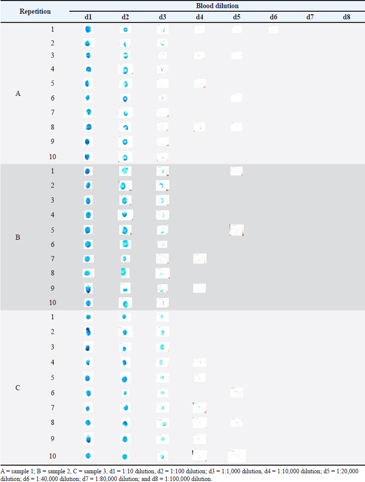
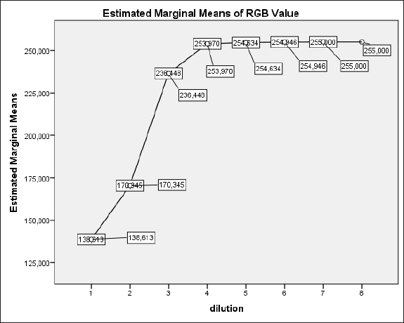
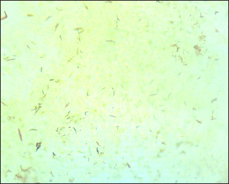
AbstractBackground: Cases of cruelty can occur in wild animal, livestock, and pet animal. Cruelty to cats choose to be the background of this research because many cases of cruelty to cats have not been reported although the cases are still high. Aim: The aim of this research was to know how much dilution of cat blood can still be detected by Leucomalachite Green (LMG) and Takayama reagent. Methods: Three samples of domestic cat blood were diluted with ratios 1:10; 1:100; 1:1,000; 1:10,000; and 1:100,000. Bloodstain is made by dropping each blood dilution on the filter paper for the LMG test and on the object glass for the Takayama test. Bloodstain for the LMG test was done with ten repetitions from each sample and Duplo for the Takayama test. Positive results from the LMG test are presented as a bluish-green discoloration in stains. A positive result from the Takayama test is the formation of hemochromogen crystals under a microscope with 400× magnification. Results: Based on this research, cat bloodstain can be detected with LMG reagent until 1:40,000 dilution, while Takayama reagent only can detect cat bloodstain until 1:1,000. Conclusion: LMG and Takayama reagents are reagents that are often used in human blood spot testing. If there is a case of violence against cats and other animals, these two reagents can be relied on to help with the proof process. Keywords: Bloodstain, Cat, Leucomalachite Green, RGB value, Takayama. IntroductionPeople have begun to realize the importance of animal welfare. Cases of cruelty can occur in wild animal, livestock, and pet animals. Cruelty to animals is a violation of animal welfare. Animal violence can be in the form of actions to omission, teasing to torture, intentional to the consequences of negligence, and also including actions to fight animals, confine animals, and abandon animals (Mota-Rojas et al., 2022). Cruelty to cats choose to be the background of this research because many cases of cruelty to cats have not reported although the cases are still high (Glanville et al., 2019). Garda Satwa Indonesia (animal rescue organization, especially dogs and cats) in BBC News on June 20, 2018, said that at least every day they rescued 10 cats from persecution in major cities in Indonesia conducted in partnership with various animal lover communities. The increased number of animal lover communities is a change for having support to enforce the law of animal crime. Crime scene investigation (CSI) is the first step in revealing crime cases. CSI according to Wickenheiser (2023) is a series of investigations in which investigators along with elements of support from crime laboratories and forensic medicine try to reveal cases that have occurred from the evidence obtained at the crime scene. Forensic evidence used to support or deny the results of investigations or findings in investigations such as alibis and witness statements, help to reconstruct cases and develop investigations (Halim et al., 2022). Evidence of a crime can help to determine the alleged cause of the death or injury, the mechanism of death, and how death or injury occurs (Armstrong and Erskine, 2018). Cat cruelty cases are usually in the form of mutilation, rubber banded, hit and run, or gunshot victims. Those forms of cruelty can cause bleeding and leave bloodstain in the crime scene. Zulfiqar et al. (2021) stated that blood identification in the crime scene is used to ensure that the stain is blood, if the stain is blood then it can know the species. Furthermore, the stain can detect the DNA of the blood. Disclosure of evidence in the form of bloodstains according to Sijen and Harbison (2021) can be done by conducting a screening (presumptive test), confirmation examination, and other additional examinations. Bloodstain examination in this study is a screening (presumptive test) and a confirmation test. This study used Leucomalachite Green (LMG) as a reagent in a presumptive test and used Takayama reagent as a confirmation test. The presumptive test with LMG is sensitive to detect blood and the confirmation test with Takayama reagent is more specific to detect blood. False positive can occur in the LMG test because of contamination of strong oxidizers, such as sodium hypochlorite, the confirmation test with Takayama reagent is needed to make sure that the stain is blood (Lee et al., 2016; Sari and Prawestiningtyas, 2022). LMG reagent has been commonly used as a reagent in the blood presumptive test at the investigation of the human crime scene and the positive reaction by LMG reagent is easy to see with the naked eye (Gomes et al., 2017). Takayama reagent has a strong ability to detect human blood even though bloodstains have been exposed to non-carbolic domestic floor cleaning agents, powder detergents, and washing machine powder detergents (Sari and Prawestiningtyas, 2022). This research chooses LMG and Takayama reagents to identify bloodstains. This is because of both reagents are rarely used for bloodstain analysis in animal cases, especially for crimes against cats in forensic veterinary cases. In this experiment, cat blood stains were made with various dilutions. This adaptation to the conditions of the actual case might cause blood stains to fade because the perpetrator is trying to remove the stain using water or is naturally erased by rainwater. Materials and MethodsSample collectionThis research was conducted in July and August 2023 at the Clinical Pathology Veterinary Laboratory, Universitas Airlangga. This study used the whole blood of three cats that were taken from the cephalica vein. The blood dilution agent was NaCl 0.9%. The reagent for the presumptive test was LMG and the reagent for the confirmation test was Takayama reagent. Cat blood collectionsThe Institutional Animal Care and Use Committee (IACUC, 2014) recommends that the cat before taking a blood collection to be restrained, especially if the cat is fierce. It is best to do it by two people, one person for restraint and one person for taking blood. Blood collection should be done from the cephalica vein as much as 3 ml/cat and kept in an EDTA tube. According to IACUC (2014), the blood that can be taken is 6%–8% of the cat’s body weight. The United States Department of Agriculture in IACUC (2014) has said that the maximum amount that can be taken from a cat is 66 ml/kgBW. After blood collection done, the needle was pulled from the vein, put pressure on the part of the puncture for a few minutes to avoid the occurrence of hematoma, gave disinfectant to the blood collection area (Nexus Academic Publishers, 2013). Blood dilutionDilution of the cat blood using aquadest with a ratio of 1: 10, 1: 100, 1: 1,000, 1: 10,000, and 1: 100,000. The first dilution was done by 1 ml of blood with 9 ml NaCl 0.9%. The second dilution was done by taking 1 ml from the first dilution and then mixing with 9 ml NaCl 0.9%. The third dilution was done by taking 1 ml from the second dilution and then mixing with 9 ml NaCl 0.9%. The fourth dilution was done by taking 1 ml from the third dilution and then mixing with 9 ml NaCl 0.9%. The fifth dilution was done by taking 1 ml from the fourth dilution and then mixing with 9 ml NaCl 0.9%. The results of the last dilution that detected positively on the dilution series had made higher dilutions with multiples of two to close to the level of dilution that was detected negatively. This dilution was made by mixed 5 ml from the last dilution series that detected LMG test positive with 5 ml NaCl 0.9% and so on until it approaches the previous dilution level which detected negatively on the LMG test. These dilution series are used for LMG test. The bloodstains makingBloodstains are made by taking two drops of each dilution on the filter paper using a Pasteur pipette (Pereira et al., 2017). The blood drop method means that blood is dropped at a distance of 5 cm from the filter paper and then allowed to dry at room temperature for at least 1 hour (Börsch-Supan et al., 2021). Each blood dilution is made 10 repetitions. Presumptive test of bloodstain using LMG reagentThe use of the presumptive test with LMG was by dropping the LMG reagent on the bloodstain then followed by dropping H2O2. Each stain on the filter paper was given a drop of LMG reagent and followed by a drop of H2O2. The color changes that occurred were observed, color changes were the indicator of a positive reaction. The color shift captured using Sony Cyber-Shot DSC-W570 with ISO200 10 cm distance from the object. All images taken from the same position and lighting. The digital image was processed with Adobe Potoshop® to change the background to lift the bluish-green color as the center of attention of the image. The adjustment which has been done using Adobe Potoshop® are brightness 50%, details 100%, and contrast 50%. The image was then analyzed with red, green, and blue (RGB). Measure of the NIH ImageJ version 1.52a software to know the RGB value. The adjustment of the image was based on Gunawan et al. (2017) who wrote that some image adjustment techniques can be done to eliminate unnecessary artifacts or image details, improve image content for the disclosure of cases, and changing the background of the image to lift objects as the center of attention of the image. Martin and Blatt (2013) also wrote that image files can manipulated using image processing programs. Confirmation test of bloodstain using the Takayama reagentThe hemochromogen crystal examination procedure used the Takayama test adapted from Veeraraghavan and Lukose (2010). The procedure was by giving one drop of diluted blood that has positive reaction on LMG test into the object glass; add 1 drop of Takayama reagent (mixture of 7 ml of Aquadest, 3 ml of pyridine, 3 ml of NaOH, and 3 ml of glucose) then cover with a cover glass; heat the bloodstained slide at a temperature of ±65°C using bunsen for 10–15 seconds; take one drop of Takayama reagent; and after cooldown, observe the formation of crystals under the microscope with 400× magnification. The positive result of this test was when the crystal hemochromogen seen under the microscope with 400× magnification. The positive result means that the sample was contains blood. The negative reaction seen nothing under the microscope with 400× magnification, it means that the blood is not detected from the sample. Data analysisThe score of color shifts as an LMG test reaction will be analyzed with Statistical Package for the Social Sciences (SPSS) 20 program for Windows using repeated measures ANOVA and continued with Greenhouse-Greisser test to determine the differences between each group if the result showed significant difference (p < 0.05). The Takayama test reaction to blood dilution is analyzed descriptively. Ethical approvalCats obtained from healthy cats that intensively cared, with previous approval from ACUC, Faculty of Veterinary Medicine, Universitas Airlangga, Surabaya. Certificate number: 1.KEH.118.09.2022. ResultsLMG testThe result of LMG reaction on bloodstain with various dilution series of three cats’ blood (A, B, and C). Cat A and B are female cat, while cat C is a male. Three cat blood samples with 10 repetitions in all samples at dilution of 1:10 (d1); 1:100 (d2); and 1:1,000 (d3) showed color changes to be bluish-green. Bluish-green color expression is converted into RGB values through computer software, the NIH ImageJ version 1.52a program. At a dilution of 1:10,000 (d4) sample that had a color change to be bluish-green in samples A1, A3, A5, A8, B7, B9, C5, C7, C8, C9, and C10 with average RGB value 253.970 ± 0.418. At a dilution of 1:100,000 (d8) there was no color change to bluish-green in all samples with an average RGB value of 255 (Table 1). Based on the results of the serial dilution above, a higher series of dilutions between 1: 10,000 and 1:100,000 with a multiple of two times higher is made; 1:20,000 (d5); 1:40,000 (d6) and 1:80,000 (d7). At a dilution of 1:20,000 there was a color change to be bluish-green in samples A1, A3, A6, B1, B10, C6, and C8 with the average RGB value 254.633 ± 0.207. At a dilution of 1:40,000 color changes were obtained only in sample C10 with an RGB value of 253.386 and the average RGB value of sample C was 254.946 ± 0.054. At a dilution of 1:80,000 there was no color change to bluish-green in all samples with RGB value 255. The color changes of bloodstain from all samples and repetition in filter paper are shown in Table 1. Furthermore, the mean score from all dilutions in each sample result is shown in Table 2. The number in Table 2 processed with SPSS 20 program for Windows using repeated measures ANOVA and continued with the Greenhouse-Greisser test to determine the differences between each group if the result showed significant difference (p < 0.05). The result of Within-Subjects Effects test Greenhouse-Greisser showed significant differences (p < 0.05). This means that there is a significant difference in the RGB value of LMG reaction to stains from the blood with different levels of dilution. RGB values d1 significant different (p < 0.05) with d4, d5, d6, d7, and d8. RGB values at d2 are significantly different (p < 0.05) with d3. RGB d5 values are significantly different (p < 0.05) with d7 and d8. RGB d6 values significant different with d1, d2, d3, d4, d7, and d8. Figure 1 shows the sharpness of the average increase in RGB values of various levels of dilution of cat blood from the lowest dilution (1:10) to the highest dilution (1:100,000). Figure 1 places a plot of RGB values 1:10,000; 1:20,000; and 1:40,000 blood dilution in almost the same line with plots of RGB values 1:80,000 and 1:100,000 blood dilution. Takayama reagent testThe result of Takayama reagent test reaction on bloodstain with various dilution series of three cats blood (A, B, and C) that been observed under the microscope can be seen in Table 3. All samples had a positive result in the dilution of 1:10, 1:100, and 1:1,000. All samples in the dilution 1:10,000 had a negative result. Table 1. LMG reaction result from three samples in 8th dilution with 10 repetition from each sample.
Table 2. Average and standard error RGB value of LMG test result.
Fig. 1. Estimated marginal means of RGB value of bloodstain analysis with LMG reagent. The expression of the Takayama reagent test can be seen in Figures 2, 3, and 4. Blood dilutions 1:10, 1:100, and 1:1,000 were detected positively by the Takayama reagent. Blood dilution 1:10,000 and so on is not detected positively by the Takayama reagent under the microscope with 400× magnification. Based on this study the bloodstain that can still be detected by Takayama reagent was at 1:1,000 dilution. Hemochromogen crystals that appear under a microscope with 400× magnification are pink-colored needles. Takayama is a solution of pyridine that added on the bloodstain if there is blood detected it will appear pink crystals of a complex between pyridine and haem form as the slide is warmed. DiscussionLMG testThe change in bloodstain color to be bluish-green was the result of an oxidation reaction from LMG which is reduced by hemoglobin and peroxidase. H2O2 breaks down resulting the substance in the mixture being oxidized and producing color (Pędziwiatr et al., 2018). The darker the bluish-green color, the RGB value is lower. The lower RGB value indicates there is more hemoglobin in the stain. The white color or in this study indicates that no hemoglobin detected will get the highest RGB value, which is 255. Table 3. Takayama reagent test result based on hemochromogen crystal appearance
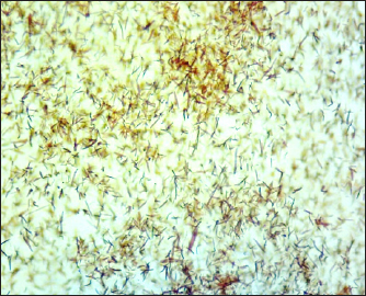
Fig. 2. Crystal hemochromogen appearence on 1:10 cat blood dilution under the microscope with 400× magnification.
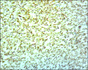
Fig. 3. Crystal hemochromogen appearence on 1:100 cat blood dilution under the microscope with 400× magnification.
Fig. 4. Crystal hemochromogen appearence on 1:1,000 cat blood dilution under the microscope with 400× magnification. Sample C was the only one sample that detected LMG test positive in the blood dilution 1:40,000 with the average RGB value 254.946 ± 0.054. It means that in sample C in dilution 1:40,000 still contained hemoglobin. It could happen because sample C was taking from a male cat, while sample A and B from female cat. Based on Masucci et al. (2022) research result male cat have a concentration of hemoglobin more higher than female cat. Male cat have hemoglobin 10.18 ± 1.52 g/dl, while female cat 9.30 ± 1.46 g/dl. Testosterone, male hormon, can increase kidney ability to produce erythropoietin, a glycoprotein hormone that stimulates the formation of erythrocytes, so the number of hemoglobin in males is higher than in females (Bachman et al., 2014). LMG was originally a colorless reagent when it was first applied to stains (Farrugia et al., 2011). LMG discoloration after added H2O2 becomes bluish-green when detecting blood appears as a result of LMG oxidation which is catalyzed by heme arising from hemoglobin. A blood stain that tested presumptively sometimes requires re-confirmation of the truth that the stain was blood. There are several categories of bloodstain confirmation tests, including microscopic tests, crystal tests, spectroscopic methods, immunological tests, and spectroscopic tests (Sandran et al., 2020). In this study bloodstain that can still be detected by LMG is in bloodstain with a blood concentration of 1:40,000. At 1:80,000 and 1:100,000 dilutions there is no color change in all samples and repetition with the proven value of RGB 255 so that there were no hemoglobin detected. This result was different with de Almeida et al. (2011) in their research of the human bloodstain analysis with LMG reagent in the filter paper, cotton cloth, and blood dilution solution gave a positive reaction at a dilution until 1:5,000 and at a dilution of 1:10,000, there is no positive reaction. This research result is also different from Arzi et al. (2016) that written in the 1:10,000 cat blood dilution there is no positive reaction. Whereas, Boyd et al. (2013) quote that LMG had a sensitivity limit of 1:100,000 dilution of human blood. This research had the same result as Tobe et al. (2007) in LMG reacted at a dilution factor of 1:10,000. The LMG reagent did not show a positive reaction at dilution factors of 1:100,000 and higher. The difference in these results can be affected by differences in the temperature of LMG reagent storage, time of making bloodstain, time of making LMG reagents, how to make bloodstain or making reagents, or because of the different amounts of hemoglobin from various species and individuals. Takayama reagent testHemochromogen crystals appeared from blood, hematin, and other derivatives of hemoglobin utilizing pyridine, piperidine, and a number of other nitrogenous compounds, with ammonium sulfide, hydrazine hydrate, and ammonium sulfide in NaOH as reductants (Ahmed et al., 2020). Takayama test reagent is one of the confirmation tests of blood-based hemochromogen crystal formation by heating dried blood stains and adding pyridine and glucose in an alkaline. Positive results in the confirmation test with Takayama reagent when hemochromogen crystals appear. Crystals are obtained from the reaction of small amounts of blood or fragments of stains with Takayama reagent in the form of shallow salmon-pink rhomboids (Gupta et al., 2021). Gupta et al. (2021) stated that the Takayama reagent is sensitive until the dilution of 1:1,000 human blood. Hossain et al. (2003) results showed the same result of this research. It can happen because between human and cat have an almost similar number of hemoglobin. Based on Bucknoff and Rolph (2024) revealed that cat hemoglobin is between 9.3 and 15.9 g/dl and Gest et al. (2015) revealed that cat hemoglobin is 11.63 ± 2.05 g/dl. While, Normal values for human hemoglobin according to Gligoroska et al. (2019) are 13–18 gm per 100 ml of blood (g/dl) in adult males and 12–16 g/dl in adult females. Benefits of both testsLMG and Takayama reagents are reagents that are often used in human blood spot testing (Charles et al., 2021). The ability of the LMG reagent to detect up to the highest dilution can be relied on for testing the presence of blood spots. However, the LMG reagent has limitations in that false positives can still occur if it is mixed with chemicals such as bleach or body fluids such as semen (Lee et al., 2016). Meanwhile, the Takayama reagent is more specific in detecting blood because the results obtained are a picture of hemochromogen crystals, but its sensitivity is low when compared to LMG (Sari and Prawestiningtyas, 2022). If there is a case of violence against cats and other animals, these two reagents can be relied on to help with the proof process. Those who commit violence against cats and other animals can be prosecuted in accordance with applicable laws, especially in terms of animal welfare. It is hoped that the results of this research can support ensuring animal welfare in society. ConclusionBased on this research, cat bloodstain can be detected with LMG reagent until 1:40,000 dilution, while Takayama reagent only can detect cat bloodstain until 1:1,000. AcknowledgmentsThe authors thank the Universitas Airlangga. Conflict of interestThe authors declare that there is no conflict of interest. FundingThis study was supported by Universitas Airlangga, Indonesia. Author’s contributionsSM and TDL: Conceived, designed, and coordinated the study. DL and TVW: Designed data collections tools, supervised the field sample and data collection, and laboratory work as well as data entry. EPH and RSW: Validation, supervision, and formal analysis. RD and ARK: Contributed reagents, materials, and analysis tools. IBM and RR: Carried out the statistical analysis and interpretation and participated in the preparation of the manuscript. All authors have read, reviewed, and approved the final manuscript. Data availabilityAll data are available in the manuscript. ReferencesAhmed, M.H., Ghatge, M.S. and Safo, M.K. 2020. Hemoglobin: structure, function and allostery. Subcell. Biochem. 94(1), 345–382. Armstrong, E.J. and Erskine, K.L. 2018. Investigation of drowning deaths: a practical review. Acad. Forensic Pathol. 8(1), 8–43. Arzi, B., Mills-Ko, E., Verstraete, F.J., Kol, A., Walker, N.J., Badgley, M.R., Fazel, N., Murphy, W.J., Vapniarsky, N. and Borjesson, D.L. 2016. Therapeutic efficacy of fresh, autologous mesenchymal stem cells for severe refractory gingivostomatitis in cats. Stem Cells Transl. Med. 5(1), 75–86. Bachman, E., Travison, T.G., Basaria, S., Davda, M.N., Guo, W., Li, M., Westfall, J.C., Bae, H., Gordeuk, V. and Bhasin, S. 2014. Testosterone induces erythrocytosis via increased erythropoietin and suppressed hepcidin: evidence for a new erythropoietin/hemoglobin set point. J. Gerontol. Ser. A: Biol. Sci. Med. Sci. 69(6), 725–735. Börsch-Supan, A., Weiss, L.M., Börsch-Supan, M., Potter, A.J., Cofferen, J. and Kerschner, E. 2021. Dried blood spot collection, sample quality, and fieldwork conditions: structural validations for conversion into standard values. Am. J. Hum. 33(4), e23517. Boyd, S., Bertino, M.F., Ye, D., White, L.S. and Seashols, S.J. 2013. Highly sensitive detection of blood by surface enhanced Raman scattering. J. Forensic Sci. 58(3), 753–756. Bucknoff, M.C. and Rolph, K.E. 2024. Splenic torsion in a cat with chronic anemia. J. Feline Med. Surg. Open Rep. 10(1), 20551169231216405. Charles, V.A., Lestari, T.D., Legowo, D., Ismudiono, I., Hidajati, N. and Wahyuni, R.S. 2021. Identification of cat (Felis catus) blood splatter on cotton fabric after periods of drying using Leucomalachite green and Takayama reagent. J. Basic Med. Vet. 10(1), 15–22. de Almeida, J.P., Glesse, N., and Bonorino, C. 2011. Effect of presumptive tests reagents on human blood confirmatory tests and DNA analysis using real time polymerase chain reaction. Forensic Sci. Int. 206(1–3), 58–61. Farrugia, K.J., Savage, K.A., Bandey, H., Ciuksza, T. and Daéid, N.N. 2011. Chemical enhancement of footwear impressions in blood on fabric—part 2: peroxidase reagents. Sci. Justice 51(3), 110–121. Gest, J., Langston, C. and Eatroff, A. 2015. Iron Status of Cats with Chronic Kidney Disease. J. Vet. Intern. Med. 29(6), 1488–1493. Glanville, C., Ford, J. and Coleman, G. 2019. Animal cruelty and neglect: prevalence and community actions in Victoria, Australia. Animals (Basel) 9(12), 1121. Gligoroska, J.P., Gontarev, S., Dejanova, B., Todorovska, L., Stojmanova D.S. and Manchevska, S. 2019. Red blood cell variables in children and adolescents regarding the age and sex. Iran. J. Public Health 48(4), 704–712. Gomes, C., López-Matayoshi, C., Palomo-Díez, S., López-Parra, A.M., Cuesta-Alvaro, P., Baeza-Richer, C., Gibaja, J.F. and Arroyo-Pardo, E. 2017. Presumptive tests: a substitute for benzidine in blood samples recognition. Forensic Sci. Int. Gen. 6(1), e546–e548. Gunawan, T.S., Hanafiah, S.A.M., Kartiwi, M., Ismail, N., Za’bah, N.F. and Nordin, A.N. 2017. Development of photo forensics algorithm by detecting photoshop manipulation using error level analysis. Indones. J. Electr. Eng. Comput. Sci. 7(1), 131–137. Gupta, S., Garg, A., Babu, S. and Yadav, D.S. 2021. Comparative study of presumptive and confirmatory tests for detection of blood on serial dilutions and washed stains. Int. J. Health Res. Medico Leg. Prac, 7(1), 59–64. Halim, A., Indarti, E. and Santoso, B. 2022. The role of forensic science in criminal acts of murder cases in Indonesia. Open Access Maced. J. Med. Sci. 10(A), 951–958. Hossain, M.A., Yamato, O., Yamasaki, M., Otsuka, Y. and Maede, Y. 2003. Relation between reticulocyte count and characteristics of erythrocyte 5’-nucleotidase in dogs, cats, cattle and humans. J. Vet. Med. Sci. 65(2), 193–197. IACUC. 2014. Policy, guideline, and standard operating procedures: blood collection. Atlanta, GA: Emory University. Lee, H., Park, M.J., Sun, S.H., Choi, D.H., Lee, Y.H., Park, K.W. and Chun, B.W. 2016. Ascorbic acid and vitamin C-containing beverages delay the leucomalachite green reaction to detect latent bloodstains. Legal Med. (Tokyo) 23(1), 79–85. Martin, C. and Blatt, M. 2013. Manipulation and misconduct in the handling of image data. Plant Cells 25(9), 3147–3148. Masucci, M., Donato, G., Persichetti, M.F., Priolo, V., Castelli, G., Bruno, F. and Pennisi, M.G. (2022). Hemogram findings in cats from an area endemic for Leishmania infantum and feline immunodeficiency virus infections. Vet. Sci. 9(9), 508. Mota-Rojas, D., Monsalve, S., Lezama-García, K., Mora-Medina, P., Domínguez-Oliva, A., Ramírez-Necoechea, R. and Garcia, R.C.M. 2022. Animal abuse as an indicator of domestic violence: one health, one welfare approach. Animals (Basel) 12(8), 977. Nexus Academic Publisher. 2013. Sample collection guide a practical approach. Lahore, Pakistan: Nexus Academic Publisher. Pędziwiatr, P., Mikołajczyk, F., Zawadzki, D., Mikołajczyk, K. and Bedka, A. 2018. Decomposition of hydrogen perodixekinetics and review of chosen catalysts. Acta Innov. 26(1), 45–52. Pereira, J., Silva, C.S., Vieira, M.J.L., Pimentel, M.F., Braz, A. and Honorato, R.S. 2017. Evaluation and identification of blood stains with handheld NIR spectrometer. Microchem. J. 133(1), 561–566. Sandran, D.D., Zakaria, Y., Md Muslim, N.Z. and Hassan, N.F.N. 2020. Species determination and discrimination of animal blood: a multi-analytical spectroscopic-chemometrics approach in forensic science. Malays. J. Med. Health Sci. 16(4), 162–169. Sari, O. and Prawestiningtyas, E. 2022. Takayama test as a means of identification of blood samples exposed to soil decomposition media. J. Sains Kes. 4(3), 251–255. Sijen, T. and Harbison, S. 2021. On the identification of body fluids and tissues: a crucial link in the investigation and solution of crime. Genes (Basel) 12(11), 1728. Tobe, S.S., Watson, N. and Daéid, N.N. 2007. Evaluation of six presumptive tests for blood, their specificity, sensitivity, and effect on high molecular-weight DNA. J. Forensic Sci. 52(1), 102–109. Veeraraghavan, V. and Lukose, S. 2010. Forensic science laboratory: practice and procedures. In: Gorea, R.K., Dogra, T.D. and Aggarwal, A.D., eds. Practical Aspects of Forensic Medicine. India: Jaypee Brothers Medical Publisher (P) Ltd., pp: 218–222. Wickenheiser, R.A. 2023. Proactive crime scene response optimizes crime investigation. Forensic Sci. Int.: Synergy 6(1), 100325. Zulfiqar, M., Ahmad, M., Sohaib, A., Mazzara, M. and Distefano, S. 2021. Hyperspectral imaging for bloodstain identification. Sensors (Basel) 21(9), 3045. | ||
| How to Cite this Article |
| Pubmed Style Meisari S, Lestari TD, Legowo D, Widiyatno TV, Hestianah EP, Wahjuni RS, Damayanti R, Khairullah AR, Moses IB, Raissa R. Identification of different blood concentration of domestic cat (Felis catus) with Leucomalachite Green and Takayama reagent. Open Vet. J.. 2024; 14(10): 2609-2617. doi:10.5455/OVJ.2024.v14.i10.10 Web Style Meisari S, Lestari TD, Legowo D, Widiyatno TV, Hestianah EP, Wahjuni RS, Damayanti R, Khairullah AR, Moses IB, Raissa R. Identification of different blood concentration of domestic cat (Felis catus) with Leucomalachite Green and Takayama reagent. https://www.openveterinaryjournal.com/?mno=207656 [Access: December 02, 2025]. doi:10.5455/OVJ.2024.v14.i10.10 AMA (American Medical Association) Style Meisari S, Lestari TD, Legowo D, Widiyatno TV, Hestianah EP, Wahjuni RS, Damayanti R, Khairullah AR, Moses IB, Raissa R. Identification of different blood concentration of domestic cat (Felis catus) with Leucomalachite Green and Takayama reagent. Open Vet. J.. 2024; 14(10): 2609-2617. doi:10.5455/OVJ.2024.v14.i10.10 Vancouver/ICMJE Style Meisari S, Lestari TD, Legowo D, Widiyatno TV, Hestianah EP, Wahjuni RS, Damayanti R, Khairullah AR, Moses IB, Raissa R. Identification of different blood concentration of domestic cat (Felis catus) with Leucomalachite Green and Takayama reagent. Open Vet. J.. (2024), [cited December 02, 2025]; 14(10): 2609-2617. doi:10.5455/OVJ.2024.v14.i10.10 Harvard Style Meisari, S., Lestari, . T. D., Legowo, . D., Widiyatno, . T. V., Hestianah, . E. P., Wahjuni, . R. S., Damayanti, . R., Khairullah, . A. R., Moses, . I. B. & Raissa, . R. (2024) Identification of different blood concentration of domestic cat (Felis catus) with Leucomalachite Green and Takayama reagent. Open Vet. J., 14 (10), 2609-2617. doi:10.5455/OVJ.2024.v14.i10.10 Turabian Style Meisari, Suraida, Tita Damayanti Lestari, Djoko Legowo, Thomas Valentinus Widiyatno, Eka Pramyrtha Hestianah, Retno Sri Wahjuni, Ratna Damayanti, Aswin Rafif Khairullah, Ikechukwu Benjamin Moses, and Ricadonna Raissa. 2024. Identification of different blood concentration of domestic cat (Felis catus) with Leucomalachite Green and Takayama reagent. Open Veterinary Journal, 14 (10), 2609-2617. doi:10.5455/OVJ.2024.v14.i10.10 Chicago Style Meisari, Suraida, Tita Damayanti Lestari, Djoko Legowo, Thomas Valentinus Widiyatno, Eka Pramyrtha Hestianah, Retno Sri Wahjuni, Ratna Damayanti, Aswin Rafif Khairullah, Ikechukwu Benjamin Moses, and Ricadonna Raissa. "Identification of different blood concentration of domestic cat (Felis catus) with Leucomalachite Green and Takayama reagent." Open Veterinary Journal 14 (2024), 2609-2617. doi:10.5455/OVJ.2024.v14.i10.10 MLA (The Modern Language Association) Style Meisari, Suraida, Tita Damayanti Lestari, Djoko Legowo, Thomas Valentinus Widiyatno, Eka Pramyrtha Hestianah, Retno Sri Wahjuni, Ratna Damayanti, Aswin Rafif Khairullah, Ikechukwu Benjamin Moses, and Ricadonna Raissa. "Identification of different blood concentration of domestic cat (Felis catus) with Leucomalachite Green and Takayama reagent." Open Veterinary Journal 14.10 (2024), 2609-2617. Print. doi:10.5455/OVJ.2024.v14.i10.10 APA (American Psychological Association) Style Meisari, S., Lestari, . T. D., Legowo, . D., Widiyatno, . T. V., Hestianah, . E. P., Wahjuni, . R. S., Damayanti, . R., Khairullah, . A. R., Moses, . I. B. & Raissa, . R. (2024) Identification of different blood concentration of domestic cat (Felis catus) with Leucomalachite Green and Takayama reagent. Open Veterinary Journal, 14 (10), 2609-2617. doi:10.5455/OVJ.2024.v14.i10.10 |


