| Research Article | ||
Open Vet. J.. 2025; 15(2): 709-713 Open Veterinary Journal, (2025), Vol. 15(2): 709-713 Research Article The effect of black cumin (Nigella sativa L.) on the number of glial cells in white rats (Rattus norvegicus) exposed to cigarette smokeMarita Wahyunengtiyas1, Amirul Amalia2, Widjiati Widjiati3*, Sunaryo Hadi Warsito4, Kadek Rachmawati5, Iwan Sahrial Hamid5, Hani Plumeriastuti6 and Viski Fitri Hendrawan71Students of Magister of Biology Reproduction, Faculty of Veterinary Medicine, University of Airlangga, Surabaya, Indonesia 2Students of Doctoral, Faculty of Medicine, University of Airlangga, Surabaya, Indonesia 3Department of Veterinary Science, Faculty of Veterinary Medicine, University of Airlangga, Surabaya, Indonesia 4Department of Animal Husbandry, Faculty of Veterinary Medicine, University of Airlangga, Surabaya, Indonesia 5Department of Basic Veterinary Science, Faculty of Veterinary Medicine, University of Airlangga, Surabaya, Indonesia 6Department of Veterinary Pathology, Faculty of Veterinary Medicine, University of Brawijaya, Malang, Indonesia 7Department of Veterinary Reproduction, Faculty of Veterinary Medicine, University of Brawijaya, Malang, Indonesia *Corresponding Author: Widjiati Widjiati. Department of Veterinary Science, Faculty of Veterinary Medicine, University of Airlangga, Surabaya, Indonesia. Email: widjiati [at] fkh.unair.ac.id Submitted: 21/10/2024 Accepted: 09/01/2025 Published: 28/02/2025 © 2025 Open Veterinary Journal
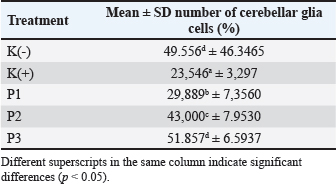
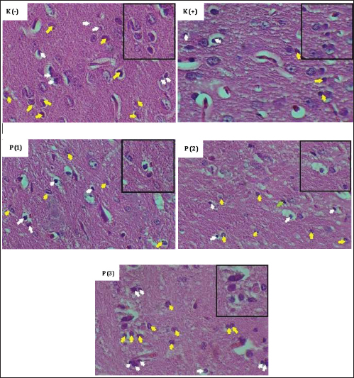
AbstractBackground: Cigarette smoke is known to have harmful effects on the brain, particularly on glial cells, which play a crucial role in maintaining the health and functionality of the central nervous system. Nigella sativa (black cumin) has been reported to have neuroprotective properties, but its effects on glial cells in the cerebellum and cerebrum when exposed to cigarette smoke are not well understood. Aim: This study aimed to determine the benefits of N. sativa on the number of glial cells in the cerebellum and cerebrum of white rats (Rattus norvegicus) exposed to cigarette smoke. Methods: Thirty white rats weighing approximately 105 g were divided into five groups: K (control group, no exposure to cigarette smoke and without given N. sativa), K+ (exposed to cigarette smoke, 2 cigarettes/day, and 0.5% CMC), and three treatment groups (P1, P2, and P3) exposed to cigarette smoke, 2 cigarettes/day, with N. sativa dosages of 300, 600, and 1200 mg/kg BW, respectively. The number of glial cells in the cerebellum and cerebrum was examined under a microscope at 400x magnification. Data were analyzed using a one-way ANOVA test, followed by the post hoc Duncan test. Results: Significant difference (p < 0.05) in the number of glial cells in both the cerebellum and cerebrum between the control and treatment groups. Conclusion: The administration of N. sativa, particularly at a dosage of 1,200 mg/kg BW/day, significantly increased and maintained the number of glial cells in the cerebellum and cerebrum of rats exposed to cigarette smoke. Keywords: Cerebellum, Cerebrum, Cigarette smoke, Good health and well-being, Nigella sativa. IntroductionCigarette smoke contains many harmful chemicals including toxic substances and free radicals that cause oxidative stress and damage nearly all organs. Oxidative stress occurs when the body cannot neutralize the harmful effects of free radicals, leading to issues such as lipid oxidation, DNA damage, and biological membrane disruption (U.S. Department of Health and Human Services, 2016). In particular, cigarette smoke significantly impacts brain health. It disrupts glial cells, such as microglia and astrocytes, which are vital for maintaining brain function. Exposure to cigarette smoke triggers inflammation in these cells, resulting in the release of proinflammatory substances such as interleukin-1 beta (IL-1β), interleukin-6 (IL-6), and tumor necrosis factor-alpha (TNF-α). Chronic inflammation can damage brain tissues and is associated with neurological and neurodegenerative disorders. In addition, cigarette smoke affects signaling pathways in glial cells associated with inflammation, oxidative stress, and cell proliferation. Prolonged exposure can impair the brain’s ability to repair itself, worsening the risk of chronic diseases and long-term health complications (Matt et al., 2019). Exposure to cigarette smoke alters the proliferation and differentiation of glial cells and triggers inflammation in glial cells, particularly microglia. In response to cigarette smoke, glial cells produce proinflammatory cytokines such as IL-1β, IL-6, and TNF-α. Chronic inflammation can damage brain tissues and contribute to neurological and neurodegenerative disorders. Free radical activity can be mitigated by antioxidants, which neutralize free radicals and prevent cell, protein, and fat damage. Various studies have shown that plants, which are rich in compounds such as carotenoids, phenols, flavonoids, anthocyanins, unsaturated fatty acids, vitamins, enzymes, and cofactors, possess significant antioxidant properties. These recommendations encourage the use of plants as preventive and curative phytoremedies (Munteanu and Apetrei, 2021). Black cumin (Nigella sativa L.) is known for its pharmacological effects, which are largely attributed to thymoquinone, its main active compound. Thymoquinone acts as a potent antioxidant that inhibits oxidative stress. While the thymoquinone content varies, black cumin consistently provides antioxidant benefits. Research has shown that N. sativa exhibits promising antioxidant activity, reducing the strength and inhibition of peroxidation (Tubesha et al., 2011). The antioxidant properties of black cumin seeds can help combat free radicals responsible for various diseases (Mahomoodally et al., 2022). A study by Stanković et al. (2017) evaluated the neuroprotective potential of black cumin and thymoquinone in preventing oxidative damage in rat brain cells. The findings revealed that both black cumin and thymoquinone reduce oxidative stress and inflammation while protecting glial cells from damage. Despite these promising results, the effects of black cumin may vary depending on the dosage, conditions, and experimental environment. This research also aligns with the United Nations Sustainable Development Goal (SDG) 3: Good Health and Well-being, addressing tobacco-related health risks. In particular, this study investigated the protective effects of black cumin on cerebral cellular structures in response to exposure to cigarette smoke. As SDG 3 aims to reduce premature mortality, including deaths linked to smoking, a leading risk factor for non-communicable diseases, this study contributes to understanding how natural substances such as black cumin can support neurological health. Based on the aforementioned background, this study sought to further explore the use of black cumin extract (N. sativa L.) as an antioxidant to preserve the number of glial cells in rats exposed to cigarette smoke. Materials and MethodsThis laboratory experiment investigated the effects of black cumin (N. sativa L.) on white rats (Rattus norvegicus) exposed to cigarette smoke from November 2022 to July 2023. The maintenance and treatment of the rats were performed in an experimental animal cage at the Faculty of Veterinary Medicine, Airlangga University. Black cumin extract preparationBlack cumin seeds were washed, dried, and ground using a blender. The ground material was macerated with 96% ethanol for 72 hours, followed by filtration through a Buchner funnel to obtain the filtrate. Maceration was repeated three times, with the filtrates combined and concentrated using a rotary evaporator vacuum (maximum 50°C) until ethanol evaporated, leaving a thick extract. The extract was stored in dark vials and weighed for further use. Experimental procedureA total of 30 white rats were divided into five groups. Rats in the K(-) group were not exposed to cigarette smoke or black cumin. The K(+) group received 0.5% CMC Na solution and was exposed to cigarette smoke twice a day for 30 minutes within the chamber. Rats in the P1, P2, and P3 groups were administered black cumin extract at specific doses and exposed to cigarette smoke. Both 0.5% CMC Na solution and black cumin extract were orally administered using a gastric probe, as described in a previous study (Shakeri et al., 2016). Cigarette smoke exposure was administered for 28 days using a custom-designed smoking chamber. This chamber, connected to a smoking pump, exposed the rats to both mainstream and sidestream smoke. Sidestream smoke from the burning end of the cigarette was introduced into the chamber, while mainstream smoke, drawn through the cigarette, was pumped into the chamber. Brain tissue preparationBrain tissue was prepared following the procedure described. On day 29, the rats were anesthetized using ketamine and xylazine, euthanized, and then necropsied. Brain samples (1 × 1 × 1 cm) were fixed in 10% neutral-buffered formalin and processed through dehydration and clearing with graded alcohol solutions (70%, 80%, 90%, 96%, absolute), toluene, and paraffin. The samples were embedded in paraffin, sectioned with a microtome at 4–5 microns thickness, stained with hematoxylin-eosin (HE), and mounted for microscopic examination. Histological changes were recorded in five microscopic fields of view at 400× magnification. Data analysisThe data consisting of the number of glial cells in the cerebellum and cerebrum of the white rats were analyzed using SPSS for Windows. A one-way ANOVA parametric test was performed to determine the statistical significance between groups (p < 0.05). If significant differences were found, a post hoc Duncan test was conducted to assess pairwise differences between the control and treatment groups. Ethical approvalEthical clearance for this research was obtained from the Animal Care and Use Committee, Faculty of Medicine, Airlangga University: No.134/EC/KEPK/FKUA/2022. ResultsThe mean and standard deviation of the number of glial cells in the cerebellum in both the control and black cumin treatment groups are presented in Table 1. Microscopic images of the cerebellum and cerebrum can be seen in Figures 1 and 2. Table 1. Mean and SD of glial cell count in the cerebellum of control and black cumin treatment groups.
Fig. 1. Preparation of counting the number of glial cells in the cerebellum cortex; HE staining, 400× and 1,000× magnification, white arrows (oligodendrocytes); yellow arrows (astrocytes).
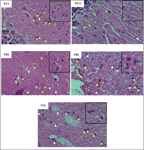
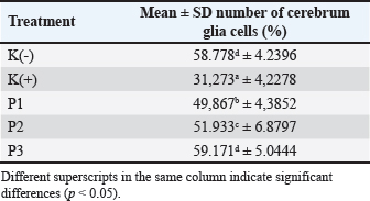
Fig. 2. Preparation of counting the number of glial cells in the cerebrum cortex; HE staining, 400x and 1,000× magnification, white arrows (oligodendrocytes); yellow arrows (astrocytes). The observation of the number of glial cells in the cerebrum revealed that the positive control group (K+) had significantly fewer glial cells compared with the negative control group (K-) and treatment groups 1 (P1), 2 (P2), and 3 (P3). However, no significant difference was observed between the negative control group (K-) and treatment group 3 (P3). The average glial cell counts in the cerebrum were 58.778% in the negative control group (K-), 31.273% in the positive control group (K+), 49.876% in treatment group 1 (P1), 51.933% in treatment group 2 (P2), and 59.171% in treatment group 3 (P3). According to Duncan’s test, the number of glial cells in the cerebellum showed that the positive control group (K+) had significantly fewer glial cells compared to the negative control group (K-) and all treatment groups (P1, P2, and P3). However, no significant difference was observed between the negative control group (K-) and Treatment 3 (P3). The mean percentages of glial cells in the cerebellum were 49.556% for the negative control group (K-), 23.546% for the positive control group (K+), 29.889% for Treatment 1 (P1), 43.000% for Treatment 2 (P2), and 51.857% for Treatment 3 (P3). The mean and standard deviation of the number of glial cells in the cerebrum in both the control and black cumin treatment groups are presented in Table 2. DiscussionThe K(+) group, which was exposed to cigarette smoke without receiving black cumin, exhibited a reduction in the number of cerebellar glial cells. Cigarette smoke contains harmful particles that can activate microglia and cause cell death (Nuha et al., 2023). As an oxidant, cigarette smoke induces oxidative stress by causing incomplete electron transfer during oxidative phosphorylation in mitochondria, resulting in the formation of highly reactive oxygen species (ROS). This leads to oxidative stress, with dangerous compounds such as tar and nicotine being the primary contributors. Table 2. Mean and standard deviation of glial cell count in the cerebrum for control and black cumin treatment groups.
The administration of black cumin at a dose of 1,200 mg/kg BW/day significantly increased the number of cerebellar glial cells compared with the K(+) group. The mean glial cell count in this treatment group was 51.86%. This increase can be attributed to the active biochemical components of black cumin, primarily thymoquinone and dithymoquinone (Hannan et al., 2021). These compounds contribute to the repair of nerve cell damage by enhancing the activity of enzymes such as acetylcholinesterase, butyrylcholinesterase, ectonucleotidase (5’-nucleotidase), lactate dehydrogenase, and monoamine oxidase (Akintunde and Irechukwu, 2016). Additionally, both in vitro and in vivo studies have shown that black cumin, with its antiviral, antioxidant, anti-inflammatory, antihistamine, antihypertensive, antidiabetic, and anti-obesity properties can be used as an adjuvant therapy in the management of infectious diseases (Maideen, 2020). Cigarette smoke is a significant source of ROS, such as superoxide anions, hydroxyl radicals, hydrogen peroxide, metal ions, and quinones. These oxidative agents elevate oxidative stress in smokers, resulting in increased lipid peroxidation and degradation of extracellular matrix proteins (Hossain, 2021). Cigarette smoke enters the bloodstream via the alveoli in the lungs and is distributed to other organs, including the brain. It is recognized by innate immune receptors that trigger an inflammatory response via the activation of NF-kB. Additionally, cigarette smoke can cause inflammation originating in the respiratory system by disrupting the epithelial barrier, thereby affecting both innate and adaptive defense mechanisms. A further investigation would strengthen the conclusions, such as longitudinal studies to evaluate the long-term neuroprotective effects of N. sativa or testing its efficacy in combination with other antioxidants. ConclusionBased on the results of this study, it can be concluded that giving black cumin to white mice exposed to cigarette smoke can maintain the number of glial cells in their cerebellum and cerebrum at a dose of 1,200 mg/kg BB/day. Conflict of interestsThe authors declare no conflict of interest. FundingInternal faculty funding. Authors’ contributionWidjiati Widjiati: Conceptualization, Methodology, Validation, Formal analysis, Investigation, Resources, Writing original draft, Visualization, Supervision, Project administration, and Funding acquisition. Marita Wahyunengtiyas and Amirul Amalia: Methodology, Validation, Investigation, and Resources. Sunaryo Hadi Warsito and Kadek Rachmawati: Methodology, Validation, Investigation, and Resources. Iwan Sahrial Hamid and Hani Plumeriastuti: Writing original draft, Visualization, and Writing review and editing. Data availabilityAll data supporting the findings of this study are available in the manuscript. ReferencesAkintunde, J.K. and Irechukwu, C.A. 2016 Differential protection of black-seed oil on econucleotidase, cholinesterases, and aminergic catabolizing enzyme in haloperidol-induced neuronal damage of male rats. Ther. Adv. Drug Saf. 7(4), 132–46. Hannan, M.A., Zahan, M.S., Sarker, P.P., Moni, A., Ha, H. and Uddin, M.J. 2021. Protective effects of black cumin (Nigella sativa) and its bioactive constituent, thymoquinone against kidney injury: an aspect on pharmacological insights. Int. J. Mol. Sci. 22(16), 9078. Hossain, M. 2021. The effect of Covid-19 on sharing economy activities. J. Clean Prod. 280, 124782. Mahomoodally, M.F., Aumeeruddy, M.Z., Legoabe, L.J., Montesano, D. and Zengin, G. 2022. Nigella sativa L. and its active compound thymoquinone in the clinical management of diabetes: a systematic review. Int. J. Mol. Sci. 23(20), 12111. Maideen, N.M.P. 2020. Prophetic medicine-Nigella sativa (black cumin seeds) - potential herb for COVID-19? J. Pharmacopuncture. 23(2), 62–70. Matt, G.E., Mahabee-Gittens, E.M., Zakarian, J.M., Quintana, P.J.E, Hoh, E. and Myers, M. 2019. Nicotine in thirdhand smoke residue predicts relapse from smoking cessation: a pilot study. Addict. Behav. 98, 106041. Munteanu, I.G. and Apetrei, C. 2021. Analytical methods used in determining antioxidant activity: a review. Int. J. Mol. Sci. 22(7), 3380. Nuha, K., Rusmil, K., Ganiem, A.R., Permadi, W. and Diah Herawati, D.M. 2023. Single-blind randomized controlled trial: comparative efficacy of dark chocolate, coconut water, and ibuprofen in managing primary dysmenorrhea. Int. J. Environ. Res. Public Health. 20(16), 6619. Shakeri, F., Gholamnezhad, Z., Mégarbane, B., Rezaee, R. and Boskabady, M.H. 2016. Gastrointestinal effects of Nigella sativa and its main constituent, thymoquinone: a review. Avicenna. J. Phytomed. 6(1), 9–20. Stanković, M.S., Niciforović, N., Mihailović, V., Topuzović, M., Solujić, S. and Mitić, V. 2017. Protective effects of Nigella sativa L. and thymoquinone against hippocampal neurodegeneration in cadmium-intoxicated rats. Bosn. J. Basic Med. Sci. 17(1), 56–63. Tubesha, Z., Iqbal, S. and Ismail, M. 2011. Effects of hydrolysis conditions on recovery of antioxidants from methanolic extracts of Nigella sativa seeds. J. Med. Plant Res. 5(22), 5393–5399. U.S. Department of Health and Human Services. 2016. E-cigarette use among youth and young adults. A report of the surgeon general. Atlanta, GA: U.S. Department of Health and Human Services, Centers For Disease. | ||
| How to Cite this Article |
| Pubmed Style Wahyunengtiyas M, Amalia A, Widjiati W, Warsito SH, Rachmawati K, Hamid IS, Plumeriastuti H, Hendrawan VF. The effect of black cumin (Nigella sativa L.) on the number of glial cells in white rats (Rattus norvegicus) exposed to cigarette smoke. Open Vet. J.. 2025; 15(2): 709-713. doi:10.5455/OVJ.2025.v15.i2.19 Web Style Wahyunengtiyas M, Amalia A, Widjiati W, Warsito SH, Rachmawati K, Hamid IS, Plumeriastuti H, Hendrawan VF. The effect of black cumin (Nigella sativa L.) on the number of glial cells in white rats (Rattus norvegicus) exposed to cigarette smoke. https://www.openveterinaryjournal.com/?mno=225558 [Access: September 03, 2025]. doi:10.5455/OVJ.2025.v15.i2.19 AMA (American Medical Association) Style Wahyunengtiyas M, Amalia A, Widjiati W, Warsito SH, Rachmawati K, Hamid IS, Plumeriastuti H, Hendrawan VF. The effect of black cumin (Nigella sativa L.) on the number of glial cells in white rats (Rattus norvegicus) exposed to cigarette smoke. Open Vet. J.. 2025; 15(2): 709-713. doi:10.5455/OVJ.2025.v15.i2.19 Vancouver/ICMJE Style Wahyunengtiyas M, Amalia A, Widjiati W, Warsito SH, Rachmawati K, Hamid IS, Plumeriastuti H, Hendrawan VF. The effect of black cumin (Nigella sativa L.) on the number of glial cells in white rats (Rattus norvegicus) exposed to cigarette smoke. Open Vet. J.. (2025), [cited September 03, 2025]; 15(2): 709-713. doi:10.5455/OVJ.2025.v15.i2.19 Harvard Style Wahyunengtiyas, M., Amalia, . A., Widjiati, . W., Warsito, . S. H., Rachmawati, . K., Hamid, . I. S., Plumeriastuti, . H. & Hendrawan, . V. F. (2025) The effect of black cumin (Nigella sativa L.) on the number of glial cells in white rats (Rattus norvegicus) exposed to cigarette smoke. Open Vet. J., 15 (2), 709-713. doi:10.5455/OVJ.2025.v15.i2.19 Turabian Style Wahyunengtiyas, Marita, Amirul Amalia, Widjiati Widjiati, Sunaryo Hadi Warsito, Kadek Rachmawati, Iwan Sahrial Hamid, Hani Plumeriastuti, and Viski Fitri Hendrawan. 2025. The effect of black cumin (Nigella sativa L.) on the number of glial cells in white rats (Rattus norvegicus) exposed to cigarette smoke. Open Veterinary Journal, 15 (2), 709-713. doi:10.5455/OVJ.2025.v15.i2.19 Chicago Style Wahyunengtiyas, Marita, Amirul Amalia, Widjiati Widjiati, Sunaryo Hadi Warsito, Kadek Rachmawati, Iwan Sahrial Hamid, Hani Plumeriastuti, and Viski Fitri Hendrawan. "The effect of black cumin (Nigella sativa L.) on the number of glial cells in white rats (Rattus norvegicus) exposed to cigarette smoke." Open Veterinary Journal 15 (2025), 709-713. doi:10.5455/OVJ.2025.v15.i2.19 MLA (The Modern Language Association) Style Wahyunengtiyas, Marita, Amirul Amalia, Widjiati Widjiati, Sunaryo Hadi Warsito, Kadek Rachmawati, Iwan Sahrial Hamid, Hani Plumeriastuti, and Viski Fitri Hendrawan. "The effect of black cumin (Nigella sativa L.) on the number of glial cells in white rats (Rattus norvegicus) exposed to cigarette smoke." Open Veterinary Journal 15.2 (2025), 709-713. Print. doi:10.5455/OVJ.2025.v15.i2.19 APA (American Psychological Association) Style Wahyunengtiyas, M., Amalia, . A., Widjiati, . W., Warsito, . S. H., Rachmawati, . K., Hamid, . I. S., Plumeriastuti, . H. & Hendrawan, . V. F. (2025) The effect of black cumin (Nigella sativa L.) on the number of glial cells in white rats (Rattus norvegicus) exposed to cigarette smoke. Open Veterinary Journal, 15 (2), 709-713. doi:10.5455/OVJ.2025.v15.i2.19 |


