





| Research Article | ||
Open Vet. J.. 2026; 16(7): 4274-4281 Open Veterinary Journal, (2026), Vol. 16(7): 4274-4281 Research Article TGF-β from mesenchymal stem cell secretome improves sepsis score in murine modelMutiara Indah Sari1*, Nelva Karmila Jusuf2, Delfitri Munir3, Agung Putra4, Tatang Bisri5, Syafruddin Ilyas6, Farhat Farhat3 and Adi Muradi Muhar71Department of Biochemistry, Faculty of Medicine, Universitas Sumatera Utara, Medan, Indonesia 2Department of Dermatology & Venereology, Faculty of Medicine, Universitas Sumatera Utara, Medan, Indonesia 3Department of Ear, Nose & Throat, Head & Neck, Faculty of Medicine, Universitas Sumatera Utara, Medan, Indonesia 4Stem Cell and Cancer Research, Faculty of Medicine, Universitas Islam Sultan Agung, Semarang, Indonesia 5Department of Anesthesiology and Intensive Care, Faculty of Medicine, Universitas Jendral Achmad Yani, Bandung, Indonesia 6Department of Biology, Faculty of Mathematics and Natural Sciences, Universitas Sumatera Utara, Medan, Indonesia 7Department of Surgery, Faculty of Medicine, Universitas Sumatera Utara, Medan, Indonesia *Corresponding Author: Mutiara Indah Sari. Department of Biochemistry, Faculty of Medicine, Universitas Sumatera Utara, Medan, Indonesia. Email: mutiara [at] usu.ac.id Submitted: 11/09/2025 Revised: 04/04/2026 Accepted: 27/04/2026 Published: 02/07/2026 © 2026 Open Veterinary Journal
AbstractBackground: Sepsis remains the main cause of morbidity and mortality worldwide. Early administration of appropriate antimicrobials is one of the most effective interventions for sepsis; however, antibiotic resistance in bacteria is growing continuously. Therefore, new complementary therapies are needed to treat sepsis. One of the recently studied complementary therapies is the administration of the mesenchymal stem cell (MSC) secretome. Several studies have shown that the secretome exerts immunomodulatory and anti-inflammatory effects. However, the studies did not assess the clinical changes occurring in sepsis-induced rats. Aim: This study aimed to analyze the effect of TGF-β from the MSC secretome on sepsis scores in a murine model. Methods: Forty-eight rats were divided into 4 groups: healthy rats (rats without sepsis induction and therapy), control (300 µl saline + antibiotic), T1 (150 µl TGF-β from MSC secretome + antibiotic), and T2 (300 µl TGF-β from MSC secretome + antibiotic). The murine sepsis score (MSS) was observed every 6 hours for 48 hours. Data analysis was conducted using SPSS to examine the relationship between the effect of TGF-β from MSC secretome administration and the total mean of MSS. The value of p < 0.05 was considered significant. Results: Administering 150 µl of TGF-β from MSC secretome reduced the total mean MSS compared with the control group at the 42nd hour of observation (p=0.017). Conclusion: Our research found that TGF-β from the MSC secretome improved the sepsis score in sepsis-induced rats. Keywords: Mesenchymal stem cell, Murine sepsis score, TGF-β, Umbilical cord. IntroductionSepsis is now defined as life-threatening organ dysfunction caused by a dysregulated host response to infection (Sepsis-3) (Singer et al., 2016). Sepsis is still a major cause of morbidity and mortality worldwide. A recent assessment by the Global Burden of Sepsis Collaborators reported that, in 2021, sepsis affected an estimated 166 million people worldwide in 2021 (95% UI 135–201 million). The analysis also identified approximately 21.4 10,00,000 sepsis-related deaths (20.3–22.5 10,00,000), which correspond to approximately 31.5% of all global deaths (Global Sepsis Collaborators, 2021). Early administration of appropriate antimicrobials is one of the most effective interventions for sepsis. However, antibiotic resistance in bacteria is growing and becoming a hurdle in sepsis management (Evans et al., 2021; Kumar et al., 2024). Current developments in medicine offer new, complementary therapies specifically designed for sepsis. One of the recently studied complementary therapies is the administration of mesenchymal stem cell (MSC) secretome (Harrel et al., 2019; Driscoll et al., 2019). MSCs are multipotent stem cells that originate from the mesoderm found in the bone marrow. Currently, MSCs can be isolated from various human tissues, including adipose tissue, umbilical cord, and amniotic fluid (Kobolak et al., 2016; Shang et al., 2021). Although all of these sources met the core criteria for MSCs, they did not exhibit identical biological characteristics. Umbilical-cord-derived MSCs are often considered the most advantageous because they are more primitive, show stronger proliferative and immunomodulatory capacities, and carry a lower risk of donor-related variability (Mebarki et al., 2021). In contrast, adipose-derived MSCs generally produce abundant paracrine factors, whereas amniotic fluid-derived MSCs exhibit more restricted differentiation potential and are further limited by their low initial cell yield and declining proliferative capacity with extended passaging (Conese et al., 2020; Kulus et al., 2021). MSCs secrete soluble proteins, such as interleukin (IL)-6, IL-8, transforming growth factor (TGF)-β, indoleamine 2,3-dioxygenase (IDO), and vascular endothelial growth factor (VEGF), and increase the production of IL-4 and IL-10 from T-helper 2 (Th2). MSCs also express cyclooxygenase-2 (COX-2/PTGS2), an intracellular enzyme that drives prostaglandin E₂ (PGE₂) synthesis, although COX-2 is not secreted (Abolhasani et al., 2018; Ankrum et al. 2014; Shang et al., 2021; Trigo et al., 2025). Collectively referred to as the secretome, these factors suppress pro-inflammatory cytokines (TNF-α, IL-1β, IL-6) and enhance regulatory mediators that restore immune balance (Trigo et al., 2025). Their mechanism parallels toxin-induced inflammatory cascades, such as paraquat-related renal injury, where excessive ROS activates NF-κB, elevates COX-2 activity, and drives downstream inflammatory and apoptotic responses (Ijaz et al., 2023). The secretome contributes to broader immunoregulatory protection by modulating these same cytokine networks and oxidative stress signals. Secretome derivatives have several advantages. Safety, dosage, and potency can be evaluated in a manner similar to that of conventional pharmaceutical materials. Secretome-based products offer practical advantages over live-cell therapy, particularly in terms of scalability and the potential for standardized manufacturing. However, to maintain bioactivity, long-term storage still requires optimized formulation strategies, including lyophilization or stabilizers (Vizoso et al., 2017; Trigo et al., 2025). Several studies have proven the anti-inflammatory effects of cytokines contained in secretomes. A previous study reported that administering adipose-derived stem/stromal cells’ conditioned medium (ASC-CM) secretome significantly reduced MMP activity and PGE2 expression (Giannasi et al., 2020). Another study found that the administration of MSC TGF-β1 derived from bone marrow reduced the levels of pro-inflammatory cytokines and inhibited macrophage infiltration into tissues (Liu et al., 2020). However, that study did not clinically assess the changes in rats with sepsis. Therefore, this study aimed to analyze the effect of TGF-β from the MSC secretome on the sepsis score in a murine model. The MSS is used to assess the severity or clinical degree of sepsis in rats and mice based on observational characteristics. This scoring system is similar to clinical EWS as a tool for assessing the development of sepsis using a mouse model induced by intraperitoneal injection of fecal slurry (Shrum et al., 2014; Mai et al., 2018; Sulzbacher et al., 2022). Materials and MethodsStudy designMSC secretome creation standards, maintenance, and treatment of test animals, and examination of dependent variables were performed at the Stem Cell and Cancer Research (SCCR) Laboratory and the Animal House facility of its Integrated Biomedical Laboratory in Universitas Islam Sultan Agung (Unissula) Faculty of Medicine, Semarang, Indonesia. MSCs preparationUmbilical cord tissues were collected from 19-day pregnant rats for MSC isolation. The sterilized umbilical cord was placed in the biological safety cabinet (BSC) and washed with phosphate-buffered saline (PBS) until clean. A 1 mm piece of umbilical cord was placed in a T25 flask. Then, it was cultured in a complete medium containing low-glucose Dulbecco's Modified Eagle Medium (DMEM), 10% Fetal Bovine Serum (FBS), 1% penicillin/streptomycin, and 0.5% fungizone. The incubation process was carried out at 37°C and 5% CO2, and the culture was observed with an inverted microscope every 24 hours. The medium was replaced every 2 to 3 days (Putra, 2019). Cells successfully cultured from the umbilical cord underwent validation and characterization tests. Umbilical cord-derived mesenchymal stem cells (UC-MSC) were validated using flow cytometry with CD90-APC, CD29-PE, CD31-PerCP, and CD45-FITC markers. Cells were released at a concentration of 1 × 10⁷ cells/ml from the flask using BDTM AccutaseTM cell detachment solution (cat. no. 561527). Cells were then washed with PBS for further reading using flow cytometry (Putra, 2019). To create a hypoxic MSC secretome, MSCs that had reached 80% confluence were added to complete medium up to 10 ml and then placed in a hypoxic chamber and incubated for 24 hours at 37°C and 5% O2. After 24 hours, the culture medium was taken and filtered using Tangential Flow Filtration (TFF) to obtain TGF-β, which was then prepared according to the administration dose. Calculations were performed using a commercial enzyme-linked immunosorbent (ELISA) assay kit following the manufacturer’s instructions (Putra, 2019). Preparation of animal modelsForty-eight male Rattus norvegicus rats with an average body weight of 200 to 300 grams (aged 10–12 weeks) were obtained from the Animal House facility of the Integrated Biomedical Laboratory in Unissula Faculty of Medicine, Semarang, Indonesia. Each rat was maintained under controlled environmental conditions throughout the study to ensure physiological stability. Temperature was maintained between 20°C and 24°C, humidity was maintained within 55% to 60%, and animals were exposed to a consistent 12 hours light-dark cycle (08:00–20:00). Each rat was housed individually in a pathogen-free room with unrestricted access to standard laboratory feed and clean drinking water. This controlled housing approach minimized environmental stressors and ensured uniform baseline conditions for all experimental procedures (Yelena et al., 2021; Nguyen et al., 2024). Rats that were sick or infected during the adaptation period and those that died before sepsis induction were excluded from the study. The research samples were divided into 4 groups, with 12 rats per group: - Healthy rats: rats without sepsis induction and therapy. - Control: rats with sepsis induction and 25 mg/kg BW imipenem antibiotics in 150 µl physiologic saline administered intraperitoneally. - T1: rats with sepsis induction + 25 mg/kg BW imipenem antibiotics in 150 µl physiologic saline administered intraperitoneally + 150 µl (low dose) of TGF-β from MSC secretome injected intravenously into the rat’s tail. - T2: rats with sepsis induction + 25 mg/kgBW imipenem antibiotics in 150 µl physiologic saline administered intraperitoneally + 300 µl (high dose) of TGF-β from MSC secretome injected intravenously into the rat’s tail. Rats were adapted for 7 days. On the eighth day, sepsis induction was performed on the control, T1, and T2 groups. Fecal slurry at a dose of 1 g/kg BW was used to induce sepsis. The stool was suspended in a saline solution with a concentration of 90 mg/ml, and then as much as 198 to 297 mg was injected intraperitoneally in 2.2 to 3.3 ml of sterile saline. Injection was performed in the lower right quadrant of the abdomen using a 21-G cannula, and the abdominal wall was gently lifted with forceps to avoid trauma from needle insertion (Fang et al., 2020; Yao et al., 2021). TGF-β administrationAdministration of TGF-β from MSC secretome in this study used two doses: 1 × empirical dose and 2 × empirical dose, which consisted of 150 µl (T1) and 300 µl (T2) via venous injection in the rat’s tail 4 hours after sepsis induction (Xia et al., 2018; Yao et al., 2021). Intraperitoneal administration of imipenem antibiotics at a dose of 25 mg/kg body weight (BW) in 150 µl of physiological saline was performed (Seymour et al., 2019). MSS assesses 7 components consisting of appearance, awareness, activity, stimulus response, eyes, respiratory rate, and respiratory quality. Each component is assigned a score between 0 and 4. The score is calculated by assessing the average of the 7 components for each treatment group, as described by Shrum et al. (2014). MSS was observed every 6 hours for 48 hours. Data analysisData were analyzed using a statistical application. The effect of TGF-β from the MSC secretome on MSS was tested using the Kruskal-Wallis test and then analyzed using the Mann-Whitney test. A p value < 0.05 was considered significant. Ethical approvalAll experimental procedures were approved by the Health Research Ethics Committee of Universitas Sumatera Utara (USU; approval number 541/2022). ResultsMSC validation and secretome contentIn the validation test by flow cytometry, UC-MSCs expressed CD90.1 and CD29 (99.4% and 96.9%, respectively) and a little CD45 and CD31 (1.3% and 6.6%), as shown in Fig. 1. UC-MSC culture under 5% O2 hypoxia for 24 hours produced a secretome containing 138.15 ± 6.12 pg/ml of TGF-β.
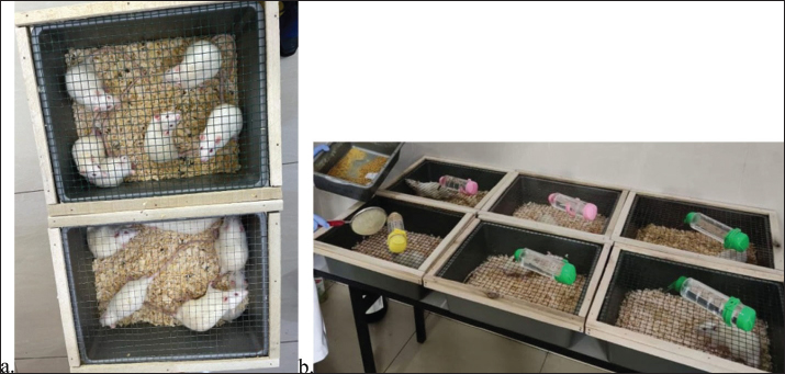
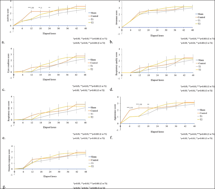
Fig. 1. Experimental rats before (a) and after (b) sepsis induction. Panel (b) shows visible clinical changes following induction, including a more hunched posture, unkempt fur, and a clear lethargic appearance compared with the baseline condition in panel (a). Sepsis inductionThe rats developed rough, unkempt fur after sepsis induction. Activity parameters revealed that the rats were passive, lethargic, and slept (Fig. 1). MSSThe mean MSS of activity, awareness, eye condition, respiratory quality, respiratory rate, appearance, and stimulus response in the sepsis-induced rat groups are shown in Figure 2. At the 48th hour of observation, the sepsis-induced rat group given 150 µl secretome had the lowest mean MSS for all categories compared with the control group and the sepsis-induced rat group given 300 µl secretome.
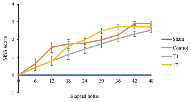
Fig. 2. Graphs depicting the mean MSS of activity (a), awareness (b), eye condition (c), respiratory quality (d), respiratory rate (e), appearance (f), and response to stimulus (g) in sepsis-induced rats. Sham (no treatment); Control (300 µl saline + antibiotics); T1 (150 µl secretome + antibiotics); T2 (300 µl secretome + antibiotics). The total mean of MSS in the sepsis-induced rat groups is shown in Figure 3, with the sepsis-induced rat group receiving 150 µl of secretome having the lowest total mean of MSS compared with the control group and the sepsis-induced rat group receiving 300 µl of secretome. Kruskal-Wallis analysis was performed to determine the relationship between the secretome administration effect and the total mean MSS. The results showed that there was a significant difference in the total mean of MSS in the sepsis-induced rat groups given secretome, with p < 0.05 at the 6th (p=0.015), 12th (p=0.001), 18th (p=0.001), 24th (p=0.001), 30th (p=0.000), 36th (p=0.000), 42nd (p=0.000), and 48th (p=0.000) hours of observation.
Fig. 3. Graph depicting the total mean MSS in the sepsis-induced rat groups. Sham (no treatment); Control (300 µl saline + antibiotics); T1 (150 µl secretome + antibiotics); T2 (300 µl secretome + antibiotics). To see the difference between the groups, a Mann-Whitney analysis was performed. The results showed a significant difference between the T1 and control groups at the 42nd hour of observation (p=0.017). Nevertheless, the distinction between T1 and T2 remained minimal throughout the observation period, suggesting that the apparent advantage of the 150 µl dose reflects a subtle trend rather than a substantial or strongly dose-dependent effect. DiscussionMSCs are multipotent adult stem cells capable of proliferation, self-regeneration, and differentiation into several cell lineages (Montero-Vilchez et al., 2021; Rocca et al., 2025). MSCs can be isolated from various sources, including bone marrow (BM-MSC), adipose tissue (AT-MSC), umbilical cord (UC-MSC), amnion, placenta, or dental pulp (Bogatcheva and Coleman, 2019; Montero-Vilchez et al., 2021). MSC can also improve ischemic injury by increasing vascularity and reducing inflammation in the hind limbs, lungs, heart, and brain (Liu et al., 2015; Sun et al., 2015; Han et al., 2022; Guan et al., 2025). In this study, we investigated the effect of administering TGF-β from the MSC secretome on the mean MSS of sepsis-induced rats. Because the MSS is based on simple observations, it can be easily implemented in any laboratory using an animal sepsis model. Additionally, this method is well-suited for any investigative timeline because of its noninvasive nature. MSS can accurately predict sepsis development and mortality. An MSS value above 3 in any variable was used as the cut-off point for sepsis development in mice with fecal-induced peritonitis (FIP), with a sensitivity of 57% (95% CI: 47%–67%) and specificity of 100% (95% CI: 82%–100%) (Yao et al., 2021). Mai et al. (2018) showed that compared with other scoring methods, namely the Mouse Clinical Assessment Score for Sepsis (M-CASS) and the modified Mouse Grimace Scale (MGS), the MSS is the most effective scoring system for differentiating moderate and severe sepsis in cecal ligation puncture (CLP) models and predicting mortality in sepsis. MGS is effective for assessing the degree of disease severity but is less effective in predicting mortality. The modified M-CASS is effective in predicting mortality but more variable than other scores in differentiating sepsis severity (Mai et al., 2018; Yao et al., 2021). In this study, administering 150 µl (T1) of TGF-β from UC-MSC secretome resulted in a significant reduction in the mean total MSS compared with that in the other groups of rats at the 42nd hour of observation. Within the first 12 hours of observation, no difference was observed in the total mean MSS of the rat group that received 150 µl (T1) and 300 µl (T2) of secretome. However, T2 shows an increasing trend in the total mean of the MSS graph with each passing time. Throughout the observation, the total mean MSS of T1 was lower than that of the control group. Although the difference in the total mean MSS between T1 and T2 was minimal, T1 consistently showed a lower trend. TGF-β is a critical anti-inflammatory cytokine for resolving inflammation during tissue injury. In particular, TGF-β inhibits the production of TNF and IL-1β from monocytes (Ohta et al., 2018; Senousy et al., 2022). TGF-β has 3 isoforms: TGF-β1, -β2, and -β3. Among these 3 isoforms, TGF-β1 is the most widely expressed isoform in the immune system and is considered one of the key immunoregulatory cytokines. Several mechanisms underlying the regulation of TGF-β include: (i) suppressing effector Th cell differentiation, (ii) promoting naive T cell differentiation into regulatory T cells, (iii) inhibiting T and B cell proliferation, (iv) inhibiting effector cytokine production, and (v) suppressing macrophages, dendritic cells, and natural killer cells (Yoshimura et al., 2010; Salmond, 2023). These broad immunosuppressive activities create a systemic environment that strongly favors inflammation resolution and tissue recovery. TGF-β signaling is initiated when activated TGF-β binds to transforming growth factor-β receptor-2 (TβRII) with high affinity. This binding requires the participation of transforming growth factor-β receptor-3 (TβRIII) or β-glycan, which causes a conformational change in TβRII that facilitates receptor-ligand binding. These ligand-receptors translocate to the nucleus to interact with various transcription factors, leading to cellular responses (Haque and Morris, 2017; Tzavlaki and Moustakas, 2020). This canonical pathway activates downstream signaling that regulates numerous genes involved in immune suppression, extracellular matrix repair, and macrophage polarization, making TGF-β a central orchestrator of the injury response. Through this transcription factor, TGF-β can regulate NF-κB and support the differentiation and polarization of M2 macrophages and the anti-inflammatory and immunosuppressive subtype of macrophages (Zhang et al., 2016; Aglan et al., 2024; Abd-Rabou et al., 2025). In vitro studies have demonstrated that TGF-β suppresses the release of pro-inflammatory mediators, including high mobility group box 1 protein (HMGB1), IL-1, and TNF-α, from monocytes and macrophages. Recent data indicate that TGF-β reverses the depression of cardiac myocyte contraction induced by pro-inflammatory cytokines, such as TNF-α and IL-1, in the serum of patients with septic shock. This suggests that TGF-β may have a cardioprotective effect on cardiac injury due to sepsis (Schulte et al., 2013; Li et al., 2023). ConclusionOur research found that TGF-β from the MSC secretome improves the sepsis score of sepsis-induced rats. Further studies are needed to confirm the effects of TGF-β from MSC secretome on sepsis biomarkers and the survival of patients with sepsis. AcknowledgmentsThe authors would like to express their gratitude to the Chancellor of USU, the Dean of the USU Faculty of Medicine, and the SCCR of the Unissula Faculty of Medicine. FundingThis study received no external funding. Authors’ ContributionsMutiara Indah Sari, Nelva Karmila Jusuf, Delfitri Munir, and Agung Putra designed and conducted the experiment, collected and analyzed the data, and drafted the manuscript. Critical revision and final approval were carried out by Tatang Bisri, Syafruddin Ilyas, Farhat Farhat, and Adi Muradi Muhar. All authors have read and approved the final version of the manuscript. Conflict of InterestThe authors declare no conflicts of interest. Data AvailabilityThe data are available upon reasonable request from the corresponding author. ReferencesAbd-Rabou, A.A., Kamal, M., Alharbi, H.Y., Aljohani, M.S., El-Atawy, M.A. and Kishta, M.S. 2025. Modulation of PI3K/AKT signaling and DFT modeling via selected pharmaceutical compounds attenuates carrageenan-induced inflammation and oxidative stress in rats. Naunyn. Schmiedebergs. Archiv. Für. Pharmazie. 398(6), 7583–7603; doi:10.1016/j.nspharma.2025.15.016 Abolhasani, M., Rezaee, M.A., Mohammadi, M., Ghadimi, T., Mohammadi, M. and Rahmani, M.R. 2018. Influence of gestational age and in vitro expansion on the immunomodulatory properties of umbilical cord vein mesenchymal stromal cells. Immunol. Lett. 194, 62–68. Aglan, H.A., Ahmed, H.H., Beherei, H.H., Abdel-Hady, B.M., Ekram, B. and Kishta, M.S. 2024. Generation of cardiomyocytes from stem cells cultured on nanofibrous scaffold: an experimental approach for attenuating myocardial infarction. Tissue Cell 89, 102461. Ankrum, J.A., Dastidar, R.G., Ong, J.F., Levy, O. and Karp, J.M. 2014. Performance-enhanced MSCs via intracellular delivery of steroids. Sci. Rep. 4, 4645. Bogatcheva, N.V. and Coleman, M.E. 2019. Conditioned mesenchymal stromal cell medium: a new class of therapeutics. Biochem. (Mosc). 84, 1375–1389. Conese, M., Annacontini, L., Carbone, A., Beccia, E., Cecchino, L.R., Parisi, D., Di Gioia, S., Lembo, F., Angiolillo, A., Mastrangelo, F., Lo Muzio, L. and Portincasa, A. 2020. Role of adipose-derived stem cells, dermal regenerative templates, and platelet-rich plasma in tissue engineering–based treatments of chronic skin wounds. Stem. Cells. Int. 2020, 7056261. Della Rocca, Y., Mazzone, A., Marconi, G.D., Trubiani, O., Pizzicannella, J. and Diomede, F. 2025. Stem Cells in Regenerative Medicine: a Journey from Adult Stem Cells to Induced Pluripotent Cells. Int. J. Mol. Sci. 26(17), 8255. Driscoll, J. and Patel, T. 2019. The mesenchymal stem cell secretome as an acellular regenerative therapy for liver disease. J. Gastroenterol. 54, 763–773. Evans, L., Rhodes, A., Alhazzani, W., Antonelli, M., Coopersmith, C.M., French, C., Machado, F.R., Mcintyre, L., Ostermann, M., Prescott, H.C., Schorr, C., Simpson, S., Wiersinga, W.J., Alshamsi, F., Angus, D.C., Arabi, Y., Azevedo, L., Beale, R., Beilman, G., Belley-Cote, E., Burry, L., Cecconi, M., Centofanti, J., Coz Yataco, A., De Waele, J., Dellinger, R.P., Doi, K., Du, B., Estenssoro, E., Ferrer, R., Gomersall, C., Hodgson, C., Møller, M.H., Iwashyna, T., Jacob, S., Kleinpell, R., Klompas, M., Koh, Y., Kumar, A., Kwizera, A., Lobo, S., Masur, H., Mcgloughlin, S., Mehta, S., Mehta, Y., Mer, M., Nunnally, M., Oczkowski, S., Osborn, T., Papathanassoglou, E., Perner, A., Puskarich, M., Roberts, J., Schweickert, W., Seckel, M., Sevransky, J., Sprung, C.L., Welte, T., Zimmerman, J. and Levy, M. 2021. Surviving sepsis campaign: international guidelines for management of sepsis and septic shock 2021. Intensive Care Med. 47(11), 1181–1247. Fang, H., Gong, C., Fu, J., Liu, X., Bi, H., Cheng, Y., Liu, Y., Tang, Y. and Wang, D. 2020. Evaluation of 2 rat models for sepsis developed by improved cecal ligation/puncture or feces intraperitoneal injection. Med. Sci. Monit. 26, 919054. GBD 2021: Global Sepsis Collaborators 2025. 2021. Global, regional, and national sepsis incidence and mortality, 1990–2021: a systematic analysis. Lancet Glob. Health 13(12), e2013–e2026. Giannasi, C., Niada, S., Magagnotti, C., Ragni, E., Andolfo, A. and Brini, A.T. 2020. Comparison of 2 ASC-derived therapeutics in an in vitro OA model: secretome vs extracellular vesicles. Stem. Cell. Res. Ther. 11(11), 521. Guan, A., Alibrandi, L., Verma, E., Sareen, N., Guan, Q., Lionetti, V. and Dhingra, S. 2025. Clinical translation of mesenchymal stem cells in ischemic heart failure: challenges and future perspectives. Vascul. Pharmacol. 159, 107491; doi:10.1016/j.vaspharm.2025.01.07491 Han, Y., Yang, J., Fang, J., Zhou, Y., Candi, E., Wang, J., Hua, D., Shao, C. and Shi, Y. 2022. Secretion profile of mesenchymal stem cells and potential applications in treating human diseases. Signal. Transduct. Target. Ther. 7, 92; doi: 10.1038/s41392-022-00932-0 Haque, S. and Morris, J.C. 2017. Transforming growth factor-β: a therapeutic target for cancer. Hum. Vaccin. Immunother. 13(8), 1741–1750. Harrell, C., Fellabaum, C., Jovicic, N., Djonov, V., Arsenijevic, N. and Volarevic, V. 2019. Molecular mechanisms responsible for therapeutic potential of mesenchymal stem cell-derived secretome. Cells 8(5), 467; doi:10.1016/j.cells.2019.04.013 Ijaz, M.U., Kalsoom, A., Hamza, A. and Ehsan, N. 2023. Sciadopitysin attenuates paraquat-induced renal toxicity by modulating the Nrf-2/Keap-1 pathway in male albino rats. Asian J. Agric. Biol. 4(4), 2023110. Kobolak, J., Dinnyes, A., Memic, A., Khademhosseini, A. and Mobasheri, A. 2016. Mesenchymal stem cells: identification, phenotypic characterization, biological properties and potential for regenerative medicine through biomaterial micro-engineering of their niche. Methods 99, 62–68. Kulus, M., Sibiak, R., Stefańska, K., Zdun, M., Wieczorkiewicz, M., Piotrowska-Kempisty, H., Jaśkowski, J.M., Bukowska, D., Ratajczak, K., Zabel, M., Mozdziak, P. and Kempisty, B. 2021. Mesenchymal stem/stromal cells derived from human and animal perinatal tissues: origins, characteristics, signaling pathways, and clinical trials. Cells 10(12), 3278. Kumar NR Balraj., Kempegowda, T.A. and Prashant, A. 2024. Multidrug-Resistant Sepsis: a Critical Health Care Challenge. Antibiotics. (Basel). 13, 46. Li R Ye., Gan, J.J., Zhang, L., Sun, M., Li, D., Wang, Y. and Chang, P. 2024. Traumatic inflammatory response: pathophysiological role and clinical value of cytokines. Eur. J. Trauma Emerg. Surg. 50(4), 1313–1330. Liu, F., Xie, J., Zhang, X., Wu, Z., Zhang, S., Xue, M., Chen, J., Yang, Y. and Qiu, H. 2020. Overexpressing TGF-β1 in mesenchymal stem cells attenuates organ dysfunction during CLP-induced septic mice by reducing macrophage-driven inflammation. Stem Cell Res. Therapeutics 11, 378; doi:10.1016/j.stemcr.2019.01.003 Liu, J., Hao, H., Xia, L., Ti, D., Huang, H., Dong, L., Tong, C., Hou, Q., Zhao, Y., Liu, H., Fu, X. and Han, W. 2015. Hypoxia pretreatment of bone marrow mesenchymal stem cells facilitates angiogenesis by improving the function of endothelial cells in diabetic rats with lower ischemia. PLos One. 10(5), 126715. Mai, S.H.C., Sharma, N., Kwong, A.C., Dwivedi, D.J., Khan, M., Grin, P.M., Fox-Robichaud, A.E. and Liaw, P.C. 2018. Body temperature and mouse scoring systems as surrogate markers of death following cecal ligation and puncture sepsis. Intensive Care Med. Exp. 6, 20. Mebarki, M., Abadie, C., Larghero, J. and Cras, A. 2021. Human umbilical cord-derived mesenchymal stem/stromal cells: a promising candidate for the development of advanced therapy medicinal products. Stem Cell Res. Therapeutics 12, 152. Montero-Vilchez, T., Sierra-Sánchez., Sanchez-Diaz, M., Quiñones-Vico, M.I., Sanabria-de-la-Torre, R., Martinez-Lopez, A. and Arias-Santiago, S. 2021. Mesenchymal stromal cell-conditioned medium for skin diseases: a systematic review. Front. Cell. Dev. Biol. 9, 654210. Nguyen, D.H., Ta, N.A.T., Van, H.G., Chu, D.T., Nguyen, T.S., Can, V.M., Nguyen, Q.U. and Vinh, H.V. 2024. Effects of the multi-strain probiotic preparation LabMix on immune indices and intestinal microbiota in an antibiotic-associated diarrhea rat model. Asian J. Agric. Biol. 2024(2), 2023241; doi:10.35495/ajab.2023.241 Ohta, M., Chosa, N., Kyakumoto, S., Yokota, S., Okubo, N., Nemoto, A., Kamo, M., Joh, S., Satoh, K. and Ishisaki, A. 2018. IL-1β and TNF-α suppress TGF-β-promoted NGF expression in periodontal ligament-derived fibroblasts through inactivation of TGF-β-induced Smad2/3- and p38 MAPK-mediated signals. Int. J. Mol. Med. 42, 1484–1494; doi:10.1016/j.ijm.2018.02.004 Putra, A. 2019. Basic molecular stem cell. Unissula Press. Salmond, R.J. 2023. Regulation of T cell activation and metabolism by transforming growth factor-beta. Biol. (Basel). 12, 297. Salomão, R., Ferreira, B.L., Salomão, M.C., Santos, S.S., Azevedo, L.C.P. and Brunialti, M.K.C. 2019. Sepsis: evolving concepts and challenges. Braz. J. Med. Biol. Res. 52(4), e8595. Schulte, W., Bernhagen, J. and Bucala, R. 2013. Cytokines in sepsis: potent immunoregulators and potential therapeutic targets—an updated view. Mediators. Inflamm. 2013, 165974. Senousy, S., Ahmed, A., El-Daly, M. and Khalifa, M. 2022. Cytokines in sepsis: friend or enemy?. J. Adv. Biomed. Pharm. Sci. 5(1), 29–39. Seymour, C.W., Kerti, S.J., Lewis, A.J., Kennedy, J., Brant, E., Griepentrog, J.E., Zhang, X., Angus, D.C., Chang, C.C.H. and Rosengart, M.R. 2019. Murine sepsis phenotypes and differential treatment effects in a randomized trial of prompt antibiotics and fluids. Crit. Care 23(1), 1–9. Shang Y Guan. and Zhou, F. 2021. Biological Characteristics of Umbilical Cord Mesenchymal Stem Cells and Its Therapeutic Potential for Hematological Disorders. Front. Cell. Dev. Biol. 9, 570179. Shrum, B., Anantha, R.V., Xu, S.X., Donnelly, M., Haeryfar, S.M., Mccormick, J.K. and Mele, T. 2014. A robust scoring system to evaluate sepsis severity in an animal model. BMC. Res. Notes. 7(1), 1–11. Singer, M., Deutschman, C.S., Seymour, C.W., Shankar-Hari, M., Annane, D., Bauer, M., Bellomo, R., Bernard, G.R., Chiche, J.D., Coopersmith, C.M., Hotchkiss, R.S., Levy, M.M., Marshall, J.C., Martin, G.S., Opal, S.M., Rubenfeld, G.D., Van Der Poll, T., Vincent, J.L. and Angus, D.C. 2016. The third international consensus definitions for sepsis and septic shock (Sepsis-3). JAMA 315(8), 801–810. Sulzbacher, M.M., Sulzbacher, L.M., Passos, F.R., Bilibio, B.L.E., de Oliveira, K., Althaus, W.F., Frizzo, M.N., Ludwig, M.S., Cruz, I.B.M.D. and Heck, T.G. 2022. Adapted Murine Sepsis Score: Improving the Research on Experimental Mouse Sepsis Hindawi Biomed. Res. Int. 2022, 5700853. Sun, C.K., Leu, S., Hsu, S.Y., Zhen, Y.Y., Chang, L.T., Tsai, C.Y., Chen, Y.L., Chen, Y.T., Tsai, T.H., Lee, F.Y., Sheu, J.J., Chang, H.W. and Yip, H.K. 2015. Mixed serum-derived and normal adipose-derived mesenchymal stem cells against acute lung ischemia–reperfusion injury in rats. Am. J. Transl. Res. 7, 209–231. Trigo, C.M., Rodrigues, J.S., Camões, S.P., South, S. and Miranda, J.P. 2025. Mesenchymal stem cell secretome for regenerative medicine: where do we stand?. Res 70, 103–124. Tzavlaki, K. and Moustakas, A. 2020. TGF-β Signaling. Biomolecules 10(3), 487. Vizoso, F., Eiro, N., Cid, S., Schneider, J. and Perez-Fernandez, R. 2017. Mesenchymal stem cell secretome: toward cell-free therapeutic strategies in regenerative medicine. Int. J. Mol. Sci. 18(9), 1852. Xia, X., Chiu, P.W.Y., Lam, P.K., Chin, W.C., Ng, E.K.W. and Lau, J.Y.W. 2018. Secretome from hypoxia-conditioned adipose-derived mesenchymal stem cells promotes the healing of gastric mucosal injury in a rodent model. Biochimica. Et. Biophysica. Acta. -. Mol. Basis. Dis. 1864(1), 178–188. Yao, M., Cui, B., Zhang, W., Ma, W., Zhao, G. and Xing, L. 2021. Exosomal miR-21 secreted by IL-1β-primed-mesenchymal stem cells induces macrophage M2 polarization and ameliorates sepsis. Life. Sci. 264, 118658; doi:10.1016/j.lifescience.2021.118658 Yelena, P., Dmitry, S., Yelena, T., Valeriy, B., Nazgul, O. and Marina, K. 2024. Influence of Stachys sieboldii Miq. root powder on changes in neural system parameters in growing male rats on a high fat and sucrose diet. Asian. J. Agricult. Biol. 2024(4), 2023243; doi: 10.35495/ajab.2023.243 Yoshimura, A., Wakabayashi, Y. and Mori, T. 2010. Cellular and molecular basis for the regulation of inflammation by TGF-beta. J. Biochem. 147(6), 781–792. Zhang, F., Wang, H., Wang, X., Jiang, G., Liu, H., Zhang, G., Wang, H., Fang, R., Bu, X., Cai, S. and Du, J. 2016. TGF-β induces M2-like macrophage polarization via SNAIL-mediated suppression of a pro-inflammatory phenotype. Oncotarget 7(32), 52294–52306. | ||
| How to Cite this Article |
| Pubmed Style Sari MI, Jusuf NK, Munir D, Putra A, Bisri T, Ilyas S, Farhat F, Muhar AM. TGF-β from mesenchymal stem cell secretome improves sepsis score in murine model. Open Vet. J.. 2026; 16(7): 4274-4281. doi:10.5455/OVJ.2026.v16.i7.14 Web Style Sari MI, Jusuf NK, Munir D, Putra A, Bisri T, Ilyas S, Farhat F, Muhar AM. TGF-β from mesenchymal stem cell secretome improves sepsis score in murine model. https://www.openveterinaryjournal.com/?mno=283198 [Access: June 30, 2026]. doi:10.5455/OVJ.2026.v16.i7.14 AMA (American Medical Association) Style Sari MI, Jusuf NK, Munir D, Putra A, Bisri T, Ilyas S, Farhat F, Muhar AM. TGF-β from mesenchymal stem cell secretome improves sepsis score in murine model. Open Vet. J.. 2026; 16(7): 4274-4281. doi:10.5455/OVJ.2026.v16.i7.14 Vancouver/ICMJE Style Sari MI, Jusuf NK, Munir D, Putra A, Bisri T, Ilyas S, Farhat F, Muhar AM. TGF-β from mesenchymal stem cell secretome improves sepsis score in murine model. Open Vet. J.. (2026), [cited June 30, 2026]; 16(7): 4274-4281. doi:10.5455/OVJ.2026.v16.i7.14 Harvard Style Sari, M. I., Jusuf, . N. K., Munir, . D., Putra, . A., Bisri, . T., Ilyas, . S., Farhat, . F. & Muhar, . A. M. (2026) TGF-β from mesenchymal stem cell secretome improves sepsis score in murine model. Open Vet. J., 16 (7), 4274-4281. doi:10.5455/OVJ.2026.v16.i7.14 Turabian Style Sari, Mutiara Indah, Nelva Karmila Jusuf, Delfitri Munir, Agung Putra, Tatang Bisri, Syafruddin Ilyas, Farhat Farhat, and Adi Muradi Muhar. 2026. TGF-β from mesenchymal stem cell secretome improves sepsis score in murine model. Open Veterinary Journal, 16 (7), 4274-4281. doi:10.5455/OVJ.2026.v16.i7.14 Chicago Style Sari, Mutiara Indah, Nelva Karmila Jusuf, Delfitri Munir, Agung Putra, Tatang Bisri, Syafruddin Ilyas, Farhat Farhat, and Adi Muradi Muhar. "TGF-β from mesenchymal stem cell secretome improves sepsis score in murine model." Open Veterinary Journal 16 (2026), 4274-4281. doi:10.5455/OVJ.2026.v16.i7.14 MLA (The Modern Language Association) Style Sari, Mutiara Indah, Nelva Karmila Jusuf, Delfitri Munir, Agung Putra, Tatang Bisri, Syafruddin Ilyas, Farhat Farhat, and Adi Muradi Muhar. "TGF-β from mesenchymal stem cell secretome improves sepsis score in murine model." Open Veterinary Journal 16.7 (2026), 4274-4281. Print. doi:10.5455/OVJ.2026.v16.i7.14 APA (American Psychological Association) Style Sari, M. I., Jusuf, . N. K., Munir, . D., Putra, . A., Bisri, . T., Ilyas, . S., Farhat, . F. & Muhar, . A. M. (2026) TGF-β from mesenchymal stem cell secretome improves sepsis score in murine model. Open Veterinary Journal, 16 (7), 4274-4281. doi:10.5455/OVJ.2026.v16.i7.14 |


