





| Research Article | ||
Open Vet. J.. 2026; 16(7): 4999-5008
Open Veterinary Journal, (2026), Vol. 16(7): 4999-5008 Research Article Protective effects of folic acid against ciprofloxacin toxicity in male ratsWaleed K. Albahadly1*, Mukhallad A. Ramadhan2 and Abbas C. Mraisel31Department of Pathology and Forensic Medicine, College of Medicine, University of Misan, Maysan, Iraq 2Department of Pharmacology, College of Pharmacy, University of Karbala, Karbala, Iraq 3Clinical Laboratory Science Department, College of Pharmacy, University of Misan, Maysan, Iraq *Corresponding Author: Mukhallad A. Ramadhan. Department of Pathology and Forensic Medicine, College of Medicine, University of Misan, Maysan, Iraq. Email: mukalad.mcm [at] uomisan.edu.iq Submitted: 19/09/2026 Revised:04/04/2026 Accepted: 13/04/2026 Published: 27/07/2026 © 2025 Open Veterinary Journal
ABSTRACTBackground: Ciprofloxacin, a commonly prescribed antibiotic, has been linked to possible side effects and lasting health issues. The use of a supplement that minimizes the side effects of ciprofloxacin is necessary to augment its activity. Aim: This study aimed to evaluate the protective effects of folic acid against ciprofloxacin-induced toxicity in male rats, focusing on hematological parameters and cardiac histological changes. Methods: Forty Albino Wistar male rats were divided into four groups (n=10): control, ciprofloxacin-treated (250 mg/kg bw), folic acid-treated (100 mcg/kg bw), and a combination of folic acid and ciprofloxacin. The study lasted 20 days. Blood samples were collected for hematological analysis, and cardiac tissues were inspected histopathologically. The data of the study were analyzed by one-way analysis of variance. Results: Ciprofloxacin administration resulted in significant decreases in red blood cellcount, hemoglobin (Hb) concentration, platelet count, packed cell volume, mean corpuscular volume, and mean corpuscular Hb concentration. White blood cells count was significantly elevated. Histopathological examination revealed disrupted cardiac muscle architecture, loss of muscular striations, degeneration, focal apoptosis, coagulative necrosis, congestion, and inflammatory cell infiltration in the ciprofloxacin-treated group. Co-administration of folic acid with ciprofloxacin showed significant improvement in hematological parameters and milder histopathological changes in cardiac tissue. Conclusion: The study revealed that ciprofloxacin can induce significant hematological and cardiac muscle changes in rats. Folic acid supplementation appears to mitigate these adverse effects, potentially through its antioxidant properties, support of DNA stability and repair, and preservation of cellular function. Further research, especially clinical studies in humans, is necessary to confirm these protective effects and establish appropriate dosing regimens. Keywords: Antioxidant, Cardiotoxicity, Ciprofloxacin, Folic acid, Hematological parameters. IntroductionAntibiotics play a crucial role in modern medicine, combating bacterial infections and saving countless lives. However, their widespread use has led to increasing concerns about potential side effects and long-term health impacts. Among these antibiotics, ciprofloxacin, a second-generation fluoroquinolone, has garnered particular attention due to its broad-spectrum antibacterial activity and its ability to effectively penetrate tissues when administered orally (Millanao et al., 2021). Ciprofloxacin is widely used to treat various bacterial infections, including those affecting the urinary tract, soft tissues, bones, respiratory tract, and gastrointestinal system (Cao et al., 2021). Its mechanism of action involves inhibiting DNA gyrase and topoisomerase IV activities, which are essential for bacterial DNA replication and cell division (Von Keutz et al., 2004). Ciprofloxacin, like many potent medications, is effective against both gram-positive and gram-negative bacteria, but it is not without its drawbacks. Khalee et al. (2021)found that ciprofloxacin can induce chondrotoxicity and tendinopathy. The risks of joint injury mean that this medication should not be used as a first-line treatment in children. Necrosis of chondrocytes, degeneration of the extracellular matrix, and formation of clefts in the middle of reticular cartilage. These processes can result in cartilage detachment and erosion. The various forms of drug-induced cardiac tissue damage can be explained as either a direct toxic effect or an idiosyncratic effect. A direct toxic effect is dose-dependent, whereas idiosyncratic effects—metabolic or immune-derived—can occur at any dose. Short latency, immuno-allergic features, and severe recurrent exposure suggest possible idiosyncratic hypersensitivity damage (Al-faris et al., 2012). The impact of ciprofloxacin on the cardiovascular system is believed to involve oxidative stress pathways. Studies have demonstrated that cardiac muscle cells exhibit low levels of active oxidative stress-destroying enzymes, which makes the heart more susceptible to reactive oxygen species (ROS)-induced injury (Pispirigos and Chrysanthopoulos, 2001). Folic acid, also known as vitamin B9, is one such promising protective agent. Folic acid plays a crucial role in various cellular metabolic activities, including hematopoiesis and DNA synthesis, as a water-soluble vitamin. Its antioxidant properties have made it an interesting subject for research into its potential to reduce oxidative stress and lipid peroxidation, thereby maintaining cellular health (Shalabi et al., 2019). Folic acid supplementation may reduce the risk of cardiovascular diseases and blood disorders and improve oxidative-induced kidney damage by reducing lipid peroxidation levels and stimulating the antioxidant defense system (Ebaid et al., 2020). Given the widespread use of ciprofloxacin and its potential for adverse effects, particularly on cardiac tissue, there is a clear need for strategies to mitigate these risks. The antioxidant properties of folic acid present a promising avenue for investigation in this context. However, the specific protective effects of folic acid against ciprofloxacin-induced toxicity, particularly in cardiac tissue, remain under studied, therefore aim to investigate the effects of ciprofloxacin administration on hematological parameters and cardiac tissue structure in male rats. The study aimed to evaluate the protective effects of folic acid on ciprofloxacin-induced hematological and cardiac changes, including cardiac histopathology. Materials and MethodsChemicals and reagentsCiprofloxacin tablets (CIPRONEER, 500 mg/kg bw) were obtained from the Pioneer Company for Pharmaceutical Industries, Iraq. Folic acid tablets (500 mcg/kg) were purchased from ITAL-FARMACO S.P.A Company, Milan, Italy, and sourced from Altoot Pharmacy, Missan Province, Italy. The folic acid dose administered orally at 100 g/200 µg body weight per day was calculated using equivalent therapeutic dosage conversion factors (Paget and Barnes, 1964). Experimental animalsForty male Albino Wistar rats, weighing 200–250 g, were obtained from the animal house at the Faculty of Medicine, Almustanseria University. All animals were handled in accordance with the principles of laboratory animal care outlined in the National Institutes of Health Guide for the Care and Use of Laboratory Animals. The Local Ethics Committee for Animal Research at the College of Nursing, University of Misan, approved the experimental protocol (Approval No. 379, dated 22/3/2024). Housing conditionsRats were housed in stainless steel-bottomed wire cages maintained under controlled environmental conditions: temperature of 220C ± 2°C, relative humidity of 40%–60%, and a 12-hour light/dark cycle. The participants had ad libitum access to standard laboratory food and water throughout the study period. Experimental designThe rats were divided into four groups (n=10 per group) as follows:
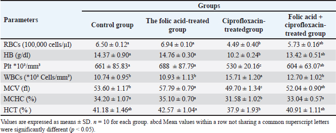
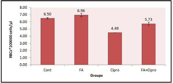
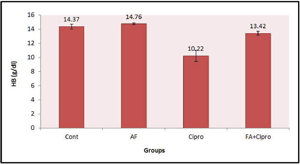
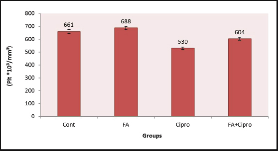
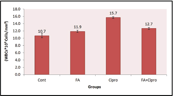
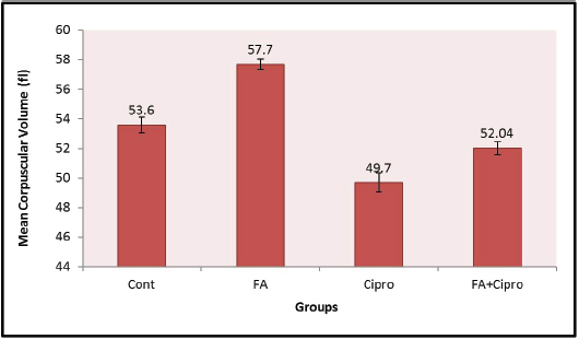
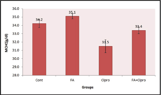
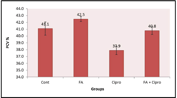
Sample collectionAt the end of the experimental period, the rats were fasted for a period, after which they were euthanized and dissected. Fasting was performed overnight, and blood was drawn from the inferior vena cava using glass tubes containing the anticoagulant EDTA. Complete blood counts, including hemoglobin (Hb), red blood cells (RBCs), white blood cells (WBCs), platelets (Plt), and packed cell volume (PCV), were analyzed using a Celltac X kx 021n automated hematology analyzer from Japan CARE Co. Ltd. Histopathological analysisA pathology specialist conducted the histopathological study in the Department of Pathology and Forensic Medicine, College of Medicine, University of Misan. The heart tissue samples were immediately removed, gently handled to minimize trauma, weighed, and fixed in 10% neutral buffered formalin for 24 hours. The fixed tissues were then dehydrated through a graded series of ethanol, embedded in paraffin wax, sectioned according to Luna’s method (1968)and stained with hematoxylin and eosin for histopathological examination. Statistical analysisThe study results are expressed as mean ± standard error. Statistical analyses were performed with one-way analysis of variance and Tukey test using SPSS version 7. The means of the groups were compared for significance in (p < 0.05) by using the T test for independent samples. Ethical approvalThe Bioethical Committee at the College of Medicine/University of Misan approved the study procedures (approval reference No. 170, 2024). ResultsThe hematological findings in Table 1 and Figures 1–7 of this study exhibited significant decreased (p < 0.05) in erythrocytes (RBCs) count, Hb concentration, Plt count, PCV, mean corpuscular volume (MCV), and mean corpuscular hemoglobin concentration (MCHC). On the other hand, a significantly elevated (p < 0.05) WBC count was observed in Group 3 compared with Groups 1 and 2. Group 4 showed a significant improvement in hematological parameters compared with the other groups. Table 1. Effect of ciprofloxacin, folic acid, and their combination on rat hematological parameters.
Fig. 1. Changes in RBC levels in rats administered with folic acid, ciprofloxacin, or their combination
Fig. 2. Changes in Hb concentration in rats administered with folicacid, ciprofloxacin, or their combination
Fig. 3. Changes in Plt counts in rats administered with folic acid, ciprofloxacin, or a combination of both (FA + Cipro). Values are presented as mean ± standard deviation (SD) for 10 rats per group.
Fig. 4. Changes in WBC counts in rats administered with folic acid, ciprofloxacin, and their combination (FA + Cipro). Values are presented as mean ± standard deviation (SD) for 10 rats per group.
Fig. 5. Changes in the MCV in rats administered with folic acid, ciprofloxacin, or their combination (FA + Cipro). Values are presented as mean ± standard deviation (SD) for 10 rats per group.
Fig. 6. Changes in MCHC in rats treated with folic acid, ciprofloxacin, or their combination (FA + Cipro). Values are presented as mean ± standard deviation (SD) for 10 rats per group.
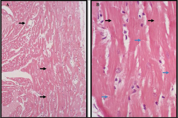
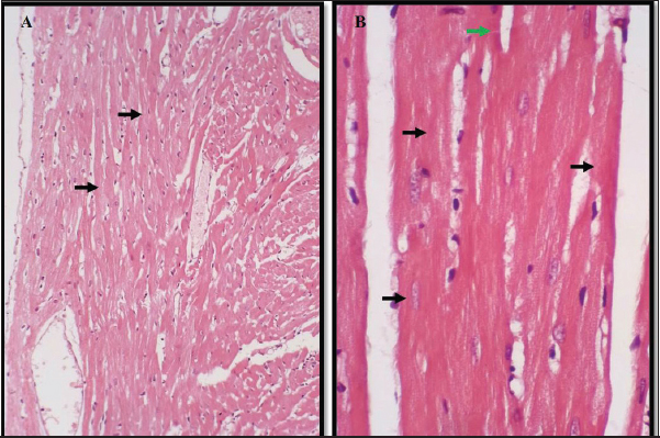
Fig. 7. Changes in PCV in rats administered with folic acid, ciprofloxacin, or their combination In the control group (G1) and folic acid group (G2), the myocardium exhibited normal histological appearance; however, the myocardial muscle fibers appeared cylindrical with centrally located nuclei in the longitudinal section and obvious striation; however, they joined to each other by intercalated discs (Figs. 8 and 9).
Fig. 8. Heart tissue section of the control group shows normal cardiomyocytes (black arrow) and normal connective tissue containing intercalated discs (blue arrow). H&E staining: A) 10×, B) 40×.
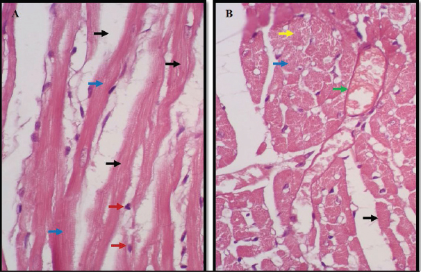
Fig. 9. The heart tissue section of the folic acid-treated group shows a normal histological appearance with normal cardiomyocytes (black arrow) and connective tissue containing intercalated discs (green arrow). H&E staining: A) 10×, B) 40×. On the other hand, the heart section in (G3) exhibited coagulative necrosis of some cardiac muscle fibers; however, it showed increased cytoplasmic eosinophilia in the absence of nuclear staining. In addition to the loss of muscular striations of some other muscle fibers, some degenerated muscle fibers and infiltration of inflammatory cells were observed (Fig. 10).
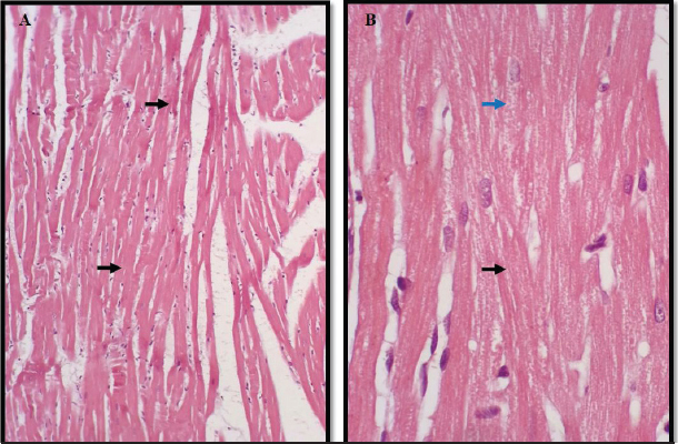
Fig. 10. The heart tissue section of the ciprofloxacin-treated group shows coagulative necrosis in some cardiac muscle fibers (black arrow), loss of muscular striations (blue arrow), congestion (green arrow), vacuolation (yellow arrow), and infiltration of inflammatory cells (red arrow). H&E staining: A) and B) 40×. While some myocardial muscle fibers showed mild degeneration, others showed mild loss of muscle striation (Fig. 11).
Fig. 11. The heart tissue section of the combination-treated group shows mild cardiac muscle architecture degeneration (black arrow) and muscular striation loss (blue arrow). H&E staining: (A) and (B) 40×. DiscussionThe experimental outcomes demonstrate that 250 mg/kg ciprofloxacin induces marked toxicity in male rats. This observation aligns with previous studies reporting that high doses of FQs promote oxidative stress, impair mitochondrial function, and disrupt cellular homeostasis. Ciprofloxacin-induced toxicity has been associated with the excessive generation of ROS, depletion of endogenous antioxidants, and damage to lipids, proteins, and DNA, ultimately leading to functional and structural alterations in vital organs, including the liver, kidneys, and reproductive tissues (Halliwell and Gutteridge, 2015). Administration of folic acid at 100 µg/kg appeared to attenuate several of the toxic effects induced by ciprofloxacin. Folic acid plays a central role in one-carbon metabolism, nucleotide synthesis, and DNA repair, processes that are particularly vulnerable to stress (Crider et al., 2012). Folic acid exerts indirect antioxidant effects by enhancing cellular antioxidant capacity and reducing lipid peroxidation. Even at a low dose, folic acid may stabilize cellular membranes, support mitochondrial integrity, and facilitate the recovery of damaged tissues, thereby limiting the extent of ciprofloxacin-induced injury (Asbaghi et al., 2021). ElBoshy et al. (2012)mentioned that the observed protective effect of folic acid, despite the relatively high ciprofloxacin dose, suggests that the cytoprotective mechanisms of folic acid are effective but not fully compensatory. Protection is likely partial, reducing the severity of biochemical and histological alterations rather than completely preventing toxicity. This finding highlights the importance of dose-response relationships; higher folic acid doses or different treatment schedules might provide greater protection, whereas lower ciprofloxacin doses could yield results more relevant to clinical practice. Male rats were used in this study because of their hormonal stability, consistent drug metabolism, avoidance of estrogen-related protection, and historical precedent. Increasing evidence shows that sex differences significantly influence toxic responses (Clayton and Collins, 2014). The study observed significant changes in various hematological parameters in rats treated with ciprofloxacin, including significantly decreased RBC count, Hb, Plt, PCV, MCV, and MCHC, and significantly increased WBC count. These findings are consistent with those of previous studies on the hematological effects of ciprofloxacin, where Owolabi and Omogbai (2017) reported similar decreases in RBC count and Hb levels in rats treated with ciprofloxacin. The decrease in these parameters suggests that ciprofloxacin suppresses erythropoietin. Talla and Veerareddy (2011) reported that ciprofloxacin may induce oxidative stress, leading to increased RBC destruction. Ciprofloxacin can induce oxidative stress through several mechanisms, including the generation of ROS due to mitochondrial dysfunction, depletion of antioxidant defenses, and the direct pro-oxidant effects of the drug or its metabolites. This oxidative stress can contribute to cellular damage in heart tissue, hemolysis leading to decreased RBC count and Hb levels, and DNA damage, potentially affecting HSC (Talla and Veerareddy, 2011). These findings help explain the myocardial muscle damage observed in the ciprofloxacin-treated group. The increase in WBC count observed in the ciprofloxacin-treated group could be indicative of an inflammatory response. This aligns with the findings of Yigitturk et al. (2018)who reported that ciprofloxacin can induce an inflammatory response in various tissues. The study found that co-administration of folic acid with ciprofloxacin (Group G4) resulted in a significant improvement in hematological parameters, bringing them closer to normal levels. This protective effect of folic acid is supported by previous research, such as Ebaid et al. (2020)who demonstrated that folic acid supplementation could improve oxidative damage and stimulate the antioxidant defense system. The histopathological examination revealed significant alterations in the heart tissue of rats treated with ciprofloxacin (Group G3), including disrupted cardiac muscle architecture, loss of muscular striations, degeneration and focal apoptosis, coagulative necrosis and congestion, and inflammatory cell infiltration. These findings are consistent with the cardiotoxic effects of ciprofloxacin reported in other studies. For instance, Al-Faris et al. (2012)observed similar histopathological changes in rat heart tissue after ciprofloxacin administration. The study also found that co-administration of folic acid with ciprofloxacin (Group G4) resulted in milder histopathological changes compared with the ciprofloxacin-only group. This suggests that folic acid has a protective effect against ciprofloxacin-induced cardiotoxicity. As suggested by Shalabi et al. (2019)this protective effect could be attributed to the antioxidant properties of folic acid. Kalghatgi et al. (2013)reported that ciprofloxacin impairs mitochondrial function and demonstrated that fluoroquinolones, including ciprofloxacin, inhibit mitochondrial DNA synthesis and disrupt electron transport chain complexes. This mitochondrial dysfunction may lead to cardiotoxicity because cardiomyocytes are highly dependent on mitochondrial function. These findings are consistent with the present study’s results. Folic acid plays a crucial role in one-carbon metabolism, which is essential for DNA synthesis and repair, methylation reactions, and amino acid metabolism. Folic acid may enhance the body’s antioxidant defenses by increasing glutathione levels and supporting the synthesis of NADPH, a crucial cofactor for many antioxidant enzymes (Ebaid et al., 2020). Folic acid plays a crucial role in DNA synthesis and repair and may help counteract ciprofloxacin-induced DNA damage, particularly in rapidly dividing cells such as those in the bone marrow. Additionally, some studies have suggested that FA possesses anti-inflammatory properties. For instance, Feng et al. (2011)demonstrated that folic acid supplementation could reduce inflammatory markers in patients with hypertension. The improvement in heart tissue histology in the folic acid + ciprofloxacin group suggests specific cardioprotective effects. This aligns with the findings of Moens et al. (2008)who reported that FA could improve cardiac function in animal models of heart failure. ConclusionCiprofloxacin induces significant hematological and cardiac histopathological changes in rats. Co-administration of folic acid partially mitigates these effects through its antioxidant properties, DNA stability support, and cellular function preservation. Further research, especially clinical studies, is needed to confirm these protective effects, determine appropriate dosing, explore underlying molecular mechanisms, assess other antioxidants, and evaluate long-term outcomes of chronic ciprofloxacin use and FA supplementation. AcknowledgmentsThe authors wish to thank the staff of the Department of Pathology and Forensic Medicine and the Department of Microbiology (College of Medicine, University of Misan) for their essential support. Conflict of interestThe authors declare no conflict of interest. FundingThe researchers supported all the costs of the study, and no financial support was received from any government or private institute. Authors’ contributionsThe author contributions were as follows: Mukhallad A. Ramadhan: designed the experiment and performed data collection. Waleed Khaled Y. Albahadly: conducted the data analysis. Abbas C. Mraisel: drafted the manuscript. Data availabilityAll data supporting the findings of this study are available within the article. ReferencesAkhtara, N. and Bharali, M.K. 2025. Genotoxicity assessment after sub-chronic exposure to amoxicillin, azithromycin, and ciprofloxacin in male albino mice. Mol. Cell. Toxicol. 21, 1151–1160; doi:10.1007/s13273-025-00524-3 Al-Faris, E.A., Alkhawajah, A.M., Alsheikh, A. and Al-Kurdi, A.D. 2012. Antibiotic resistance pattern in gram-negative urinary pathogens: impact on empiric therapy. Saudi Med. J. 33(7), 817–820. Asbaghi, O., Ghanavati, M., Ashtary-Larky, D., Bagheri, R., Rezaei Kelishadi, M., Nazarian, B., Nordvall, M., Wong, A., Dutheil, F., Suzuki, K. and Alavi Naeini, A. 2021. Effects of folic acid supplementation on oxidative stress markers: a systematic review and meta-analysis of randomized controlled trials. Antioxidants 10(6), 871; doi:10.3390/antiox10060871 Cao, Y., Ding, W., Zhang, J., Gao, Y. and Qiu, L. 2021. Molecular mechanism and physiological role of pkcδ-abilty2-rack1 complex in the regulation of neutrophil chemotaxis. J. Leukocyte Biol. 109(2), 343–355. Clayton, J.A. and Collins, F.S. 2014. NIH to balance sex in cell and animal studies. Nature 509(7500), 282–283. Crider, K.S., Yang, T.P., Berry, R.J. and Bailey, L.B. 2012. Folate and DNA methylation: a review of molecular mechanisms and the evidence for folate’s role. Adv. Nutr. 3(1), 21–38. Ebaid, H., Bashandy, S.A., Alhazza, I.M., Rady, A. and El-Shehry, S. 2020. Folic acid and melatonin ameliorate carbon tetrachloride-induced hepatic injury, oxidative stress and inflammation in rats. Nutr. Metab. 17, 1. ElBoshy, M.M., Ahmed, H.H. and Salem, H.A. 2012. Protective role of folic acid against doxorubicin-induced oxidative stress, DNA damage, and testicular injury in rats. Int. J. Pharmacol. 8(7), 562–570. Feng, D., Zhou, Y., Xia, M. and Ma, J. 2011. Folic acid inhibits lipopolysaccharide-induced inflammatory response in RAW264.7 macrophages by suppressing MAPKs and NF-κB activation. Inflammation Res. 60(9), 817–822. Halliwell, B. and Gutteridge, J.M.C. 2015. Free radicals in biology and medicine, 5th ed. Oxford, UK: Oxford University Press, p: 978. Kalghatgi, S., Spina, C.S., Costello, J.C., Liesa, M., Morones-Ramirez, J.R., Slomovic, S., Molina, A., Shirihai, O.S. and Collins, J.J. 2013. Bactericidal antibiotics induce mitochondrial dysfunction and oxidative damage in mammalian cells. Sci. Transl. Med. 5, 192. Khalee, Z.H., Khudair, A.N. and Abood, S.J. 2021. Protective effects of melatonin against ciprofloxacin-induced chondrotoxicity in rats. Drug Chem. Toxicol. 44(3), 303–310. Luna, L.G. 1968. Manual of histologic staining methods of the Armed Forces Institute of Pathology. Blakiston Division, McGraw-Hill. Millanao, A.R., Barrientos-Schaffeld, C., Siegel-Tike, C.D., Tomova, A., Ivanova, L., Godfrey, H.P., Dölz, H.J., Buschmann, A.H. and Cabello, F.C. 2021. Antimicrobial resistance in Chile and the One Health paradigm: dealing with threats to human and veterinary health resulting from antimicrobial use in salmon aquaculture and the clinic. Rev. Chilena Infectol. Organo. Oficial. De La. Soc. Chilena Infectol. 38(1), 69. Moens, A.L., Takimoto, E., Tocchetti, C.G., Chakir, K., Bedja, D., Cormaci, G., Ketner, E.A., Majmudar, M., Gabrielson, K., Halushka, M.K., Mitchell, J.B., Biswal, S., Channon, K.M., Wolin, M.S., Alp, N.J., Paolocci, N., Champion, H.C. and Kass, D.A. 2008. Reversal of cardiac hypertrophy and fibrosis from pressure overload by tetrahydrobiopterin: efficacy of recoupling nitric oxide synthase as a therapeutic strategy. Circulation 117(20), 2626–2636. Owolabi, J.O. and Omogbai, E.K.I. 2017. Evaluation of the potentials of some selected medicinal plants in Southern Nigeria as antimalarial and immunomodulatory agents. Afr. J. Pharmacol. Therapeutics 6(1), doi:10.1016/j.ajpt.2017.09.010 Paget, G.E. and Barnes, J.M. 1964. Toxicity tests. Eval. Drug. Activities. Pharmacometrics. 1, 135–166. Pispirigos, K. and Chrysanthopoulos, K. 2001. Ciprofloxacin-induced myocardial infarction and cerebral ischemia. Clin. Cardiol. Int. Indexed Peer-Reviewed. J. For Adv. Treatment Cardiovasc. Dis. 24(12), 798–800. Shalabi, N., Alakilli, S., Massoud, M. and Al-Hazmi, N. 2019. Protective effect of folic acid on DNA damage induced by methotrexate in human peripheral lymphocytes. Saudi J. Biol. Sci. 26(7), 1668–1673. Talla, V. and Veerareddy, P.R. 2011. Oxidative stress induced by fluoroquinolones on treatment for complicated urinary tract infections in Indian patients. J. Young Pharmacists 3, 304–309. Von Keutz, E., Rühl-Fehlert, C., Drommer, W. and Rosenbruch, M. 2004. Effects of ciprofloxacin on joint cartilage in immature dogs immediately after dosing and after a 5-month treatment-free period. Arch. Toxicol. 78(7), 418–424; doi:10.1007/s00204-004-0551-6 Yigitturk, E.H., Cavusoglu, G., Baygar, T., Ozgul, T., Onal, M. and Ozturk, F. 2018. Comparison of ultrastructural changes and the expression of apoptotic proteins in the rat testis following ciprofloxacin and gemifloxacin treatments. Ultrastruct. Pathol. 42(1), 41–54. | ||
| How to Cite this Article |
| Pubmed Style Albahadly WK, Ramadhan MA, Mraisel AC. Protective effects of folic acid against ciprofloxacin toxicity in male rats. Open Vet. J.. 2026; 16(7): 4999-5008. doi:10.5455/OVJ.2026.v16.i7.79 Web Style Albahadly WK, Ramadhan MA, Mraisel AC. Protective effects of folic acid against ciprofloxacin toxicity in male rats. https://www.openveterinaryjournal.com/?mno=285098 [Access: July 27, 2026]. doi:10.5455/OVJ.2026.v16.i7.79 AMA (American Medical Association) Style Albahadly WK, Ramadhan MA, Mraisel AC. Protective effects of folic acid against ciprofloxacin toxicity in male rats. Open Vet. J.. 2026; 16(7): 4999-5008. doi:10.5455/OVJ.2026.v16.i7.79 Vancouver/ICMJE Style Albahadly WK, Ramadhan MA, Mraisel AC. Protective effects of folic acid against ciprofloxacin toxicity in male rats. Open Vet. J.. (2026), [cited July 27, 2026]; 16(7): 4999-5008. doi:10.5455/OVJ.2026.v16.i7.79 Harvard Style Albahadly, W. K., Ramadhan, . M. A. & Mraisel, . A. C. (2026) Protective effects of folic acid against ciprofloxacin toxicity in male rats. Open Vet. J., 16 (7), 4999-5008. doi:10.5455/OVJ.2026.v16.i7.79 Turabian Style Albahadly, Waleed K., Mukhallad A. Ramadhan, and Abbas C. Mraisel. 2026. Protective effects of folic acid against ciprofloxacin toxicity in male rats. Open Veterinary Journal, 16 (7), 4999-5008. doi:10.5455/OVJ.2026.v16.i7.79 Chicago Style Albahadly, Waleed K., Mukhallad A. Ramadhan, and Abbas C. Mraisel. "Protective effects of folic acid against ciprofloxacin toxicity in male rats." Open Veterinary Journal 16 (2026), 4999-5008. doi:10.5455/OVJ.2026.v16.i7.79 MLA (The Modern Language Association) Style Albahadly, Waleed K., Mukhallad A. Ramadhan, and Abbas C. Mraisel. "Protective effects of folic acid against ciprofloxacin toxicity in male rats." Open Veterinary Journal 16.7 (2026), 4999-5008. Print. doi:10.5455/OVJ.2026.v16.i7.79 APA (American Psychological Association) Style Albahadly, W. K., Ramadhan, . M. A. & Mraisel, . A. C. (2026) Protective effects of folic acid against ciprofloxacin toxicity in male rats. Open Veterinary Journal, 16 (7), 4999-5008. doi:10.5455/OVJ.2026.v16.i7.79 |


