Open Veterinary Journal, (2026), Vol. 16(7): 4642-4660
Research Article
10.5455/OVJ.2026.v16.i7.45
Effects of butternut squash seed extract (Cucurbita moschata Duchesne) on antioxidants and kidney damage in diabetic mice
Suwanto Suwanto1,2, Slamet Widiyanto1*, Budi Setiadi Daryono1, Bambang Retnoaji1
and Harto Widodo3
1Biology Doctoral Study Program, Faculty of Biology, Universitas Gadjah Mada, Yogyakarta, Indonesia
2Sekolah Tinggi Keguruan dan Ilmu Pendidikan (STIKIP) Catur Sakti, Yogyakarta, Indonesia
3Research Center for Pharmaceutical Ingredients and Traditional Medicines, National Research and Innovation Agency, Bogor, Indonesia
*Corresponding Author: Slamet Widiyanto. Faculty of Biology, Universitas Gadjah Mada, Yogyakarta, Indonesia.
Email: slametbio [at] ugm.ac.id
Submitted: 23/10/2025 Revised: 05/05/2026 Accepted: 23/05/2026 Published: 20/07/2026
© 2025 Open Veterinary Journal
This is an Open Access article distributed under the terms of the Creative Commons Attribution-Non Commercial-No Derivatives License (http://creativecommons.org/licenses/by-nc-nd/4.0/), which permits non-commercial re-use, distribution, and reproduction in any medium, provided the original work is properly cited, and is not altered, transformed, or built upon in any way.
Abstract
Background: Diabetic mellitus (DM) is a chronic metabolic disorder associated with increased oxidative stress and vascular complications. This disease is managed with natural ingredients. Butternut squash is a natural ingredient with therapeutic effects due to its antioxidant content in the form of fatty acids.
Aim: To determine the effects of butternut squash seed extract (Cucurbita moschata Duchesne) on antioxidants and kidney damage improvement in a diabetic mice model.
Methods: The research used 30 male mice divided into 6 groups consisting of CN, C-, C+, P1 at a dose of 100 mg/kg BW. P2 at a dose of 150 mg/kg BW, and P3 at a dose of 200 mg/kg BW. Treatment was administered according to the dose in each group for 28 days. The research results were described with mean ± standard deviation, and one-way analysis of variance was followed by Duncan’s multiple range test to observe differences at a significance level of < 0.05.
Results: Butternut squash seed extract exerted a therapeutic effect on weight gain on days 0 (p= 0.017), 7 (p=0.000), 14 (p=0.000), 21 (p=0.000), and 28 (p=0.000), decreased fasting blood glucose levels on days 0 (p=0.000), 7 (p=0.004), 14 (p=0.003), 21 (p=0.002), and 28 (p=0.000), increased superoxide dismutase (SOD) levels (p=0.007), decreased tubular cell necrosis (p=0.000), and decreased tubular cell degeneration (p=0.000). Additionally, the extract did not have a therapeutic effect on the increase in catalase levels (p=0.074) and did not reduce the widening of the Bowman space (p=0.395).
Conclusion: Butternut squash seed extract has therapeutic effects on weight gain, fasting blood glucose levels, SOD levels, tubular cell necrosis, and tubular cell degeneration, thus demonstrating that the extract is beneficial for improving oxidative stress and microvascular complications due to hyperglycemia. Further clinical trials are needed to ensure the safety and effectiveness of butternut squash seed extract as a therapy for DM.
Keywords: Butternut squash seed, Catalase, Diabetic mellitus, Kidney, Superoxide dismutase.
Introduction
Nowadays, human lifestyle exhibits disparities compared to ancient times, attributable to advancements in the contemporary era, which facilitated rapid access to resources. The lifestyle in this case is unhealthy food containing food additives and instant food preparation. Food additives are substances that are added to food products to affect their properties or forms (Setyawati and Mahmudiono, 2023). Food additives include flavor enhancers, sweeteners, colorants, preservatives, thickeners, and emulsifiers (Dunford et al., 2023). The use of food additives has been regulated in accordance with the applicable provisions by the Minister of Health of the Republic of Indonesia, Number 033 of 2012, covering the types, categories, and quantities used in food additives. Children, adolescents, and adults prefer food products containing additives as a form of food consumption at all times. Consumption of food that does not comply with applicable regulations has low nutritional content and can adversely affect the body’s health (Seto et al., 2025). In addition to unhealthy food factors, the lack of adequate physical activity triggers the emergence of degenerative diseases, one of which is diabetic mellitus (DM).
DM is is a chronic metabolic disease characterized by disturbances in the metabolism of carbohydrates, proteins, and fats caused by impaired insulin secretion, insulin activity, or both, leading to increased blood glucose concentration (hyperglycemia) (Chike-Ekwughe et al., 2024). Several studies have revealed that the prevalence of DM is increasing worldwide. The International Diabetes Federation predicts that the global incidence of DM is 10.4% in high-income countries, 9.5% in middle-income countries, and 4.03% in low-income countries (Saeedi et al., 2019). According to Cho et al. (2018), in 2017, there were 451 million DM sufferers worldwide in 2017, which is projected to increase to 693 million by 2045. The prevalence of DM in Indonesia is estimated to increase from 9.19% in 2020 (18.69 million cases) to 16.09% in 2045 (40.7 million cases) (Wahidin et al., 2024).
Hyperglycemia is associated with increased oxidative stress and vascular complications (Adeshara et al., 2024). Oxidative stress in DM is caused by a shift in the balance of redox reactions due to changes in the metabolism of carbohydrates, proteins, and fats, which will increase the formation of reactive oxygen species (ROS) from glycation and lipid oxidation reactions, resulting in a decrease in endogenous antioxidants such as superoxide dismutase (SOD) and catalase (CAT) (Caturano et al., 2023). A decrease in the antioxidants SOD and CAT occurs because they are extensively used to counteract high levels of ROS (Jena et al., 2023). DM not only causes hyperglycemia but also leads to diabetic nephropathy, which is a microvascular complication of hyperglycemia (Kumar et al., 2023). Diabetic nephropathy is a clinical syndrome in patients with DM characterized by an increase in albuminuria > 300 mg/24 hour, leading to end-stage renal failure (Amelia et al., 2021). Diabetic nephropathy is based on the mechanism of oxidative stress, which is the imbalance between the amounts of free radicals and antioxidants in the body, leading to glomerular atrophy and damage to the proximal tubules in the kidney. Kidney enlargement and hyperfiltration are also observed (Malini et al., 2021).
Controlling blood glucose levels and slowing the onset of microvascular complications is done by patients with DM by consuming oral synthetic drugs. Metformin is a synthetic drug for DM from the biguanide class (Yerevanian and Soukas, 2019). Metformin reduces insulin resistance, lowers blood glucose levels by inhibiting gluconeogenesis, and suppresses liver glucose production by increasing insulin sensitivity (Yang et al., 2017). The long-term consumption of synthetic drugs can have negative effects on body organs; therefore, there is a need for natural ingredients that are safer than synthetic drugs (Blahova et al., 2021). Butternut squash, a species of C. moschata Duchesne, a member of the Cucurbitaceae family, is one such natural treatment.
The plant has been cultivated for over 5,000–6,000 years before Christ, and it is an annual plant widely grown around the world (Huang et al., 2023). The characteristics of the butternut squash plant are as follows: vine-like, herbaceous stem that can grow more than 10 m, monoecious, and short-lived (Hosen et al., 2021). Several countries, such as Austria, Hungary, Mexico, Slovenia, China, Spain, and some countries in Asia and Africa, use butternut squash as a functional food, as it has health benefits (Batool et al., 2022). The health benefits of butternut squash include anti-diabetic, antioxidant, and antiinflammatory properties (Dotto and Chacha, 2020). The health benefits of butternut squash arise from the presence of fatty acids, including linoleic, oleic, palmitic, stearic, linolenic, myristic, palmitoleic, arachidic, and behenic acids (Suwanto et al., 2025).
Based on in vivo research, the alcohol extract of pumpkin seeds (Cucurbita pepo L.) can reduce HbA1c and blood glucose levels, increase insulin, SOD, CAT, and glutathione (GSH) levels, and improve the liver organs affected by streptozotocin induction in rats (Lateef et al., 2024). Chenni et al. (2022) showed that C. pepo seed can improve metabolic parameters and play a role in protecting against oxidative stress caused by a high-glucose diet in mice. Similarly, studies on C. maxima leaves have shown potential for protection against hyperglycemia and nephrotoxicity in rats treated with streptozotocin (Onuche et al., 2023). The results of this research can be used as a basis for further studies; however, the treatment using butternut squash seed has not been extensively studied. Therefore, research on the effects of butternut squash seed extract (Cucurbita moschata Duchesne) on antioxidants and kidney damage improvement in a diabetic mice model is needed. This research provides scientific evidence of the effects of butternut squash seed extract on oxidative stress and microvascular complications due to hyperglycemia, making it an alternative to synthetic drugs.
Materials and Methods
Collection and identification of plants
Butternut squash seeds were obtained from farmers in Butuh village, Mojosongo district, Boyolali regency, Central Java (7°32'58"S 110°37'54"E 296 m asl). Butternut squash was identified at the Plant Systematics Laboratory, Faculty of Biology, Universitas Gadjah Mada (ref. no. 00595/S.Tb./III/2024).
Materials
Streptozotocin (Santa Cruz Biotechnology Inc, USA), n-hexane, citric acid monohydrate, sodium citrate, sodium carboxymethyl cellulose (Na-CMC), chloroform, xylene, liquid paraffin, physiological saline (Sigma Aldrich, USA), 10% formalin buffer, 70% alcohol (PT One Medika, IDN), standard feed (Citra feed, IDN), male mice BALB-C strain, zeolite sand (Chee hamster, IDN), distilled water, 500 mg of metformin HCl (Hexpharm Jaya, IDN), 5% dextrose infusion (PT Otsuka, IDN), total SOD and CAT kit (Elabscience, USA), and hematoxylin and eosin (Santa Cruz Biotechnology Inc, USA).
Tools
The tools used in this research were a grinder (Maspion, IDN), oven UN75 (Memmert, DEU), micro pipette 200 μl, 1,000 μl, microtome HM 310 (Thermo Fisher Scientific, USA), syringe (1 ml), urine pot, and vacutainer ethylenediaminetetraacetic acid (PT One Medika, IDN), glucometer, and test strips (Gluko Dr AGM-2100 PT Medisindo Bahana, IDN) 50 ml conical tube (PT Iwaki Glass, Indonesia), analytical balance ADJ 200-4 (KERN, IDN), semi-analytical balance BC-500 (ACIS, IDN ), filter paper, dark bottle, 300 mesh sieve, microscope (Olympus BX-51, JPN), Optilab camera (Miconos, IDN), scalpel, scissors, needle, paraffin mold, measuring cup and stirrer, animal testing cage, mouse drinking bottle, and UV-VIS spectrophotometer 1800 (Shimadzu, JPN).
Preparation of the butternut squash seed extract
Butternut squash seeds were cleaned with water and then drained. The seeds were then oven-dried at a temperature of 65°C for 5–6 hours, cleaned again, and then ground using a grinder. The seeds were then sifted using a 300-mesh sieve, placed in a dark bottle, protected from light, and stored at room temperature (Suwanto et al., 2020; Halim et al., 2024). After obtaining a fine simplicia, the simplicia was extracted by soaking it in n-hexane solvent in a 1:10 (w/v) ratio for 5 days in a dark glass bottle, with occasional stirring. The residue and filtrate were separated using a filter paper. The filtrate was evaporated for 24 hours in an oven at 40°C until a thick extract was obtained, which was subsequently placed in a dark glass bottle (Suwanto et al., 2025).
Preparation of streptozotocin formulation
Streptozotocin at a dose of 50 mg/kg BW was administered intraperitoneally to make the mice diabetic. It was prepared by weighing 45-mg streptozotocin for mice weighing 30 g, with 30 mice. The weighted streptozotocin was placed into a 50-ml conical tube, dissolved in 9 ml of 0.1 M citrate buffer (pH 4.5), and mixed until homogeneous.
Preparation of the metformin solution
The preparation of a metformin solution at a dose of 1.95 mg/kg BW was conducted by weighing 68.25 mg of metformin dissolved in 10.5 ml of 1% Na-CMC. The solution was administered orally to 5 mice for 7 days. Metformin production was performed weekly to ensure a fresh supply. Oral metformin administration was performed for 28 days. The metformin dosage of 1.95 mg/kg body weight (BW) was obtained by converting the human dose for a 70 kg individual to a 20 g mouse, which was 0.0026 (Poerwanagara et al., 2025). The metformin dose for adults is 500 mg, whereas the dose for a 20-g-mice (500 × 0.0026) is 1.3 mg/kg BW. Because the mice weight was 30 g (30/20 × 1.3), the metformin dosage was 1.95 mg/kg BW.
Preparation of the butternut squash seed extract test solution
The preparation of a test solution of butternut squash seed extract at a dose of 100 mg/kg BW was conducted by weighing 105 mg of butternut squash seed extract dissolved in 10.5 ml of 1% Na-CMC. The solution was needed for 5 mice with a BW of 30 g for 7 days. Every 7 days, a test extract solution was prepared in the same way.
The preparation of a test solution of butternut squash seed extract at a dose of 150 mg/kg BW was conducted by weighing 157.5 mg of butternut squash seed extract dissolved in 10.5 ml of 1% Na-CMC. The solution was needed for 5 mice with a BW of 30 g for 7 days. Every 7 days, a test extract solution was prepared in the same way.
The preparation of a test solution of butternut squash seed extract at a dose of 200 mg/kg BW was conducted by weighing 210 mg of butternut squash seed extract dissolved in 10.5 ml of 1% Na-CMC. The solution was needed for 5 mice with a BW of 30 g for 7 days. Every 7 days, a test extract solution was prepared in the same way.
Experimental animals
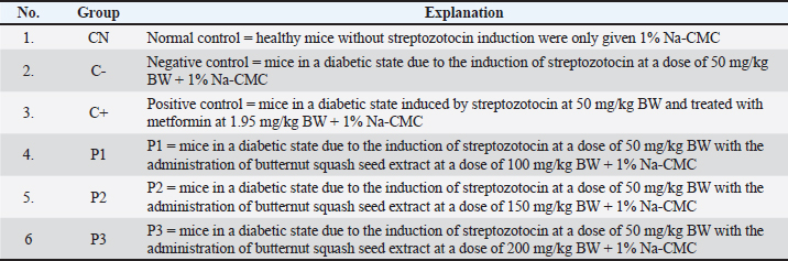
A total of 30 male mice were obtained from the Basic Pharmacology Laboratory at the Faculty of Pharmacy, Universitas Gadjah Mada. Mice were acclimatized for 7 days with adequate food and water. The mice were weighed, and their fasting blood glucose levels were measured. Subsequently, mice were induced once with a dose of 50 mg/kg BW of streptozotocin and given 5% dextrose through oral gavage for 7 days. On the eighth day after streptozotocin induction, the BW and fasting blood glucose levels of the mice were measured. Fasting blood glucose levels of >126 mg/dl indicate that the mice are in a hyperglycemia condition (Shi et al., 2021). In addition to blood glucose level measurement, clinical symptoms such as polyuria, polyphagia, and polydipsia were observed to ensure that the model animals were in a diabetic condition (Poznyak et al., 2020). After the mice were in a state of DM, they were grouped into 6 groups, each of which consisted of 5 animals. The treatment groups are shown in Table 1.
Table 1. Research Group.
Treatment of the model animals
After being conditioned to hyperglycemia, the mice were given treatment according to their respective groups. Treatment was administered for 28 days using an oral probe. During the treatment, the BW of the mice was measured every 7 days over 28 days using a semiemi scale. Body weight was used to determine the weight of butternut squash seed extract and metformin administered to mice.
Measurement of blood glucose levels
Blood glucose levels in mice were measured using a glucometer (Gluko Dr AGM-2100). Blood sampling was performed by cleaning the tail with a cotton swab moistened with water to remove any dirt, and then cleaning again with 70% alcohol. The tip of the tail was cut, and the blood came out and touched the glucose meter strip. Blood glucose level would be displayed on the screen after 10 seconds and expressed in mg/dl. Blood glucose measurements were taken in mice before and after streptozotocin induction on days 0, 7, 14, 21, and 28 after treatment administration.
Antioxidant measurement
Antioxidant measurements were conducted in all groups on day 28. Mice were euthanized by inhalation using chloroform, and then the chest cavity was opened, and the diaphragm was incised. Chloroform is used as an anesthetic agent because it interacts with gamma-aminobutyric acid receptors in the brain, causing hyperpolarization of nerve cells and reducing their activity (Haryanto. et al., 2025). The use of chloroform must be performed carefully and under strict supervision to minimize health risks. After opening the thoracic cavity and incising the diaphragm, blood was taken from the heart using a syringe and placed in a microtube before being centrifuged to obtain plasma. The SOD and CAT levels in the plasma samples were analyzed using the enzyme-linked immunosorbent assay method according to the manufacturer’s instructions. The microplate was read using a UV-Vis spectrophotometer at 540 nm (Shimadzu UV-1800).
Renal histopathology
Kidney histopathological samples were prepared in accordance with Tunnur et al. (2023). Kidney organ removal was performed on all mice after they were sacrificed by inhalation using chloroform, followed by necropsy of mice and then removal of the kidneys, which were washed with physiological saline (NaCl), fixed in 10% buffered formalin for 18–24 hours, and then dehydrated with 80%, 90%, 95%, and absolute alcohols. The specimen was impregnated in xylene, placed in pure xylene, and then embedded in liquid paraffin for 1 hour.
The specimen within the paraffin block was cut transversely to a thickness of 5 microns using a microtome. Slices were placed on a poly-L-lysine-coated glass slide. Incubation is done to remove the paraffin, then stained with Hematoxylin and Eosin. The histopathological preparation was observed using a microscope with 400x magnification across five fields of view, and tubular cell necrosis, tubular cell degeneration, and Bowman's space widening were observed.
Data analysis
The obtained research data were then described with the mean ± standard deviation. Analysis of differences was performed using one-way analysis of variance (ANOVA) followed by Duncan’s multiple range test at a significance level of < 0.05 using Statistical Package for the Social Sciences (SPSS) version 16.0 (2019) (SPSS Inc., Chicago, Illinois).
Ethical approval
The stages of research implementation were carried out in accordance with ethical standards and have been approved by the Chair of the Ethics Commission for Research at the Faculty of Veterinary Medicine, Universitas Gadjah Mada, No. 13/EC-FKH/int./2024.
Results
Body weight in the diabetic mice model
Table 2 shows the results of the study on the average BW of the diabetic mice model in the treatment groups CN, C-, C+, P1, P2, and P3 on days 0, 7, 14, 21, and 28.
Table 2. Body weight in a diabetic mice model.
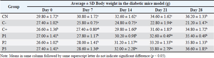
The results of the ANOVA test (p < 0.05) on days 0, 7, 14, 21, and 28 showed p-values of 0.017, 0.000, 0.000, 0.000, 0.000, and 0.000, respectively, meaning that butternut squash seed extract had an effect on weight gain in the diabetic mice model. The significant difference test on days 0 and 7 showed that the CN group was significantly different from the C-, C+, P1, P2, and P3 groups. On days 14, 21, and 28, the CN group was not significantly different from the C+, P1, P2, and P3 groups, but it was significantly different from the C- group.
Fasting blood glucose levels in a diabetic mice model
Table 3 shows the average fasting blood glucose levels in the diabetic mice model of CN, C-, C+, P1, P2, and P3 groups on days 0, 7, 14, 21, and 28.
Table 3. Fasting blood glucose levels in diabetic mice.
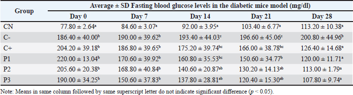
Results of the ANOVA test (p < 0.05) on days 0, 7, 14, 21, and 28 showed p-values of 0.000, 0.004, 0.003, 0.002, and 0.000, respectively, meaning that butternut squash seed extract has an effect on reducing fasting blood glucose levels in the diabetic mice model. Statistical test on days 0 and 7 showed that the CN group was significantly different from the C-, C+, P1, P2, and P3 groups. On days 14 and 21, the CN group was not significantly different from P2 and P3 but significantly different from C-, C+, and P1. On day 28, the CN group was not significantly different from the C+, P1, P2, and P3 groups but was significantly different from the C- group.
SOD profile in the diabetic mice model
The average SOD levels in the diabetic mice model of groups CN, C-, C+, P1, P2, and P3 are shown in Figure 1.
Results of the ANOVA test (p < 0.05) show that butternut squash seed extract has an effect on increasing SOD levels in the diabetic mice model (p=0.007). The results of the significant difference test show that the average SOD levels in the diabetic mice model group CN are not significantly different from P2 and P3 but are significantly different from C-, C+, and P1.
CAT profile in a diabetic mice model
Average CAT levels in the diabetic mice model of groups CN, C-, C+, P1, P2, and P3 are shown in Figure 2.
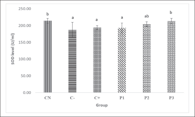
Fig. 1. SOD levels in the diabetic mice model.
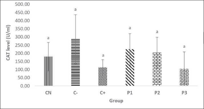
Fig. 2. CAT levels in the diabetic mice model.
Results of the ANOVA test (p < 0.05) indicated that the butternut squash seed extract did not affect the increase in CAT levels in the diabetic mice model (p=0.074).
Kidney histopathology in the diabetic mice model
The average kidney damage in the diabetic mice model of the CN, C-, C+, P1, P2, and P3 groups is shown in Table 4.
Table 4. Kidney damage in a diabetic mice model.
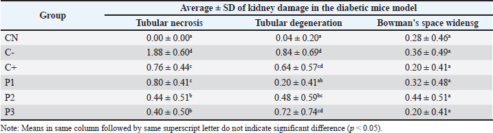
Results of the ANOVA test (p < 0.05) showed that butternut squash seed extract had an effect on reducing damage (necrosis) of tubular cells in the diabetic mice model, with p-value of 0.000. The results of the significance difference test showed that P1 had the same effect in reducing tubular cell necrosis as the C+ group, whereas the P2 and P3 groups had lower tubular cell necrosis, although it had not yet reached the level of the CN group.
Results of the ANOVA test (p < 0.05) showed that butternut squash seed extract has an effect in reducing of tubular cell degeneration in a diabetic mice model, with a p-value of 0.000. The results of the significant difference test show that the P3 group has an effect on reducing tubular degeneration similar to the C+ group, but the P1 group showed lower tubular cell degeneration, although it has not yet reached the level of the CN group.
Results of the ANOVA test (p < 0.05) showed that butternut squash seed extract does not affect the widening of the Bowman’s space in a diabetic mice model (p=0.395).
Discussion
Body weight in the diabetic mice model
The average BW of mice on day 0 in the CN group was higher than that in the C-, C+, P1, P2, and P3 groups (Table 2). The nutritional elements in the daily standard feed influenced the weight gain in the CN group. Nutrition refers to nutrients or vitamins found in the feed that enter the mice’s body through consumption (Mardiati and Sitasiwi, 2016). Deng et al. (2023) reported an increase in the BW of mice in the normal control group, with BW on day 1=26.75 g, while BW on day 31=31.34 g. In addition to nutritional factors, acclimatization affects the mice so that they do not experience stress. Stress can affect eating patterns, and mice in a stressed state may reduce food intake, leading to weight loss (Theresia et al., 2017). Qiao et al. (2024) showed that stress conditions in mice result in a significant decrease in BW and appetite.
Groups C-, C+, P1, P2, and P3 experienced weight loss on day 0 due to the inability of mice to utilize glucose as an energy source due to insulin deficiency. Streptozotocin induction can cause dysfunction of the insulin-producing pancreatic β-cell (Chen et al., 2019). Insulin deficiency prevents glucose from entering the cells, causing the body to switch to obtaining energy from fat and muscle, leading to weight loss (Ukratalo et al., 2024). According to Rinawati et al. (2020), an increase in the glycolysis process also triggers weight loss in test animals suffering from DM. Glucose expression activates pancreatic cells, which increases glucagon activity, resulting in the use of stored body fat as an energy source. These results are consistent with those of previous research (Mathias Akinlade et al., 2021; Hendarto et al., 2022), which showed that streptozotocin induction at a dose of 50 mg/kg BW causes weight loss in rats compared with the normal control group.
On day 7, groups C+, P1, P2, and P3 did not exhibit any influence on weight gain; this was due to the short treatment duration, which had not yet produced observable reaction effects. On day 14, groups P2 and P3 exhibited weight gain approaching that of the CN group. On days 21 and 28, groups P1, P2, and P3 exhibited weight gain comparable to that of group CN. The weight gain in groups P1, P2, and P3 was attributed to the butternut squash seed extract, which contains fatty acids such as linoleic, oleic, lauric, stearic, and palmitic acids. The fatty acid content of butternut squash seed extract plays a role in stimulating insulin production and enhancing glucose uptake by adipose tissue, thereby reducing blood glucose levels by activating pancreatic β-cell to produce insulin, maintaining normal insulin levels, and ensuring that cells receive adequate energy. This allows glucose to be effectively stored in the muscles and liver, resulting in increased weight in mice. This study aligns with Suwanto et al. (2019), who showed that feeding pumpkin seed extract affects weight gain in subjects with diabetes exposed to streptozotocin. Suwannapong et al. (2023) found that the BW of diabetic rats treated with C. Moschata extract (354.16 ± 7.44 g) and C. Maxima extract (359.66 ± 7.74 g) significantly increased compared with the diabetic control group and the diabetic rats treated with glibenclamide.
On day 28, the C+ group experienced weight loss compared with the P1, P2, and P3 groups. Metformin, an oral pharmacological agent that acts as an insulin-sensitizing and anti-hyperglycemic agent, was attributed to this weight loss. In addition to its use in treating diabetes and reducing cardiovascular risk, it leads to modest and sustained weight loss (Kosnayani et al., 2021). Metformin reduces BW by decreasing carbohydrate absorption in the intestines, reducing glucose 6-phosphate production and hepatic glycogenesis, and reducing hepatic gluconeogenesis (Zhou et al., 2018; Madiraju et al., 2018). Metformin enhances mitochondrial longevity similar to leptin. Leptin is an adipocyte-derived hormone that influences appetite, increases energy expenditure, and regulates autonomic nervous control, thereby regulating BW (Tang et al., 2016).
Based on the ANOVA results from days 0, 7, 14, 21, and 28, it is evident that butternut squash seed extract has an effect on weight gain in a diabetic mice model, with p-values of 0.017, 0.000, 0.000, 0.000, and 0.000, respectively. The significant difference test results on days 0 and 7 indicate that the CN group is significantly different from the C-, C+, P1, P2, and P3 groups; this is due to good glucose metabolism and nutrition-rich feed intake, resulting in proportional weight gain along with growth. Similarly, Xie et al. (2023) found that the control group exhibited increased weight in type 2 diabetic mice.
On days 14, 21, and 28, the CN group was not significantly different from the C+, P1, P2, and P3 groups but showed a significant difference compared with the C- group. This outcome is attributed to the treatment administered to diabetic model mice using metformin and butternut squash seed extract, which resulted in weight gain similar to that of the CN group. The weight gain observed in mice treated with butternut squash seed extract is due to its linoleic, oleic, lauric, stearic, and palmitic acid content. Fatty acids improve glucose metabolism disorders by regulating the PI3K/Akt pathway and glycogen synthesis, as well as modulating glucose metabolism through the gut microbiota, thereby promoting weight gain in mice with diabetic. Chen et al. (2022) stated that fatty acids can inhibit weight loss in rats with type 2 diabetes and show no significant difference in weight compared to the normal group. In contrast, the C- group experienced weight loss because the diabetic model mice did not receive any drug treatment and were only given 1% Na-CMC, which had no therapeutic effect on weight loss.
Fasting blood glucose levels in a diabetic mice model
Table 3 shows that the average fasting blood glucose levels in mice on day 0 for groups C-, C+, P1, P2, and P3 experienced an increase in fasting blood glucose levels compared to group CN without induction of streptozotocin at a dose of 50 mg/kg BW via intraperitoneal injection. The increase in fasting blood glucose levels is influenced by the induction of streptozotocin entry into pancreatic β-cell through glucose transporter 2 (GLUT2), causing mitochondrial depolarization due to the entry of Ca2+ ions, followed by excessive energy consumption, which results in energy deficiency within the cell (Munjiati, 2021). This condition disrupts insulin production, causing insulin deficiency that impairs glucose metabolism and leads to elevated glucose levels in the body.
The reaction of streptozotocin on pancreatic β-cell inhibits insulin secretion, resulting in hyperglycemia and decreased blood insulin levels, and altered blood glucose properties (Hahn et al., 2020). Increased insulin resistance is caused by decreased sensitivity of peripheral insulin receptors after induction with streptozotocin in mice. In addition, the restriction of adenosine triphosphate (ATP) synthesis by the mitochondria increases xanthine oxidase activity, and the inhibition of the Krebs cycle contributes to elevated blood glucose levels in mice (Mardiana et al., 2022). This will lead to decreased mitochondrial oxygen consumption, resulting in deoxyribonucleic acid (DNA) damage that can activate poly-ADP-ribosylation, which then leads to the suppression of cellular Nicotinamide Adenine Dinucleotide, subsequent decrease in ATP levels, and ultimately inhibit insulin secretion and synthesis. Graniel-Amador et al. (2022) showed that a dose of 50 mg/kg BW of streptozotocin significantly induced hyperglycemia in male C57BL/6J mice compared with the control group.
On day 7, groups C+, P1, P2, and P3 did not show any effect, resulting in a decrease in fasting blood glucose levels. This was likely due to the short treatment duration. The effects of these interventions had not yet manifested. On days 14 and 21, groups P2 and P3 experienced a decrease in fasting blood glucose levels approaching the levels observed in group CN. On day 28, groups C+, P1, P2, and P3 exhibited decreased fasting blood glucose levels near those of group CN. The decrease in glucose levels in group C+ was attributed to metformin administration. Metformin works by reducing glucose production in the liver and increasing muscle and adipose tissue sensitivity to insulin through the activation of adenosine monophosphate-activated protein kinase (AMPK) within cells via a process mediated by liver kinase B1 (LKB1) (Kusuma et al., 2022; Jasim and Abdul-Razzaq, 2024). Liver cells take up metformin, disrupting mitochondrial activities and reducing ATP levels. Metformin facilitates the binding of LKB1 to AMPK, leading to AMPK activation by LKB1 through Thr172 phosphorylation. AMPK activation shifts the cellular state from anabolic to catabolic, resulting in increased glucose uptake and decreased activity in biosynthetic pathways, including glucose, glycogen, and lipid synthesis in the liver (Ukratalo et al., 2024). Zhang et al. (2021) showed that metformin treatment significantly reduced the symptoms of diabetes mellitus (DM) in streptozotocin-induced diabetic rats.
Groups P1, P2, and P3 experienced a decrease in fasting blood glucose levels approaching those of the CN group. This is attributed to the natural antioxidant content of butternut squash seeds. Antioxidants play a role in preventing the oxidation of pancreatic β-cell, thereby minimizing damage. The natural antioxidants found in butternut squash seeds include linoleic, oleic, palmitic, stearic, linolenic, myristic, palmitoleic, arachidic, and behenic acids (Suwanto et al., 2025). The decrease in fasting blood glucose levels was caused by the antioxidants in butternut squash seed through increased cellular glucose uptake and enhanced insulin secretion, leading the body to utilize blood glucose in normal metabolic processes such as glycolysis, lipogenesis, and glucose metabolism, all regulated by insulin. Antioxidants also work by inhibiting glucose absorption in the small intestine through mechanisms involving glucose transporters, such as sodium-glucose transporter 1, glucose transporter 5, and GLUT2. Huang et al. (2023) supported this research, who found that the administration of pumpkin polysaccharides significantly reduced blood glucose, triglyceride, and total cholesterol levels in diabetic rats (p < 0.05).
The administration of butternut squash seed extract for 28 days was optimal in reducing fasting blood glucose levels. The use of synthetic antidiabetic drugs for 4 weeks resulted in a decrease in blood glucose levels, indicating that the duration of treatment with butternut squash seed extract for 28 days is comparable to the use of synthetic antidiabetic drugs (Dowidar et al., 2020).
Based on the results of ANOVA on days 0, 7, 14, 21, and 28, the results showed that butternut squash seed extract affects the decrease in fasting blood glucose levels in diabetic model mice, with respective p-values of 0.000, 0.004, 0.003, 0.002, and 0.000. The results of the significant difference test on days 0 and 7 indicate that CN was significantly different from C-, C+, P1, P2, and P3 groups. This is due to the induction of streptozotocin in mice entering the pancreatic β-cell through GLUT2, which leads to β-cell necrosis and loss of insulin production, resulting in an increase in fasting blood glucose levels in mice (Marino et al., 2023). Additionally, the short duration of treatment administration in each group may have affected this, which has not yet shown any observable effects.
On days 14 and 21, the CN group did not show significant differences compared with groups P2 and P3 but showed significant differences compared with groups C-, C+, and P1. This is because the fasting blood glucose levels in diabetic model mice decreased to levels close to those of the CN group due to the butternut squash seed extract, which includes fatty acids. The types of fatty acids present in the butternut squash seed extract include linoleic, oleic, palmitic, stearic, linolenic, myristic, palmitoleic, arachidic, and behenic acids (Suwanto et al., 2025). Fatty acids are involved in regulating gene expression through epigenetic mechanisms, thereby affecting the metabolism of the body in conditions such as type 2 diabetes by modifying insulin signaling, insulin sensitivity, oxidative stress, and glycemic control (Jiang et al., 2023).
On day 28, the CN group did not show a significant difference from groups C+, P1, P2, and P3 but showed a significant difference from group C-. The lack of a significant difference in the decrease in fasting blood glucose levels was affected by the presence of metformin as a therapeutic drug in the diabetic mouse model. Metformin exerts an antihyperglycemic effect by inhibiting liver gluconeogenesis and increasing insulin sensitivity in muscle and fat cells, thereby enhancing glucose uptake from the blood (Cho et al., 2015). Metformin can also enhance glucose uptake in the intestines and help control blood glucose levels (Koffert et al., 2017). Furthermore, the decrease in fasting blood glucose levels is influenced by the fatty acid content of butternut squash seed extract. The C- group still experienced an increase in fasting blood glucose levels due to streptozotocin induction in mice, which can damage pancreatic β-cell, thereby affecting insulin production and resulting in insulin deficiency and increased blood glucose levels.
SOD profile in the diabetic mice model
Figure 1 shows an increase in SOD levels in the CN group compared with those in the C- group. The high SOD levels in the CN group are due to the absence of streptozotocin induction in healthy mice, which does not trigger the excessive production of free radicals. Conversely, low SOD levels in the C-group were associated with oxidative stress conditions due to streptozotocin induction. Patients with diabetes experience sustained oxidative stress due to high blood glucose levels. High blood glucose levels increase ROS formation through redox reactions, thereby driving greater amounts of the electron donors nicotinamide adenine dinucleotide (NADH) and flavin adenine dinucleotide into the electron transport chain (Strugała et al., 2019). The increased rate of electron transport also contributes to the formation of superoxide anions.
Oxidative stress can lead to an increased rate of lipid peroxidation, contributing to the production of free radicals, including the formation of superoxide anions, which results in oxidative modifications that cause the inactivation of SOD (Taso et al., 2019; Masenga et al., 2023). Lipid peroxidation is the rapid oxidation of lipids. Oxidized lipids are part of the phospholipids of the cell membrane in pancreatic β-cell of diabetic patients, which can affect the stability of its structure. The vital function of the cell as the producer of the insulin hormone can be disrupted because lipid peroxidation causes an increase in cell membrane permeability (Sadžak et al., 2020), insulin resistance, and β-cell dysfunction (Shabalala et al., 2022). This condition causes hyperglycemia, which contributes to the decrease of SOD by inducing alternative glucose metabolism and generating more free radicals.
The C+ group showed an increase in SOD levels, and the increase in SOD levels observed in the diabetic mice model resulted from metformin treatment. Metformin activates intracellular signaling pathways in response to changes in energy levels in skeletal muscles to enhance glucose uptake (Horakova et al., 2019). Several studies have indicated that metformin inhibits NADH (Agius et al., 2020). NADH contributes to the production of ROS NADH inhibition can decrease ROS levels, as this results from reduced electron transport from NADH plus H+. Therefore, metformin can reduce the levels of endogenous mitochondrial ROS (Wang et al., 2022). Sun et al. (2021) indicated that metformin can reduce oxidative damage in diabetic mice. Liu et al. (2022) found that metformin can prevent insulin resistance, whereas Zhang et al. (2021) showed that metformin treatment can increase SOD activity and decrease MDA levels in diabetic mice.
Groups P2 and P3 showed increased SOD levels approaching those of the CN group, which may be attributed to the butternut squash seed fatty acid content. Fatty acids identified in butternut squash seed include linoleic, oleic, palmitic, stearic, linolenic, myristic, palmitoleic, arachidic, and behenic acids (Suwanto et al., 2025). The fatty acids in butternut squash seed possess antioxidant properties that are involved in the regeneration of damaged extracellular matrix proteins and cell growth enhancement. Therefore, the antioxidant activity of fatty acids in butternut squash seed may play a role in the management of DM. The results of this study are consistent with those reported by Gurumallu et al. (2022), who demonstrated that flaxseed oil and sesame seed oil significantly increased CAT, SOD, and GSH levels in rats with streptozotocin. Meanwhile, the P1 group showed low SOD levels, not approaching the CN group, due to differences in secondary metabolite levels in each dose of butternut squash seed extract administered. The dose administered to the P1 group was 100 mg/kg BW, which was less effective; hence, the compounds in butternut squash seed may not have exhibited optimal activity in the body. Additionally, physiological reactions to anxiety influenced differences in secondary metabolite levels, which affected the function of the hypothalamic-pituitary-adrenal endocrine system, leading to increased cortisol levels that antagonize insulin action, resulting in poor blood glucose control (Muttaqien and Purnama, 2024).
Results of ANOVA showed that butternut squash seed extract significantly increased SOD levels in mice with DM (p=0.007). The results of the mean difference test for SOD levels in the diabetic model mice indicated that group CN did not differ significantly from P2 and P3 but did differ significantly from C-, C+, and P1. The lack of a significant difference is caused by the content of butternut squash seed extract at doses of 150 and 200 mg/kg BW that were given to the diabetic model mice, resulting in SOD levels approaching those of group CN, whereas the content of the butternut squash seed extract includes fatty acids. The types of fatty acids included in this study are linoleic, oleic, palmitic, stearic, linolenic, myristic, palmitoleic, arachidic, and behenic (Suwanto et al., 2025). Linoleic acid plays a role in maintaining redox homeostasis in pancreatic β-cell and increases cytoprotective antioxidant compounds such as vitamin E, SOD, and glutathione reductase (Mohan and Das, 2001). Meanwhile, the significant difference between CN and C-, C+, and P1 is due to the diabetic state of C- mice as a result of streptozotocin induction, leading to damage to β-cell that produce insulin. Group P1 is treated with butternut squash seed extract at a dose of 100 mg/kg BW, which has not shown an increase in SOD levels approaching that of CN due to the lower dose given, thus affecting the pharmacological effect because it does not reach the minimum effective dose.
CAT profile in a diabetic mice model
Figure 2 shows that the average CAT level in the CN group was 178.51 U/ml, which is sufficient to neutralize free radicals under normal conditions. Free radicals are a logical consequence of biochemical reactions in aerobic cell metabolism under normal physiological conditions. The body naturally has a defense system against free radicals, which consists of endogenous intracellular antioxidants made up of enzymes synthesized by the body, such as SOD, CAT, and glutathione peroxidase. Antioxidants in the body must be present in adequate amounts. In pathological conditions, such as those caused by the excessive formation of free radicals, the activity of enzymes that function as endogenous antioxidants may decrease.
Group C- had a higher CAT level than the other groups, which was 286.346 U/ml. The increased CAT levels in group C- is a response of the body to high oxidative stress. Under normal conditions, a balance exists between free radicals and antioxidants. However, this balance can shift when free radical production increases. Oxidative stress is caused by an imbalance between free radical production and decreased antioxidant defense activity. Additionally, increased CAT levels often reflect the biological response of the body against elevated oxidative stress levels due to high blood glucose levels. The findings of this study agree with those of Marefati et al. (2023), who showed increased CAT activity in the livers of streptozotocin-induced diabetic rats compared with the normal group. Furthermore, Gilani et al. (2021) observed an increase in CAT levels in rats with diabetes. The increase in antioxidant CAT levels is a response to oxidative stress during diabetes. The activity of antioxidant enzymes that are sensitive to oxidative stress causes the elevation of CAT levels, thereby impacting various pathological conditions with ROS accumulation.
Groups P1 and P2 showed an average increase in CAT levels compared with those in group C+. The increase in CAT levels is due to the fatty acid content in butternut squash seed, which triggers the production of the hormone glucagon-like peptide-1 (GLP-1). GLP-1 slows gastric emptying and stimulates insulin secretion. There is a connection between glycemia response and gastric emptying. Plasma insulin will increase with increasing plasma glucose, but plasma glucose will decrease when GLP-1 is present.
GLP-1 is a potent antihyperglycemic hormone that stimulates insulin when blood glucose levels increase. However, when blood glucose levels are normal or low, GLP-1 stops or withdraws its role in stimulating insulin. The active and inactive mechanisms of GLP-1 are influenced by glucagon suppression and stimulation. This mechanism is interesting because GLP-1 appears to play a role in restoring glucose sensitivity in pancreatic β-cell, with mechanisms that may involve increasing GLUT2 and glucokinase expression. The GLP-1 hormone can inhibit apoptosis in pancreatic β-cell, stimulate proliferation and secretion of insulin from pancreatic β-cell; in addition, GLP-1 plays a role in delaying gastric emptying (Tudurí et al., 2016; Zheng et al., 2024). This is crucial for delaying or slowing carbohydrate absorption and contributes to satiety. The results of this study align with the research of Huang et al. (2023) that pumpkin polysaccharides significantly increase the activity of SOD, CAT, and GSH (p < 0.05) in diabetic mice.
The C+ group had a lower average CAT level than the P1 and P2 groups. This indicates that the use of metformin as a treatment in the diabetic mice model does not affect the increase in CAT levels. Metformin is an antidiabetic medication from the biguanide class that can lower blood glucose levels, thereby controlling hyperglycemia. Blood glucose levels are lowered by stimulating insulin hormone secretion, increasing glucose uptake from the blood into tissues, oxidizing glucose, and activating glycogen synthesis in the liver and adipose tissue. It is hoped that the administration of metformin can enhance CAT activity in the diabetic mice model, considering its ability to lower glucose levels. However, CAT activity in diabetic mice treated with metformin did not increase. The action of metformin may be more focused on stimulating the insulin hormone to lower blood glucose levels in the early stages, thus inhibiting alternative pathways such as autoxidation, glycation, or polyol. Okesola et al. (2025) showed that the administration of metformin at a dose of 5 mg/kg in diabetic rats did not increase CAT levels compared with the treatment group given 12.45, 24.9, and 49.8 mg/kg of Solanum macrocarpon leaf extract. Meanwhile, Kashyap and Gupta (2019) found that metformin can lower antioxidant CAT levels in the brain tissue of diabetic rats compared with the group of diabetic rats not receiving drug therapy (C-).
The P3 group had a lower average CAT concentration than the C+, P1, and P2 groups. The decrease in CAT concentration is due to the high dose of butternut squash seed extract at 200 mg/kg BW given to the diabetic model mice, resulting in a higher concentration of active compounds in the extract and leading to a stronger ability to suppress or anti-activity (Suwanto. et al., 2019).
Based on the results of ANOVA, it shows that butternut squash seed extract has no effect on the increase of CAT levels in diabetic model mice, with a p-value of 0.074. Based on the average CAT levels in each group, the highest CAT levels were observed in groups P1 and P2. This is due to the presence of fatty acids, specifically linoleic acid, in butternut squash seed extract, which can reduce oxidative stress and inflammation by neutralizing free radicals caused by hyperglycemia in mice (Putera et al., 2023).
Kidney histopathology in the diabetic mice model
Tubular cell necrosis
Based on the results in Table 4 and Fig. 3, the CN group had an average tubular cell necrosis of 0.00, indicating that the cells were in normal condition, as the healthy mice were not in a state of DM. The C- group has a higher average amount of necrosis than the C+, P1, P2, and P3 groups, which is due to the presence of hyperglycemia. Hyperglycemia resulting from streptozotocin induction in mice can increase oxidative stress and trigger inflammatory reactions that lead to the release of proinflammatory cytokines, such as tumor necrosis factor-alpha (TNF-α). TNF-α is the main inducer of renal microvascular inflammation and is involved in cell necrosis (Budiastuti et al., 2025). Cell necrosis results in blood not being filtered, causing the elimination of metabolic waste from the body. If tubule cell death continues, chronic disease and kidney failure will occur.
The average number of tubular cell necrosis in Group C+ is lower than that in Group C-, which is due to the presence of metformin as a therapeutic drug in streptozotocin-induced mice. Metformin works by reducing oxidative stress production during diabetic kidney disease through the AMPK/SIRT1-FoxO1 pathway, enhancing autophagy response in the early stages of diabetic kidney disease, and reducing renal tubulointerstitial fibrosis (Wang et al., 2021). Zhang et al. (2017) showed that metformin exerts nephroprotective effects in streptozotocin-induced diabetic rat models.
The P1 group had an average tubular cell damage score of 0.80, which was higher than that of the C+, P2, and P3 treatment groups. The higher incidence of tubular cell damage is due to the low dosage of 100 mg/kg BW, which means that the antioxidants in butternut squash seed extract are unable to balance the amount of ROS in the kidneys of the mice as a result of streptozotocin induction, leading to oxidative stress that results in tubular cell damage. Oxidative stress triggers an inflammatory response that prompts the release of proinflammatory cytokines, such as TNF-α, present in renal cells (mesangial, glomerular, and endothelial). TNF-α, along with its receptor tumor necrosis factor receptor 1, activates RIP Kinase-1, leading to cell death processes that damage kidney tissue (Nurasidah et al., 2020).
Groups P2 and P3 had an average tubular cell necrosis of 0.44 and 0.40, respectively, lower than that of other groups. Butternut squash seed extract contains fatty acids as an energy source for the body and is an important component of cell membranes (Li, 2020; Frydrych et al., 2025). The types of fatty acids present in the butternut squash seed extract include linoleic, oleic, palmitic, stearic, linolenic, myristic, palmitoleic, arachidic, and behenic acids (Suwanto et al., 2025). Fatty acids in butternut squash seed can induce lipotoxicity by inhibiting endoplasmic reticulum stress, reducing the synthesis of C/EBP analog proteins, and causing kidney cell apoptosis (Feng et al., 2025). Jeong et al. (2023) demonstrated that docosahexaenoic acid, an omega-3 fatty acid, can inhibit inflammatory factors and oxidative stress.
Fatty acids also influence glycemia control by reducing plasma adipokin disturbances, increasing insulin sensitivity by maintaining normal insulin signaling, suppressing pro-inflammatory signaling, and enhancing AMPK activation in skeletal muscle (Sasongko et al., 2024). Furthermore, omega-3 fatty acids are associated with nephroprotective effects by reducing oxidative stress and inflammation in kidney tissue (El-Boshy et al., 2021). The results of this study align with those of Uljević et al. (2019), who reported that omega-3 fatty acids, as unsaturated fatty acids, have anti-diabetic, anti-oxidative stress, and anti-inflammatory properties, thereby reducing albuminuria and delaying the progression of diabetic nephropathy in rats. Xu et al. (2024) clinically indicated that polyunsaturated fatty acids (docosahexaenoic acid and eicosapentaenoic acid) can lower the risk of diabetic nephropathy.
As shown by the ANOVA results, butternut squash seed extract affects the reduction of tubular cell damage (necrosis) in diabetic model mice, with a p-value of 0.000. Moreover, the results of the significant difference test indicate that P1 has the same effect on tubular cell necrosis as the C+ group due to the administration of a dose of 100 mg/kg BW of butternut squash seed extract and a dose of 1.95 mg/kg BW of metformin given to diabetic model mice. However, the P2 and P3 groups showed lower tubular cell necrosis, although they were not yet at the level of the CN group. The lower tubular cell necrosis in the P2 and P3 groups is due to the content of butternut squash seed extract at doses of 150 and 200 mg/kg BW given to diabetic model mice as therapy. Butternut squash seed extract contains omega-3 fatty acids, which play a role in protecting against cell dysfunction and damage to β-cell and improving insulin sensitivity, thus helping to repair hyperglycemic tubular damage (Lee et al., 2015).
Degeneration of tubular cells
The results shown in Table 4 and Fig. 3 indicate that the CN group experienced low tubular cell degeneration (0.04), which is suspected to be due to the presence of free radicals from external factors, such as environmental and psychological stress. Endogenous antioxidants in mice are believed to still be capable of balancing the levels of free radicals in the body, resulting in minimal damage (Purnomo et al., 2020). The C- group had the highest average tubular cell degeneration (0.84) compared with the other groups. Mice in this group were in a state of DM due to streptozotocin induction without drug therapy. Streptozotocin induction damages insulin-producing pancreatic β-cell, leading to insulin deficiency and hyperglycemia, which can affect cell permeability and the formation of tubular cell degeneration lesions (Sedik et al., 2025). According to Cobbs et al. (2019), hyperglycemic conditions can worsen kidney function through insulin resistance, cell apoptosis, and inflammation, ultimately leading to kidney dysfunction.
The C+ group had an average tubular cell degeneration of 0.64, with the decrease in tubular cell degeneration being influenced by the effect of metformin as a therapy given to diabetic model mice. Metformin is an antidiabetic agent that is used to treat patients with type 2 diabetes mellitus. It activates AMPK through increased phosphorylation of AMPK at Thr172, which in turn induces mitophagy and macroautophagy, providing protection against diabetic nephropathy (Sun et al., 2021). The average tubular cell degeneration in the P1 (0.20), P2 (0.48), and P3 (0.72) groups also experienced a decrease compared to the C- group (0.84). This is due to the presence of butternut squash seed extract given to the diabetic model mice, where oleic acid can help protect diabetic kidney cells by increasing insulin sensitivity, reducing oxidative stress by lowering ROS production, decreasing inflammation by inhibiting the nuclear factor-κβ pathway, reducing cytokine regulation and pro-inflammatory enzymes, and alleviating endoplasmic reticulum stress by reducing stress markers caused by insulin resistance, thereby preventing cell damage in diabetic kidneys (Munteanu et al., 2025).
The results of ANOVA reveal that butternut squash seed extract has an effect on reducing tubular cell degeneration in diabetic model mice, with a p-value of 0.000. Meanwhile, the results of the significant difference test indicate that P3 has a similar effect on reducing tubular cell degeneration as the C+ group. This is due to the administration of butternut squash seed extract at a dose of 200 mg/kg BW and metformin at a dose of 1.95 mg/kg BW given to the diabetic model mice. The reduction in tubular cell degeneration is attributed to the butternut squash seed content, specifically linoleic acid, which can help reduce oxidative stress (Putera et al., 2023). Oxidative stress contributes to cellular protein, lipid, and DNA damage and increases inflammation, leading to damage to renal tubular cells and ultimately resulting in chronic kidney failure (Frąk et al., 2024). Linoleic acid also modulates inflammation by acting as an anti-inflammatory agent that controls the inflammatory response, thus supporting the repair from tubular cell degeneration (Prommaban et al., 2021). The C+ group affects reducing tubular cell degeneration due to the metformin content administered to the diabetic mouse model. Metformin induces AMPK, inhibits the epithelial-mesenchymal transition of renal tubular epithelial cells, and reduces renal interstitial tubular fibrosis, thereby effectively decreasing tubular cell degeneration in diabetic kidneys (Wang et al., 2021).
Bowman’s widening space
The average widening of Bowman’s space in group C- is wider than that in groups CN, C+, P1, and P3 (Table 4 and Fig. 3). This is due to the induction of streptozotocin, leading to increased blood glucose levels or hyperglycemia in the mice. Hyperglycemia results in increased oxidative stress, which ultimately leads to proinflammatory cytokine accumulation. The presence of pro-inflammatory cytokines triggers the inflammatory process, increases extracellular matrix synthesis, boosts collagen production, thickens the basement membrane, causes arterial hyalinosis, glomerulosclerosis, and tubulointerstitial fibrosis, resulting in damage to the histological structure of the kidneys, particularly the glomeruli. Almousa et al. (2022) reported that streptozotocin can cause kidney damage and dysfunction, as indicated by a decrease in the glomerular filtration rate. This study aligns with Mestry et al. (2017), who found an increase in glomerular space in diabetic rats due to streptozotocin induction.
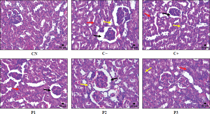
Fig. 3. Microscopic depiction of necrosis, degeneration of tubular cells, and widening of Bowman’s space in the kidney (hematoxylin and eosin staining, magnification 400 X). Description. Black line: Bowman’s space widening; red line: cell necrosis; yellow line: cell degeneration.
The C+, P1, P2, and P3 groups reduced the average widening of Bowman’s space due to streptozotocin induction in diabetic mice. The C+ group is the metformin therapy group. Metformin is an oral hypoglycemia drug approved by the Food and Drug Administration for the treatment of patients with type 2 diabetes mellitus (Sanchez-Rangel and Inzucchi, 2017). Metformin decreases liver glucose production, reduces glucose absorption in the small intestine, and improves insulin sensitivity by increasing peripheral blood glucose uptake (Mohammad et al., 2023). Hong et al. (2023) showed improvements in glomerular morphology and kidney function. Additionally, Lehtonen (2020) reported that metformin affects podocytes, which are glomerular epithelial cells that play a crucial role in maintaining glomerular ultrafiltration function.
Groups P1, P2, and P3 received therapy using active ingredients from butternut squash seed extract, which is a source of polyunsaturated fatty acids, monounsaturated fatty acids, and antioxidants that are beneficial for body health (Batool et al., 2022). The types of fatty acids in butternut squash seed extract include linoleic, oleic, palmitic, stearic, linolenic, myristic, palmitoleic, arachidic, and behenic (Suwanto et al., 2025). The fatty acids in butternut squash seeds have the potential to prevent and treat diabetic nephropathy due to their protective properties, such as antioxidant, anti-inflammatory, and neuroprotective effects (Fathima et al., 2023). Chewcharat et al. (2020) stated that fatty acids can help improve proteinuria in patients with type 2 diabetes mellitus over 24 weeks without affecting HbA1c levels. Meanwhile, Sharma et al. (2022) indicated that fatty acids have the potential to minimize or treat glomerular damage due to hyperfiltration.
Based on the results of ANOVA, it is shown that butternut squash seed extract does not affect the decrease of the Bowman’s space widening in a diabetic mice model, with a p-value of 0.395. The average widening of the Bowman’s space in the P3 and C+ groups showed a lower decree of widening of the Bowman’s space, which is attributed to the fatty acid content in butternut squash seed extract, such as omega-3 fatty acids that play a role in increasing the expression of transforming growth factor-β, reducing levels of interleukin-6, and the expression of monocyte chemoattractant protein-1 (Han et al., 2016). The reduction of the Bowman’s space widening in the C+ group is due to the metformin content, which plays a role in reducing oxidative stress due to hyperglycemia conditions and increasing AMPK phosphorylation (Rogacka and Piwkowska, 2021; Jin et al., 2023). Xu et al. (2019) indicated that metformin can protect kidney function and pathological changes in glomerular structure in diabetic rats.
Conclusion
The use of natural materials as therapy is safer than synthetic drugs and has effects on the management of DM and its complications. Butternut squash is a plant-based natural material that has been utilized as a functional food and has health benefits such as antidiabetic properties; however, research on its effects on endogenous antioxidants and the repair of kidney damage due to diabetes has not been extensively conducted. Research findings indicate that butternut squash seed extract has therapeutic effects on weight gain, reduced fasting blood glucose levels, increased SOD levels, decreased tubular cell necrosis, and decreased tubular cell degeneration, thereby demonstrating that the extract is beneficial for improving oxidative stress and microvascular complications due to hyperglycemia. Further clinical trials are needed to ensure the safety and effectiveness of butternut squash seed extract as a therapy for DM.
Acknowledgments
The author expresses gratitude to the Deputy for Human Resource Development in Science and Technology of the National Research and Innovation Agency (BRIN) for providing a Degree by Research scholarship for the odd semester of the 2023/2024 academic year (grant no. 111/II/HK.2023). The author also thanks the Directorate of Research, Technology, and Community Service of the Ministry of Education, Culture, Research, and Technology for providing operational assistance for the 2024 budget year of the doctoral dissertation research program (grant no. 048/E5/PG.02.00.PL/2024).
Conflict of interest
The author declares no conflicts of interest.
Funding
Received operational research assistance from BRIN through the Degree by Research scholarship program in 2023 and operational assistance for the doctoral dissertation research program 2024 fiscal year 2024 from the Directorate of Research, Technology, and Community Service, Ministry of Education, Culture, Research, and Technology.
Authors' contributions
S=Contributed to the preparation of the manuscript of the scientific article. S, HW=prepared data tabulation and bar diagrams. S, BR=histological preparation reading. SW, BSD, BR, HW=reviewed the draft of the
scientific article. S, HW, BSD=data analyzed using statistics. S, SW, HW=interpreted statistical data. SW, BSD, BR, HW=provided moral and material support during the research and preparation of the scientific article. S, SW, BSD, BR, HW=read and approved the manuscript for submission to the Open Veterinary Journal.
Data availability
All data used to support the research results are available in the manuscript.
References
Adeshara, K., Di Marco, E., Bordino, M., Gordin, D., Bernardi, L., Cooper, M.E. and Groop, P.H. 2024. Altered oxidant and antioxidant levels are associated with vascular stiffness and diabetic kidney disease in type 1 diabetes after exposure to acute and chronic hyperglycemia. Cardiovasc. Diabetology 23(1), 2–11.
Agius, L., Ford, B.E. and Chachra, S.S. 2020. The Metformin mechanism on gluconeogenesis and AMPK activation: the metabolite perspective. Int. J. Mol. Sci. 21(9), 2–19.
Almousa, L.A., Alfaris, N.A., Alshammari, G.M., Alsayadi, M.M., Altamimi, J.Z., Alagal, R.I. and Yahya, M.A. 2022. Rumex nervosus could alleviate streptozotocin-induced diabetic nephropathy in rats by activating Nrf2 signaling. Sci. Prog. 105(2), f2–23.
Amelia, R., Sari, D., Muzasti, R., Fujiati, I. and Wijaya, H. 2021. Early detection of diabetic nephropathy based on albumin creatinine ratio (ACR) in type 2 diabetes mellitus patients in Medan, Indonesia. Fam. Med. &. Primary. Care. Rev. 23(2), 135–138.
Batool, M., Ranjha, M.M.A.N., Roobab, U., Manzoor, M.F., Farooq, U., Nadeem, H.R., Nadeem, M., Kanwal, R., Abdelgawad, H., Al Jaouni, S.K., Selim, S. and Ibrahim, S.A. 2022. Nutritional value, phytochemical potential, and therapeutic benefits of pumpkin (Cucurbita sp.). Plants 11(11), 1–23.
Blahova, J., Martiniakova, M., Babikova, M., Kovacova, V., Mondockova, V. and Omelka, R. 2021. Pharmaceutical drugs and natural therapeutic products for the treatment of type 2 diabetes mellitus. Pharmaceuticals 14(8), 2–32.
Budiastuti, B., Noviana Thesia, N.L., Plumeriastuti, H., Effendi, M.H., Ugbo, E.N., Khairullah, A.R., Moses, I.B., Priscilia Riwu, K.H., Ahmad, R.Z., Yanestria, S.M., Wasito, W., Pratama, B.P., Awwanah, M. and Prasetyo, A. 2025. Effect of cinnamon oil (Cinnamomum burmannii) on the histological kidney of male diabetic rats (Rattus norvegicus). Open. Vet. J. 15(2), 923–930.
Caturano, A., D’Angelo, M., Mormone, A., Russo, V., Mollica, M.P., Salvatore, T., Galiero, R., Rinaldi, L., Vetrano, E., Marfella, R., Monda, M., Giordano, A. and Sasso, F.C. 2023. Oxidative stress in type 2 diabetes: impacts from pathogenesis to lifestyle modifications. Curr. Issues Mol. Biol. 45(8), 6651–6666.
Chen, L., Jiang, Q., Lu, H., Jiang, C., Hu, W., Yu, S., Xiang, X., Tan, C.P., Feng, Y., Zhang, J., Li, M. and Shen, G. 2022. Antidiabetic effect of sciadonic acid on type 2 diabetic mice through activating the PI3K-AKT signaling pathway and altering intestinal flora. Front. Nutr. 9(1), 1–15.
Chen, X., Qian, L., Wang, B., Zhang, Z., Liu, H., Zhang, Y. and Liu, J. 2019. Synergistic hypoglycemic effects of pumpkin polysaccharides and puerarin on type II diabetes mellitus mice. Molecules 24(5), 2–16.
Chenni, A., Cherif, F.Z.H., Chenni, K., Elius, E.E., Pucci, L. and Yahia, D.A. 2022. Effects of pumpkin (Cucurbita pepo L.) seed protein on blood pressure, plasma lipids, leptin, adiponectin, and oxidative stress in rats with fructose-induced metabolic syndrome. Prev. Nutr. Food Sci. 27(1), 78–88.
Chewcharat, A., Chewcharat, P., Rutirapong, A. and Papatheodorou, S. 2020. The effects of omega-3 fatty acids on diabetic nephropathy: a meta-analysis of randomized controlled trials. PLos One. 15(2), 1–18.
Chike-Ekwughe, A., John-Africa, L.B., Adebayo, A.H. and Ogunlana, O.O. 2024. Antioxidative and anti-diabetic effects of Tapinanthus cordifolius leaf extract on high-fat diet and streptozotocin-induced type 2 diabetic rats. Biomed. Pharmacother. 176(1), 1–15.
Cho, K., Chung, J.Y., Cho, S.K., Shin, H.W., Jang, I.J., Park, J.W., Yu, K.S. and Cho, J.Y. 2015. Antihyperglycemic mechanism of metformin occurs via the AMPK/LXRα/POMC pathway. Scientific. Rep. 5(1), 1–7.
Cho, N.H., Shaw, J.E., Karuranga, S., Huang, Y., Da Rocha Fernandes, J.D., Ohlrogge, A.W. and Malanda, B. 2018. IDF diabetes atlas: global estimates of diabetes prevalence for 2017 and projections for 2045. Diabetes. Res. Clin. Pract. 138(1), 271–281.
Cobbs, A., Chen, X., Zhang, Y., George, J., Huang, M.B., Bond, V., Thompson, W. and Zhao, X. 2019. Saturated fatty acid stimulates production of extracellular vesicles by renal tubular epithelial cells. Mol. Cell. Biochem. 458(1-2), 113–124.
Deng, Y., Zhang, Y., Liu, G., Zhou, P., Li, P., Zhao, Z., Zhang, R., Tang, X., Wang, Z., Wei, Z. and Zhang, M. 2023. Saponins from Momordica charantia exert hypoglycemic effect in diabetic mice by multiple pathways. Food. Sci. Nutr. 11(12), 7626–7637.
Dotto, J.M. and Chacha, J.S. 2020. The potential of pumpkin seeds as a functional food ingredient: a review. Scientific Afr. 10(1), 1–14.
Dowidar, M.F., Ahmed, A.I. and Mohamed, H.R. 2020. Antidiabetic effect of pumpkin seeds and gum arabic and/or vildagliptin on type 2 induced diabetes in male rats. Int. J. Vet. Sci. 9(2), 229–233.
Dunford, E.K., Miles, D.R. and Popkin, B. 2023. Food additives in ultra-processed packaged foods: an examination of US household grocery store purchases. J. Acad. Nutr. Diet. 123(6), 889–901.
El-Boshy, M., Alsaegh, A., Qasem, A.H., Sindi, R.A., Abdelghany, A.H., Gadalla, H., Reda, D., Azzeh, F., Idris, S., Ahmad, J. and Refaat, B. 2021. Enhanced renoprotective actions of Paricalcitol and omega-3 fatty acids co-therapy against diabetic nephropathy in rat. J. Adv. Res. 38(1), 119–129.
Fathima, S., Prokopiou, E. and Georgiou, T. 2023. Omega-3 polyunsaturated fatty acids and their anti-oxidant, anti-inflammatory and neuroprotective effects in diabetic retinopathy: a narrative review. Front. Biosci. (Landmark Ed). 28(7), 1–14.
Feng, A., Yin, R., Xu, R., Zhang, B. and Yang, L. 2025. An update on renal tubular injury as related to glycolipid metabolism in diabetic kidney disease. Front. Pharmacol. 16(1), 1–14.
Frąk, W., Dąbek, B., Balcerczyk-Lis, M., Motor, J., Radzioch, E., Młynarska, E., Rysz, J. and Franczyk, B. 2024. Role of uremic toxins, oxidative stress, and renal fibrosis in chronic kidney disease. Antioxidants 13(6), 2–19.
Frydrych, A., Kulita, K., Jurowski, K. and Piekoszewski, W. 2025. Lipids in clinical nutrition and health: narrative review and dietary recommendations. Foods 14(3), 473.
Gilani, S.J., Bin-Jumah, M.N., Al-Abbasi, F.A., Nadeem, M.S., Afzal, M., Sayyed, N. and Kazmi, I. 2021. Fustin ameliorates hyperglycemia in streptozotocin induced type-2 diabetes via modulating glutathione/superoxide dismutase/catalase expressions, suppress lipid peroxidation and regulates histopathological changes. Saudi J. Biol. Sci. 28(12), 6963–6971.
Graniel-Amador, M.A., Torres-Rodríguez, H.F., Martínez-Mendoza, R.E., Vargas-Muñoz, V.M., Acosta-González, R.I., Castañeda-Corral, G., Muñoz-Islas, E. and Jiménez-Andrade, J.M. 2022. Effect of chronic lithium on mechanical sensitivity and trabecular bone loss induced by type-1 diabetes mellitus in mice. BioMetal 35(1), 1033–1042.
Gurumallu, S.C., Alramadneh, T.N., Sarjan, H.N., Bhaskar, A., Pereira, C.M.F. and Javaraiah, R. 2022. Synergistic hypoglycemic and hypolipidemic effects of ω-3 and ω-6 fatty acids from Indian flax and sesame seed oils in streptozotocin-induced diabetic rats. Phytomedicine. Plus. 2(1), 1–12.
Hahn, M., Van Krieken, P.P., Nord, C., Alanentalo, T., Morini, F., Xiong, Y., Eriksson, M., Mayer, J., Kostromina, E., Ruas, J.L., Sharpe, J., Pereira, T., Berggren, P.O., Ilegems, E. and Ahlgren, U. 2020. Topologically selective islet vulnerability and self-sustained downregulation of markers for β-cell maturity in streptozotocin-induced diabetes. Communication Biol. 3(1), 1–14.
Halim, M.A., Wazed, M.A., Al Obaid, S., Ansari, M.J., Tahosin, A., Rahman, M.T., Noor, F., Mozumder, N.H.M.R. and Khatun, A.A. 2024. Effect of storage on physicochemical properties, bioactive compounds and sensory attributes of drinks powder enriched with pumpkin (Cucurbita moschata L.). J. Agriculture. Food. Res. 18(1), 1–12.
Han, E., Yun, Y., Kim, G., Lee, Y.H., Wang, H.J., Lee, B.W., Cha, B.S., Kim, B.S. and Kang, E.S. 2016. Effects of omega-3 fatty acid supplementation on diabetic nephropathy progression in patients with diabetes and hypertriglyceridemia. PLos One. 11(5), 1–11.
Haryanto, Zulkifli, Pertiwi, F.A., Amalia, S.P., Nurpinanda., Adelia, U. and Putri, A.D. 2025. Comparative study of the anesthetic effects of ether and chloroform on Wistar Rats (Rattus norvegicus): analysis of induction time and duration of anesthesia. Thermometer. Scientific J. Health Sci. Med. 3(3), 133–139.
Hendarto, H., Rifa, A., Dwita, N.C., Nurjannah, I., Sari, F.R. and Akbar, F.N. 2022. Short-term effects of Garcinia cambogia on blood glucose and body weight in streptozosin-induced diabetic rats. Malaysian J. Med. Health Sci. 18(16), 78–81.
Hong, Y., Wang, J., Sun, W., Zhang, L., Xu, X. and Zhang, K. 2023. Gallic acid improves the metformin effects on diabetic kidney disease in mice. Renal Failure 45(1), 2–10.
Horakova, O., Kroupova, P., Bardova, K., Buresova, J., Janovska, P., Kopecky, J. and Rossmeisl, M. 2019. Metformin acutely lowers blood glucose levels by inhibition of intestinal glucose transport. Scientific. Rep. 9(6156), 1–11.
Hosen, M., Rafii, M.Y., Mazlan, N., Jusoh, M., Oladosu, Y., Chowdhury, M.F.N., Muhammad, I. and Khan, M.M.H. 2021. Pumpkin (Cucurbita spp.): a crop to mitigate food and nutritional challenges. Horticulturae 7(10), 2–25.
Huang, L., Yang, Z., Yuan, J., Zuo, S., Li, Z., Yang, K., Wang, S., Li, J., Zhu, L. and Zhang, Y. 2023. Preparation and characteristics of pumpkin polysaccharides and their effects on abnormal glucose metabolism in diabetes mice. Food. Bioscience. 54(1), 2–11.
Jasim, M.E. and Abdul-Razzaq, N.E. 2024. A comparative study between metformin and insulin on biochemical variables in diabetic patients. Endocrine Metabolic Sci. 16(1), 2–6.
Jena, A.B., Samal, R.R., Bhol, N.K. and Duttaroy, A.K. 2023. Cellular red-ox system in health and disease: the latest update. Biomed. Pharmacother. 1621, 1–21.
Jeong, M., Shin, J.I., Cho, J., Jeon, Y.J., Kim, J.H., Youn, J. and Lee, K. 2023. DHA induces cell death through the production of ROS and the upregulation of CHOP in fibroblast-like synovial cells from human rheumatoid arthritis patients. Int. J. Mol. Sci. 24(2), 2–15.
Jiang, S., Yang, W., Li, Y., Feng, J., Miao, J., Shi, H. and Xue, H. 2023. Monounsaturated and polyunsaturated fatty acids concerning prediabetes and type 2 diabetes mellitus risk among participants in the National Health and Nutrition Examination Surveys (NHANES) from 2005 to March 2020. Front. Nutr. 10(1), 1–11.
Jin, Q., Liu, T., Qiao, Y., Liu, D., Yang, L., Mao, H., Ma, F., Wang, Y., Peng, L. and Zhan, Y. 2023. Oxidative stress and inflammation in diabetic nephropathy: role of polyphenols. Front. Immunol. 14(1), 1–17.
Kashyap, H. and Gupta, S. 2019. Analysis of metformin on endogenous antioxidants and oxidative stress in mice brain tissue of alloxan-induced diabetes. Int. J. Pharm. Sci. Drug Res. 11(5), 272–277.
Koffert, J.P., Mikkola, K., Virtanen, K.A., Andersson, A.M.D., Faxius, L., Hällsten, K., Heglind, M., Guiducci, L., Pham, T., Silvola, J.M.U., Virta, J., Eriksson, O., Kauhanen, S.P., Saraste, A., Enerbäck, S., Iozzo, P., Parkkola, R., Gomez, M.F. and Nuutila, P. 2017. Metformin treatment significantly enhances intestinal glucose uptake in patients with type 2 diabetes: results from a randomized clinical trial. Diabetes. Res. Clin. Pract. 131(1), 208–216.
Kosnayani, A.S., Dharmana, E., Hadisaputro, S. and Riwanto, I. 2021. Effect combination of metformin and meniran (Phyllanthus niruri Linn.) water extract on the improvement of obesity status male Sprague Dawley rats. Amerta Nutr. 5(1), 52–58.
Kumar, M., Dev, S., Khalid, M.U., Siddenthi, S.M., Noman, M., John, C., Akubuiro, C., Haider, A., Rani, R., Kashif, M., Varrassi, G., Khatri, M., Kumar, S. and Mohamad, T. 2023. The bidirectional link between diabetes and kidney disease: mechanisms and management. Cureus 15(9), 1–13.
Kusuma, I.Y., Samodra, G., Komala, Y.I., Apriliansa, E.P., Piri, J.P.A. and Fauqina, A.A. 2022. Anti-hyperglycemic effects of dapagliflozin monotherapy and combination with metformin in mice. Maritime. Pharm. Scientific. J. 13(1), 72–80.
Lateef, M.A.M., Abdulhadi, A.L. and Ali, L.H. 2024. Study of the therapeutic efficacy of pumpkin seed in improving the liver's activity in rats with stz-induced diabetes. Front. Health. Informat. 13(3), 5336–5351.
Lee, S.M., Chung, S.H., Park, Y., Park, M.K., Son, Y.K., Kim, S.E. and An, W.S. 2015. Effect of omega-3 fatty acid on the fatty acid content of the erythrocyte membrane and proteinuria in patients with diabetic nephropathy. Int. J. Endocrinol. 1(1), 1–8.
Lehtonen, S. 2020. Metformin protects against podocyte injury in diabetic kidney disease. Pharmaceuticals 13(12), 2–26.
Li, H. 2020. Evaluation of bioactivity of butternut squash (Cucurbita moschata D.) seeds and skin. Food. Sci. Nutr. 8(7), 3252–3261.
Liu, J., Aylor, K.W., Chai, W., Barrett, E.J. and Liu, Z. 2022. Metformin prevents endothelial oxidative stress and microvascular insulin resistance during obesity development in male rats. Am. J. Physiol. Endocrinol. Metab. 322(3), 293–306.
Madiraju, A.K., Qiu, Y., Perry, R.J., Rahimi, Y., Zhang, X., Zhang, D., Camporez, J., Cline, G.W., Butrico, G.M., Kemp, B.E., Casals, G., Steinberg, G.R., Vatner, D.F., Petersen, K.F. and Shulman, G.I. 2018. Metformin inhibits gluconeogenesis by a redox-dependent mechanism in vivo. Nat. Med. 24(9), 1384–1394.
Malini, D.M., Fitriani, N., Lila, A., Ratningsih, N. and Setiawati, T. 2021. Morphological and histological structure of the kidneys of diabetic model rats after being treated with ethanol extract of jengkol fruit skin (Archidendron pauciflorum). Udayana Biol. J. 25(2), 208–217.
Mardiana, Z.A., Ardiaria, M., Ayustaningwarna, F. and Rahadiyanti, A. 2022. The effect of edamame juice (Glycine max (L.) merrill) on uric acid levels in male wistar diabetic rats. J. Nutr. Coll. 11(1), 51–61.
Mardiati, S.F. and Sitasiwi, A.J. 2016. Weight gain mice (Mus musculus L.) after treatment water seed extract papaya (Carica papaya Linn.) by oral for 21 days. Bull. Anatomy Physiol. 1(1), 75–80.
Marefati, N., Navidiyan, E.S., Bahari, Z., Raei, M. and Mohammadi, M.T. 2023. The effect of hyperglycemia on catalase activity and glutathione levels of liver in streptozotocin-induced diabetes mellitus in rat. J. Pathobiol. Res. 26(3), 37–41.
Marino, F., Salerno, N., Scalise, M., Salerno, L., Torella, A., Molinaro, C., Chiefalo, A., Filardo, A., Siracusa, C., Panuccio, G., Ferravante, C., Giurato, G., Rizzo, F., Torella, M., Donniacuo, M., De Angelis, A., Viglietto, G., Urbanek, K., Weisz, A., Torella, D. and Cianflone, E. 2023. Streptozotocin-induced type 1 and 2 diabetes mellitus mouse models show different functional, cellular and molecular patterns of diabetic cardiomyopathy. Int. J. Mol. Sci. 24(2), 2–22.
Masenga, S.K., Kabwe, L.S., Chakulya, M. and Kirabo, A. 2023. Mechanisms of oxidative stress in metabolic syndrome. Int. J. Mol. Sci. 24(9), 2–28.
Mathias Akinlade, O., Victor Owoyele, B. and Olufemi Soladoye, A. 2021. Streptozotocin-induced type 1 and 2 diabetes in rodents: a model for studying diabetic cardiac autonomic neuropathy. Afr. Health Sci. 21(2), 719–727.
Mestry, S.N., Dhodi, J.B., Kumbhar, S.B. and Juvekar, A.R. 2017. Attenuation of diabetic nephropathy in streptozotocin-induced diabetic rats by Punica granatum Linn. leaves extract. J. Traditional Complementary Med. 7(3), 273–280.
Mohammad, H.M.F., Galal Gouda, S., Eladl, M.A., Elkazaz, A.Y., Elbayoumi, K.S., Farag, N.E., Elshormilisy, A., Al-Ammash, B.B., Hegazy, A., Abdelkhalig, S.M., Mohamed, A.S., El-Dosoky, M. and Zaitone, S.A. 2023. Metformin suppresses LRG1 and TGFβ1/ALK1-induced angiogenesis and protects against ultrastructural changes in rat diabetic nephropathy. Biomed. Pharmacotherapy. 158(1), 2–16.
Mohan, I.K and Das, U.N. 2001. Prevention of chemically induced diabetes mellitus in experimental animals by polyunsaturated fatty acids. Nutrition 17(2), 126–151.
Munjiati, N.E. 2021. The effect of single dose streptozotocin administration on blood glucose levels in Wistar rats (Rattus norvegicus). Medirory. J. Med. Lab. 9(1), 62–67.
Munteanu, C., Kotova, P. and Schwartz, B. 2025. Impact of olive oil components on the expression of genes related to type 2 diabetes mellitus. Nutrients 17(3), 2–56.
Muttaqien, Y.V. and Purnama, E.R. 2024. Blood glucose levels and diabetic ulcers amelioration in mice after mangrove (Bruguiera gymnorrhiza) leaves extract treatments. LenteraBio. Scientific Periodical Biol. 13(1), 55–64.
Nurasidah, R., Tangka, J., Elyani, H. and Purnomo, Y. 2020. Effect of ethanol extract of red gedi leaves (Abelmoschus manihot (L.) Medik) on the levels of tumor necrosis factor alpha (TNF-α) in the kidneys and the amount of necrosis in the renal glomerular epithelial cells in diabetic mellitus model rats. Complementary. Med. J. 7(2), 1–10.
Okesola, M.A., Ajiboye, B.O., Oyinloye, B.E., Osukoya, O.A., Owero-Ozeze, O.S., I. Ekakitie, L. and Kappo, A.P. 2025. Effect of Solanum macrocarpon Linn leaf aqueous extract on the brain of an alloxan-induced rat model of diabetes. J. Int. Med. Res. 48(5), 1–10.
Onuche, J.I., Adebisi, A.K., Ikwebe, J. and Abu, M.S. 2023. Antinephrotoxic and antihyperlipidaemic activities of Cucurbita maxima leaf supplemented diets in stz-induced diabetic rats. Toxicol. Adv. 5(4), 1–9.
Poerwanagara, I.P., Ariani, M.D., Mahati, E. and Nindita, Y. 2025. Effect of lemon peel (Citrus limon) extract administration on the macroscopic and microscopic characteristic of Wistar male rats kidneys with hyperuricemia. Diponegoro. Med. J. 14(6), 304–310.
Poznyak, A., Grechko, A.V., Poggio, P., Myasoedova, V.A., Alfieri, V. and Orekhov, A.N. 2020. The diabetes mellitus–atherosclerosis connection: the role of lipid and glucose metabolism and chronic inflammation. Int. J. Mol. Sci. 21(5), 1–13.
Prommaban, A., Kuanchoom, R., Seepuan, N. and Chaiyana, W. 2021. Evaluation of fatty acid compositions, antioxidant, and pharmacological activities of pumpkin (Cucurbita moschata) seed oil from aqueous enzymatic extraction. Plants 10(8), 2–18.
Purnomo, E., Luqman, E.M., Ratnani, H., Plumeriastuti, H., Mafruchati, M. and Dhamayanti, Y. 2020. The effect of carbofuran exposure on mice (Mus musculus) against kidney histopathology appearance of mice pups during lactation period. J. Basic Med. Vet. 9(1), 54–62.
Putera, H.D., Doewes, R.I., Shalaby, M.N., Ramírez-Coronel, A.A., Clayton, Z.S., Abdelbasset, W.K., Murtazaev, S.S., Jalil, A.T., Rahimi, P., Nattagh-Eshtivani, E., Malekahmadi, M. and Pahlavani, N. 2023. The effect of conjugated linoleic acids on inflammation, oxidative stress, body composition and physical performance: a comprehensive review of putative molecular mechanisms. Nutr. Metab. 20(35), 2–16.
Qiao, Y., Chen, H., Guo, J., Zhang, X., Liang, X., Wei, L., Wang, Q., Bi, H. and Gao, T. 2024. A study on the effects of metacinnabar (β-HgS) on weight and appetite recovery in stressed mice. J. Ethnopharmacol. 335(5), 2–16.
Regulation of the Minister of Health of the Republic of Indonesia. 2012. Regulation of the Minister of Health of the Republic of Indonesia Number 033 of 2012 concerning Food Additives.
Rinawati., Lili, E.N.N., Muhsin, S.W. and Siregar, S.M.F. 2020. The influence of sea lettuce (Ulva lactuca) water extract on body weight in diabetic rats. STIGMA. J. Math. Natural. Sci. UNIPA. 13(1), 39–46.
Rogacka, D. and Piwkowska, A. 2021. Beneficial effects of metformin on glomerular podocytes in diabetes. Biochem. Pharmacol. 192(1), 1–8.
Sadžak, A., Mravljak, J., Maltar-Strmečki, N., Arsov, Z., Baranović, G., Erceg, I., Kriechbaum, M., Strasser, V., Přibyl, J. and Šegota, S. 2020. The structural integrity of the model lipid membrane during induced lipid peroxidation: the role of flavonols in the inhibition of lipid peroxidation. Antioxidants 9(5), 2–28.
Saeedi, P., Petersohn, I., Salpea, P., Malanda, B., Karuranga, S., Unwin, N., Colagiuri, S., Guariguata, L., Motala, A.A., Ogurtsova, K., Shaw, J.E., Bright, D. and Williams, R. 2019. Global and regional diabetes prevalence estimates for 2019 and projections for 2030 and 2045: results from the international diabetes federation diabetes atlas, 9th edition. Diabetes Res. Clin. Pract. 157(1), 1–10.
Sanchez-Rangel, E. and Inzucchi, S.E. 2017. Metformin: clinical use in type 2 diabetes. Diabetologia 60(9), 1586–1593.
Sasongko, H., Endro Nugroho, A., Nurrochmad, A. and Rohman, A. 2024. Nephroprotective effect of milkfish, patin, and snakehead fish oil by suppressing inflammation and oxidative stress in diabetic rats. Indonesian J. Pharm. 35(1), 63–73.
Sedik, A.A., El-Mosallamy, A.E.M.K., Yassen, N.N. and El-Nasr, N.M.E.A. 2025. Dose dependent toxicity of streptozotocin inducing diabetic complications in male Sprague Dawley rats with metabolic biochemical histopathological effects. Scientific Rep. 15(1), 1–15.
Seto, T., Grondin, J.A. and Khan, W.I. 2025. Food additives: emerging detrimental roles on gut health. Faseb. J. 39(13), 1–13.
Setyawati, U.G. and Mahmudiono, T. 2023. Education level, duration of selling, and knowledge about food additives and methanil yellow: a study on online noodle sellers (gofood and grabfood) in East Surabaya. Indonesian Nutr. Media (Nat. Nutr. J). 18(1), 56–62.
Shabalala, S.C., Johnson, R., Basson, A.K., Ziqubu, K., Hlengwa, N., Mthembu, S.X.H., Mabhida, S.E., Mbeje, S.E.M., Hanser, S., Cirilli, I., Tiano, L. and Dludla, P.V. 2022. Detrimental effects of lipid peroxidation in type 2 diabetes: exploring the neutralizing influence of antioxidants. Antioxidants 11(10), 2–17.
Sharma, M., Singh, V., Sharma, R., Koul, A., Mccarthy, E.T., Savin, V.J., Joshi, T. and Srivastava, T. 2022. Glomerular biomechanical stress and lipid mediators during cellular changes leading to chronic kidney disease. Biomedicines 10(2), 2–33.
Shi, L.J., Tang, X., He, J. and Shi, W. 2021. Hyperlipidemia influences the accuracy of glucometer-measured blood glucose concentrations in genetically diverse mice. Am. J. Med. Sci. 362(3), 297–302.
Strugała, P., Dzydzan, O., Brodyak, I., Kucharska, A.Z., Kuropka, P., Liuta, M., Kaleta-Kuratewicz, K., Przewodowska, A., Michałowska, D., Gabrielska, J. and Sybirna, N. 2019. Antidiabetic and antioxidative potential of the blue congo variety of purple potato extract in streptozotocin-induced diabetic rats. Molecules 24(17), 3126.
Sun, T., Liu, J., Xie, C., Yang, J., Zhao, L. and Yang, J. 2021. Metformin attenuates diabetic renal injury via the AMPK-autophagy axis. Exp. Therapeutic Med. 21(6), 2–8.
Suwannapong, A., Talubmook, C. and Promprom, W. 2023. Evaluation of antidiabetic and antioxidant activities of fruit pulp extracts of Cucurbita moschata Duchesne and Cucurbita maxima Duchesne. Scientific. World. J. 1(1), 2–12.
Suwanto., Gustomi, M.P. and Kurnijasanti, R. 2020. Potential of ethanol extract of Cucurbita moschata seeds on malondialdehyde levels in diabetes model mice. Indonesian. J. Hum. Nutr. 7(1), 20–30.
Suwanto, Gustomi, M.P. and Kurnijasanti, R. Ethanol extract of pumpkin seeds (Cucurbita moschata Duch) as an antihyperglycemic in mice exposed to streptozotocin. In Proceedings of the Annual Pharmacy Conference, 2019, Surakarta, Indonesia, Pharmacy Study Program, Faculty of Mathematics and Natural Sciences, Universitas Sebelas Maret, pp 13–24.
Suwanto, Widiyanto, S., Daryono, B.S., Retnoaji, B. and Widodo, H. 2025. In Press. Diabetic ulcer healing in NIH3T3 fibroblast cells treated with butternut squash seed extract (Cucurbita moschata Duchesne). Trop. J. Natural. Product. Res.
Tang, X., Li, J., Xiang, W., Cui, Y., Xie, B., Wang, X., Xu, Z. and Gan, L. 2016. Metformin increases hepatic leptin receptor and decreases steatosis in mice. J. Endocrinol. 230(2), 227–237.
Taso, O.V., Philippou, A., Moustogiannis, A., Zevolis, E. and Koutsilieris, M. 2019. Lipid peroxidation products and their role in neurodegenerative diseases. Ann. Res. Hosp. 3(2), 2–10.
Theresia, R., Falah, S. and Safithri, M. 2017. Antihyperglycemic activity of extracts from surian (Toona sinensis) bark and leaves in diabetic rats (Sprague-Dawley) induced by Streptozotocin. J. Food. Nutr. 12(3), 187–194.
Tudurí, E., López, M., Diéguez, C., Nadal, A. and Nogueiras, R. 2016. Glucagon-like peptide 1 analogs and their effects on pancreatic islets. Trends. Endocrinol. Metab. 27(5), 304–318.
Tunnur, U., Jailani, Andalia, N. and Akmal, N. 2023. The influence of ethanol extract of kersen leaves (Muntingia calabura L.) on the histopathological profile of kidneys induced by STZ (Streptozotocin) in white rats (Rattus norvegicus). J. Learn. Sci. 2(2), 51–61.
Ukratalo, A.M., Amahoru, G., Monery, D.E., Zuneidi, T., Pangemanan, V.O. and Loilatu, M.F. 2024. Weight change in mice (Mus musculus) with type 1 diabetes mellitus treated with brown algae extract Sargassum sp. Anesthesia J. J. Health Sci. Med. 2(3), 39–47.
Uljević, M.V., Starčević, K., Mašek, T., Bočina, I., Restović, I., Kević, N., Racetin, A., Kretzschmar, G., Grobe, M., Vukojević, K., Saraga-Babić, M. and Filipović, N. 2019. Dietary DHA/EPA supplementation ameliorates diabetic nephropathy by protecting from distal tubular cell damage. Cell Tissue Res. 378(2), 301–317.
Wahidin, M., Achadi, A., Besral, B., Kosen, S., Nadjib, M., Nurwahyuni, A., Ronoatmodjo, S., Rahajeng, E., Pane, M. and Kusuma, D. 2024. Projection of diabetes morbidity and mortality till 2045 in Indonesia based on risk factors and NCD prevention and control programs. Scientific Rep. 24(1), 1–17.
Wang, F., Sun, H., Zuo, B., Shi, K., Zhang, X., Zhang, C. and Sun, D. 2021. Metformin attenuates renal tubulointerstitial fibrosis via upgrading autophagy in the early stage of diabetic nephropathy. Scientific. Rep. 11(1), 16362.
Wang, G., Wang, Y., Yang, Q., Xu, C., Zheng, Y., Wang, L., Wu, J., Zeng, M. and Luo, M. 2022. Metformin prevents methylglyoxal-induced apoptosis by suppressing oxidative stress in vitro and in vivo. Cell. Death. Dis. 13(29), 2–11.
Xie, X., Chen, C. and Fu, X. 2023. Modulation effects of Sargassum pallidum extract on Hyperglycemia and Hyperlipidemia in type 2 diabetic mice. Foods 12(24), 2–19.
Xu, J., Liu, L.Q., Xu, L.L., Xing, Y. and Ye, S. 2019. Metformin alleviates renal injury in diabetic rats by inducing Sirt1/FoxO1 autophagic signal axis. Clin. Exp. Pharmacol. Physiol. 47(4), 599–608.
Xu, S., Li, X., Hou, Q., Xu, N., Lu, Q., Wang, S. and Dai, C. 2024. Association of serum unsaturated fatty acid patterns with the risk of diabetic nephropathy. Kidney Dis. 10(2), 97–106.
Yang, X., Xu, Z., Zhang, C., Cai, Z. and Zhang, J. 2017. Metformin, beyond an insulin sensitizer, targeting heart and pancreatic β cells. Biochimica Et Biophysica Acta. (BBA)-Molecular. Basis Dis. 1863(8), 1984–1990.
Yerevanian, A. and Soukas, A.A. 2019. Metformin: mechanisms in human obesity and weight loss. Curr. Obesity Rep. 8(1), 156–164.
Zhang, S., Xu, H., Yu, X., Wu, Y. and Sui, D. 2017. Metformin ameliorates diabetic nephropathy in a rat model of low-dose streptozotocin-induced diabetes. Exp. Therapeutic. Med. 14(1), 383–390.
Zhang, W., Zhao, L., Zhang, J., Li, P. and Lv, Z. 2021. Metformin improves cognitive impairment in diabetic mice induced by a combination of streptozotocin and isoflurane anesthesia. Bioengineered 12(2), 10982–10993.
Zheng, Z., Zong, Y., Ma, Y., Tian, Y., Pang, Y., Zhang, C. and Gao, J. 2024. Glucagon-like peptide-1 receptor: mechanisms and advances in therapy. Signal. Transduction. Targeted. Therapy. 9(1), 2–29.
Zhou, T., Xu, X., Du, M., Zhao, T. and Wang, J. 2018. A preclinical overview of metformin for the treatment of type 2 diabetes. Biomed. Pharmacotherapy. 106(1), 1227–1235.








