Open Veterinary Journal, (2026), Vol. 16(7): 4670-4679
Research Article
10.5455/OVJ.2026.v16.i7.47
Prevalence, risk factors, and molecular identification of Eimeria spp. in small ruminants from three districts in Java, Indonesia
Estha Gusmalia Kustika1, Mila Putri Anggraini1, Fathur Rohman Haryadi1, Fitrine Ekawasti2,
Madarina Wasissa3 and Fajar Budi Lestari1,4*
1Department of Bioresources Technology and Veterinary, School of Applied Science, Universitas Gadjah Mada, Yogyakarta, Indonesia
2Research Center for Veterinary Science, National Research and Innovation Agency (BRIN), Bogor, Indonesia
3Department of Clinical Pathology, Faculty of Veterinary Medicine, Universitas Gadjah Mada, Yogyakarta, Indonesia
4Veterinary Technology Laboratory, School of Applied Science, Universitas Gadjah Mada, Yogyakarta, Indonesia
*Corresponding Author: Fajar Budi Lestari. Department of Bioresources Technology and Veterinary, School of Applied Science, Universitas Gadjah Mada, Yogyakarta, Indonesia. Email: fajar.budi.l [at] mail.ugm.ac.id
Submitted: 28/11/2025 Revised: 22/05/2026 Accepted: 08/06/2026 Published: 20/07/2026
© 2025 Open Veterinary Journal
This is an Open Access article distributed under the terms of the Creative Commons Attribution-Non Commercial-No Derivatives License (http://creativecommons.org/licenses/by-nc-nd/4.0/), which permits non-commercial re-use, distribution, and reproduction in any medium, provided the original work is properly cited, and is not altered, transformed, or built upon in any way.
ABSTRACT
Background: Coccidiosis caused by protozoa of the genus Eimeria remains a leading parasitic constraint in small ruminant production systems worldwide.
Aim: To determine the prevalence, risk factors, and species of Eimeria spp. that infect goats and sheep.
Methods: Fresh fecal samples were collected from 112 goats and sheep across the Sragen, Sleman, and Ngawi regencies. The samples were examined using both qualitative (direct smear and flotation) and quantitative (McMaster) methods. For molecular analysis, genomic DNA was extracted from samples with >200 oocysts per gram. The 18S rRNA gene was targeted via polymerase chain reaction, followed by sequencing representative amplicons to identify species. The data were analyzed using descriptive statistics and logistic regression.
Results: A high prevalence of 90.18% (101/112) was found. Molecular characterization identified the presence of Eimeria crandallis and Eimeria christenseni. Sex, age, and type of forage were not associated risk factors for Eimeria spp. infection (p > 0.05).
Conclusion: The prevalence of Eimeria infection in sheep and goats was high, although no clinical symptoms were observed. Eimeria crandallis and E. christenseni were identified. Multivariate analysis showed that sex, age, and forage type had no significant effect on the occurrence of Eimeria infection. Prevention strategies are needed to minimize economic losses in small ruminant production.
Keywords: Eimeria spp., Fecal samples, Goats, Java, Sheep.
Introduction
Coccidiosis, caused by protozoa of the genus Eimeria, remains a primary parasitic constraint in small ruminant production worldwide, particularly affecting goats (Capra hircus) and sheep (Ovis aries) (Ali et al., 2025). As obligatory intracellular parasites, Eimeria spp. damage the intestinal epithelium, resulting in clinical disease characterized by hemorrhagic diarrhea, severe dehydration, and mortality in young stock (Taylor et al., 2016; Weng et al., 2024). Subclinical infections impose a substantial burden on tropical farming systems by reducing feed conversion efficiency and overall growth performance (Silva et al., 2014; Ali et al., 2025).
The dynamics of these infections are closely linked to environmental conditions. Temperature and humidity are the primary determinants of oocyst sporulation and persistence in the field (Weng et al., 2024). Consequently, in tropical settings, such as Indonesia, climatic factors, combined with management stressors, such as high stocking density, may drive high prevalence rates (Khodakaram-Tafti and Hashemnia, 2017). However, despite this possibly high disease burden, there are few comprehensive reports using molecular detection and sequencing for Eimeria spp. in Indonesia. Although traditional morphological identification remains the gold standard in veterinary diagnostics, its reliance on sporulated oocysts often lacks the sensitivity required to differentiate between morphologically similar species, such as Eimeria arloingi and Eimeria ninakohlyakimovae (Hassanen, 2020). This reliance on conventional methods has created a significant diagnostic gap, limiting the accurate mapping of highly pathogenic species across local livestock in Indonesia.
To bridge this gap, molecular techniques targeting the 18S rRNA gene provide a more precise means of species classification (Kawahara et al., 2010; Liang et al., 2022). Therefore, this study aimed to determine the prevalence and identify the specific Eimeria species circulating in three districts of Java, Sragen, Sleman, and Ngawi, Indonesia while evaluating the associated risk factors. This work provides an essential framework for targeted control strategies to minimize the economic impact of coccidiosis in tropical small ruminant systems by integrating classical parasitology with molecular characterization.
Materials and Methods
Study design
This cross-sectional study was conducted to determine the prevalence of coccidiosis in domestic goats (Capra aegagrus hircus) and sheep (Ovis aries) across three subdistricts in Java, Indonesia. A purposive sampling method was used. The researchers collected fecal samples door-to-door in January 2025, and the geographic coordinates of each sampling location were recorded using a smartphone-integrated Global Positioning System with Google Maps.
The sample size was calculated using Epitools (https://epitools.ausvet.com.au/) to estimate the required proportion of samples. Based on the reported prevalence of Eimeria spp. infection in Indonesia in 2024 (37.7%) (Ninditya et al., 2024), with a 95% confidence interval (CI) and a margin of 10% error, the minimum required sample size was 91 animals. To improve the robustness of the study and account for potential data loss, additional samples were collected, resulting in a total of 112 fecal samples from three subdistricts in Java: Ngawi (40 samples), Sragen (43 samples), and Sleman (29 samples). A structured questionnaire was used to assess potential risk factors for coccidiosis, including sex, age, and type of forage.
Sample collection, processing, and examination
A total of 47 fecal samples were collected from goats and 65 from sheep. All fecal samples were collected directly from fresh feces through rectal manual evacuation, about 5–20 g per sample (Purwaningsih et al., 2024). The collected samples were stored in ziplock bags, properly labeled, kept in a cooler box, and then taken to the laboratory and stored at 4°C (Belay and Sheferaw, 2022). Conventional and molecular examinations were performed at the Laboratory of Veterinary Technology, Department of Bioresource Technology and Veterinary, School of Applied Science, Universitas Gadjah Mada.
The presence of oocysts in the fecal samples was examined using two distinct procedures: direct smear and flotation. For the direct smear method, feces were dissolved in distilled water, and a few drops were placed onto an object glass and covered with a cover glass. The morphology of coccidia oocysts was observed under a microscope at 100× and 400× magnification (Efendi et al., 2019; Hmaid et al., 2024).
The flotation method was performed as a separate concentration technique. In particular, 1 g of feces was dissolved in 14 ml of distilled water and left for 24 hours until soft, before being crushed with a pestle and mortar, homogenized, and filtered. Subsequently, the fecal suspension was mixed with Sheather’s sugar solution and incubated for 5 minutes. Oocysts were collected from the suspension surface using a Pasteur pipette, placed onto an object glass, and covered with a deck glass (Abdel-Hamied et al., 2024; Dewi et al., 2025). Finally, the morphological characteristics of oocysts and sporocysts, including shape, color, form index, micropyle, polar cap, and the presence or absence of a residual body, were used to determine Eimeria species (Eckert et al., 1995).
Oocysts per gram (OPG) were calculated using the McMaster counting technique to quantify the parasite load. A 1-g fecal sample was homogenized in 15 ml of distilled water. Subsequently, 1 ml of this suspension was diluted with an equal volume (1 ml) of Sheather’s sugar solution. The final mixture was then loaded into both chambers of a McMaster slide for microscopic examination. The total oocyst count from both chambers was multiplied by a dilution factor of 15 to estimate the OPG (OPG=n × 15), where the factor was derived from the initial 15-ml suspension volume relative to the 1-ml processed volume (Mussa et al., 2019; Zajac et al., 2021). Following the criteria set by Bangoura et al. (2011) infection severity was categorized into three levels: low (1–499 OPG), moderate (500–5,000 OPG), and high (>5,000 OPG).
Molecular identification
DNA extraction
Genomic DNA was recovered only from samples containing more than 200 OPG of each species (Ekawasti et al., 2019). DNA was extracted directly from fresh fecal matter/samples using the Zymo D6010 Quick-DNA™ Fecal/Soil Microbe Miniprep Kit (Zymo Research, Irvine, CA, USA) according to the manufacturer’s instructions.
Quantitative DNA analysis
The concentration and purity of the DNA samples used in this study met the commonly accepted spectrophotometric criteria for DNA quality. According to Sambrook and Russell (2001); Fatchiyah E Laras et al. (2011) and Lucena-Aguilar et al. (2016)DNA purity is considered acceptable when the A260/A280 ratio ranges from 1.8 to 2.0, indicating minimal protein contamination, as assessed by NanoDrop uDrop Spectrophotometer UV-VIS Multiskan SKY-S1119700DP (Thermo Fisher Scientific). In addition, DNA concentrations above 50–100 ng/µl are generally considered sufficient for downstream molecular analyses.
DNA amplification
The DNA was used for the amplification process using the polymerase chain reaction (PCR) method targeting the 18S rRNA region to identify Eimeria spp. According to the earlier study by Jinneman et al. (1999) the forward primer 5′-TACCCAATGAAAACAGTTT-3′ and reverse primer 5′-CAGGAGAAGCCAAGGTAGG-3′ were used to amplify partial sequences of the 18S rRNA gene with a 630-bp product size.
Each reaction included 1 μl of template, 1 μl (10 μM) forward and reverse primers, 0.5 μl of MyTaq HS DNA polymerase, and 5 μl of 5X MyTaq Reaction Buffer (Bioline, UK) in a 25-μl reaction mixture. We used sterile water that was free of DNA and RNA as the negative control. An initial denaturation step of 5 minutes at 95°C was used to set the PCR conditions. This was followed by 35 denaturation (30 seconds at 95°C), annealing (30 seconds at 51°C), and extension (45 seconds at 72°C) cycles. The last extension step was carried out for 5 minutes at 72°C.
DNA electrophoresis
For visualization, the PCR amplicons were run on a 1.5% agarose gel (HiMedia, India) via electrophoresis prepared in 1× TBE buffer (Tris-Base, Sigma, USA) and electrophoretically stained with FloroVue DNA stain (SMOBIO, Taiwan). Electrophoresis was performed using a Mupid-exU submarine system (Japan) at 135 V for 15 minutes. A 100-bp DNA ladder (AccuBand, SMOBIO, Taiwan) was loaded alongside the samples to determine the band size. Finally, the resulting DNA bands were visualized and documented using an light-emitting diode transilluminator (BIO-HELIX, Taiwan).
Sequencing and analysis of sequences
Positive PCR samples with high Eimeria infection loads were subjected to Sanger sequencing. The nucleotide sequence data were aligned using ClustalW in BioEdit version 7.0. The obtained sequences were compared with those from the National Center for Biotechnology Information nucleotide database (http://www.ncbi.nlm.nih.gov/). A phylogenetic tree was constructed for Eimeria at the 18S locus using additional isolates from GenBank. A maximum likelihood phylogenetic tree was inferred with MEGA 11 software under the Tamura 3-parameter model with invariant sites (T92+I) based on 1,000 bootstrap replicates. The nucleotide sequences were deposited in GenBank under accession numbers PX632036, PX632037, and PX632038.
Statistical analysis
Qualitative data for Eimeria spp. infections for each risk factor were processed into nominal variables. Sex, age, and forage type were included as independent variables to evaluate their influence as potential risk factors. The data for each variable were recorded and evaluated using the chi-square test or Fisher’s exact test for bivariate analysis (Akyüz et al., 2019; Rompo et al., 2025). Data were quantitatively analyzed using Jamovi (version 2.6.44), where odds ratios (ORs) and 95% CIs were calculated to evaluate the strength of association between the identified risk factors and Eimeria spp. infection (Rerkyusuke et al., 2024). A p-value ≤0.05 was considered significant, whereas a p-value >0.05 was considered insignificant (Ekawasti et al., 2021).
The variables were further analyzed using multivariate logistic regression to identify independent risk factors while controlling for potential confounders. This study was conducted using Jamovi software (version 2.6.44). Various regression parameters were used to determine the multicollinearity among independent variables or interactions between risk factor variables, which was assessed using the variance inflation factor (VIF). VIF values below 5 (or below 2) indicate no evidence of multicollinearity (Nuraini et al., 2023).
Model selection was conducted using forward and backward stepwise procedures, and the best-fitting model was selected based on the lowest Akaike Information Criterion (AIC) and Nagelkerke’s R-squared (R²N). Lower AIC values indicated better model fit, while R² described the proportion of variability in Eimeria spp. infection explained by the model. Model adequacy was further evaluated using the Hosmer–Lemeshow goodness-of-fit test and the Omnibus Likelihood Ratio test (Andityas et al., 2025).
Ethical approval
The use of animals in this study was approved by the Institutional Animal Care and Use Committee of the Faculty of Veterinary Medicine, Universitas Gadjah Mada (approval number 128/EC-FKH/int./2025). Fecal samples were collected via free-catch methods immediately after spontaneous defecation, ensuring that no physical restraint or invasive procedures were required. This approach was chosen to eliminate handling stress and avoid causing distress to goats and sheep.
Results
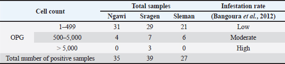
A total of 112 goat and sheep fecal samples from three locations, namely Ngawi, Sleman, and Sragen Regency, were examined. The prevalence of Eimeria spp. in goats and sheep was 90.18% (101/112). In particular, the prevalence in goats was 89.36% (42/47), whereas that in sheep was 90.77% (59/65). The prevalence of Eimeria spp. in Ngawi Regency reached 87.5% (35/40), whereas that in Sleman and Sragen was 93.1% (27/29) and 90.7% (39/43), respectively. Quantitative analysis using the McMaster method revealed that 81/101 samples had low infection, 17/101 samples had moderate infection, and only three samples had high Eimeria infection based on OPG calculation (Table 1).
Table 1. OPG in goats and sheep from three districts in Java, Indonesia.
Based on morphology identification using a microscope (Olympus CX21 Binocular, Shanghai, China) with 400× magnification, the oocysts of Eimeria were detected. The presence of a micropyle with a polar cap, four sporocysts, and two sporozoites from each sporocyst in a single sporulated oocyst were detected as morphological characters. This study also detected a single unsporulated oocyst with a polar cap micropyle. Overall, these oocysts had an ellipsoidal shape, a smooth wall, a clear yellowish color, and a micropyle with a polar cap. The measurements of an unsporulated oocyst were 16.42 × 26.53 µm, and those of a sporulated oocyst were 15.63 × 23.52 µm. Based on morphological characteristics and size, the oocysts of Eimeria spp. were detected (Fig. 1).
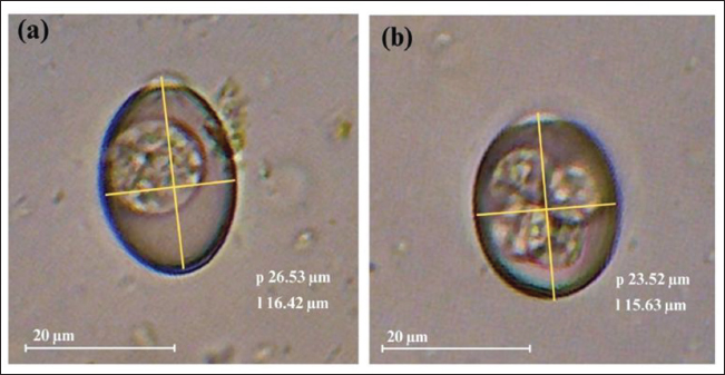
Fig. 1. Photomicrograph of oocysts of Eimeria spp. from goat feces (400× magnification): (a) single unsporulated oocyst and (b) single sporulated oocyst with visible sporocysts.
Eimeria spp. molecular characteristic
The 18S rRNA gene was targeted for PCR amplification using genomic DNA extracted from 25 fecal samples that were morphologically positive for Eimeria spp. (with counts >200 OPG). All samples yielded distinct positive bands at approximately 630 bp following the PCR protocol. No amplification was observed in the negative control, as shown in the representative gel electrophoresis image (Fig. 2).
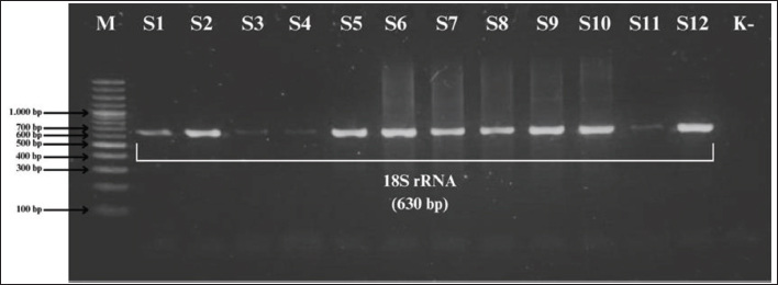
Fig. 2. PCR amplification utilizing DNA extracted from fecal samples containing Eimeria spp. oocysts targeting the 18S rRNA gene. M: marker; S1–S12: fecal samples; K−=negative control.
Three PCR-positive samples with high Eimeria spp. infection (>5,000 OPG) were sequenced. Analysis performed using the BLASTN program employing the 18S rRNA sequences obtained from the sample revealed that one sample was 100% identical to Eimeria christenseni (PX632038) and another two samples were identical to Eimeria crandallis (PX632036 and PX632037). Isolates obtained in this study are indicated by red triangles (▲) and cluster closely with E. crandallis reference sequences from Australia and E. christenseni sequences from China, indicating close genetic relatedness with these geographical groups (Fig. 3). The clustering pattern observed in the maximum likelihood tree was further supported by the analysis of pairwise genetic distances (Fig. 4).
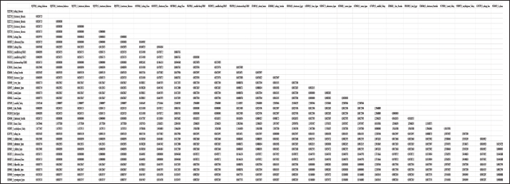
Fig. 4. Pairwise identities (%) among 18S rRNA sequences of Eimeria samples with E. arloingi, E. ahsata, E. bovis, E. auburnensis, E. zuernii, E. hirci, and other Eimeria species.
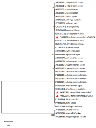
Fig. 3. Phylogenetic tree of the 18S rRNA gene sequences of E. crandallis and E. christenseni. The phylogenetic tree was generated using the maximum-likelihood method and 1,000 bootstrap values, and the best-fit model T92+I was chosen according to the BIC.
Analysis of the risk factors
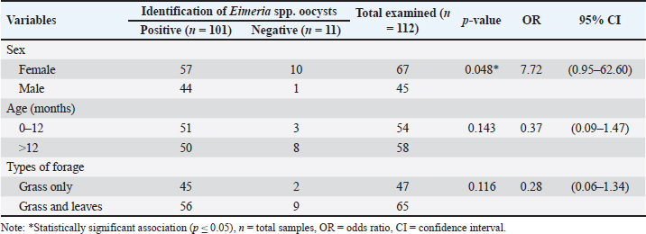
Risk factors based on sex, age, and forage type were analyzed. In this study, bivariate analysis showed that sex was a significant potential risk factor (p: 0.048) associated with Eimeria spp. infection. Furthermore, age and forage type were not significantly related to Eimeria spp. infection (p > 0.05) (Table 2).
Table 2. Bivariate analysis of Eimeria spp. infection risk factors in goats and sheep from three districts in Java, Indonesia.
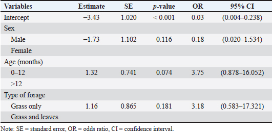
In addition, multicollinearity analysis showed the VIF values for sex (1.05), age (1.09), and forage type (1.05). This analysis indicated that all independent variables had VIF values less than 2 or did not exceed the threshold (VIF value <5), so there were no multicollinearity problems. The best decision model showed AIC values (69.5) and R²N values (18.8%), indicating that the model explained a limited proportion of the variability in Eimeria spp. infection. The R²N values indicate that 81.2% of the risk factors can be explored besides sex, age, and type of forage. The Omnibus Likelihood Ratio tests (Table 3) revealed that none of the predictors (sex, age, and type of forage) were significantly associated with the outcome at the 5% significance level (p > 0.05), although sex (χ²: 3.49; p: 0.062) and age (χ²: 3.55; p: 0.060) showed p-values close to the significance threshold. The final model from the Hosmer–Lemeshow test indicated that sex, age, and forage type were not associated (p > 0.05) with Eimeria spp. infection. However, age (p: 0.074), sex (p: 0.116), and type of forage (p: 0.181) were risk factors for Eimeria spp. infection in goats and sheep (Table 4). In conclusion, both the Omnibus Likelihood Ratio test and Hosmer–Lemeshow test indicated that sex, age, and forage type were not significantly associated with Eimeria spp. infection in the multivariable model (p > 0.05). Therefore, the observed effects should be interpreted with caution.
Table 3. Multivariate analysis using the Omnibus Likelihood Ratio test of Eimeria spp. infection risk factors in goats and sheep from three districts in Java, Indonesia.
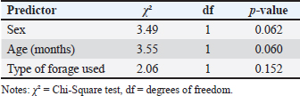
Table 4. Multivariate analysis using Hosmer–Lemeshow test of Eimeria spp. infection risk factors in goats and sheep from three districts in Java, Indonesia.
Discussion
Despite the high prevalence of coccidiosis in tropical regions, there are few comprehensive reports using molecular detection and sequencing for Eimeria spp. in Indonesia. Most previous studies have relied on traditional morphological identification, which has lower sensitivity to differentiate between highly pathogenic species. This study addresses this gap by integrating classical parasitology with 18S rRNA gene sequencing to accurately map the distribution of Eimeria species in three districts of Java.
Our results demonstrate an exceptionally high prevalence of Eimeria spp. (90.18%) among goats and sheep in Ngawi, Sleman, and Sragen. The prevalence observed in this study is significantly higher than that reported in Malang (32%) and Bone Bolango (21.1%) (Nurdianti et al., 2023; Nugroho et al., 2025), yet it aligns with findings from other parts of Java and Myanmar where rates exceed 80% (Win et al., 2020; Dewi et al., 2025). This disparity may indicate that environmental factors and semi-intensive management systems in the studied districts favor oocyst sporulation and persistence (Pamungkas et al., 2021). Despite this high infection rate, the majority of cases were subclinical, characterized by low oocyst counts (1–449 OPG) and an absence of overt clinical symptoms such as hemorrhagic diarrhea or emaciation.
Morphological characters revealed that the single unsporulated oocyst had an ellipsoidal shape and a micropyle with a polar cap present. This study also found that the single sporulated oocyst had a pear shape, a micropyle, and a polar cap. The unsporulated oocyst’s diameter was 16.42 × 26.53 µm, while the sporulated oocyst’s diameter was 15.63 × 23.52 µm. Similarly, these morphology and size results identified Eimeria spp. oocysts of goats and sheep. Based on Alsanousi et al. (2025)and Dewi et al. (2025)Eimeria spp. had the structure oocyst, including the presence of a micropyle with a polar cap, while the diameter size of oocyst had range from 16.2–26 × 24–36 µm for Eimeria spp. in goats and 17.5–18.2 × 24.2–25.3 µm for Eimeria spp. in sheep. The sporulated oocyst had four sporocysts with a two-layered wall, a colorless inner layer, while the outer layer is dark (Abdillah et al., 2021). The results found different sizes of Eimeria spp. that were observed in goats and sheep fecal samples. The variety of sizes identified was due to breeds of hosts, host immunity, stress (caused by other diseases, climate change, and location conditions), sex, body size of host, age, and nutrition circumstances or feed intake (Alsanousi et al., 2025; Haryadi et al., 2025). Historically, identifying Eimeria spp. relied on morphological analysis. Unfortunately, this method has notable limitations, including low sensitivity, long time and labor-intensive requirements, requires experienced microscopists, and difficulty in identifying morphologically similar oocysts (Carvalho et al., 2011; Khodakaram-Tafti et al., 2013). To evaluate this limitation, molecular examination with PCR and sequencing could be continued to identify and confirm the species of Eimeria spp. accurately (Dewi et al., 2025).
Molecular characterization successfully identified the presence of E. crandallis and E. christenseni, confirming the presence of specific pathogenic genotypes that were rarely reported in these local livestock populations. The phylogenetic tree generated using the maximum-likelihood method with 1,000 bootstrap values and the T92+I model clearly shows that the isolates identified in this study (marked with red triangles) cluster within the E. christenseni and E. crandallis lineages. In particular, the E. christenseni isolate from sheep (PX632038) shows a close evolutionary relationship with reference sequences from China, whereas the E. crandallis isolates (PX632036 and PX632037) closely align with isolates from Australia and Egypt. These findings confirm the circulation of specific pathogenic genotypes that were rarely previously reported in these local livestock populations. Furthermore, the detection of E. crandallis and E. christenseni indicates that subclinical infections in Java are not harmless, as these species are known to impair feed conversion and growth performance even without visible outbreaks (Ali et al., 2025).
Bivariate statistical analysis with Jamovi revealed that sex significantly impacted the occurrence of Eimeria spp. infection in goats and sheep, which was confirmed to be significant in Fisher’s exact tests (p: 0.048). Alternately, age and type of forage had an insignificant (p > 0.05) association with the occurrence of Eimeria spp. infection. In this study, male goats and sheep had a 7.72 times higher risk of Eimeria spp. infection than female goats (OR: 7.72).
The sex association with Eimeria spp. infection results align with a previous study in goats at Nakhon Si Thammarat province, Thailand (p < 0.001; OR: 9.75; CI: 2.71–27.21) (Sontigun et al., 2025), which found an association between sex and Eimeria spp. infection. Belay and Sheferaw (2022)reported a significant association between sex and Eimeria spp. infection in sheep at Gondar, Ethiopia (p < 0.001; OR: 2.8). However, Purwaningsih et al. (2024) reported an insignificant association between sex and Eimeria spp. infection in goats at Prafi District of Manokwari Regency (p > 0.05; OR: 1.1). This difference indicates that the association between sex and the incidence of Eimeria spp. infection in small ruminants across various studies is not a definitive standard.
The difference in significant association may be influenced by stress and physiological states in animals, particularly pregnancy, parturition, and lactation in females, resulting in a decline in coccidia (Verma et al., 2018; Islam et al., 2019). This condition showed that females were more susceptible to males (Mohamed et al., 2023). However, males may be more susceptible to Eimeria spp. infection because of immunosuppression brought on by increased androgen hormone, particularly testosterone, plasma levels during the reproductive period. The probability of Eimeria spp. infection in both sexes was the same, as this infection is more influenced by husbandry management systems, environmental exposure, and anthelmintic treatment (Purwaningsih et al., 2024; Awaludin et al., 2026).
Sex was marginally associated with Eimeria spp. infection in the bivariate analysis (p: 0.048). Although the p-value was close to 0.05, this finding remained statistically significant when reassessed using Fisher’s exact test to account for these relatively small sample sizes (p: 0.048). The estimated OR was accompanied by a wide 95% CI (OR: 7.72; CI: 0.95–62.60), indicating that the effect estimate had limited precision. This imprecision is likely related to the relatively small sample size and the unequal distribution of negative cases between sex categories. Under these conditions, OR estimates may vary, and the CI crossing the null value indicates that the association should be interpreted cautiously. Additional studies with larger and more balanced samples may help clarify the role of sex in Eimeria spp. infection.
The multivariate statistical analysis performed the association of sex (χ²: 3.49; df: 1; p: 0.062), age (χ²: 3.55; df: 1; p: 0.060), and type of forage (χ²: 2.06; df: 1; p: 0.060) with Eimeria spp. infection, which found an insignificant association. In conclusion, this association analysis confirmed that none of the risk factors were independent risk factors (p > 0.05) for Eimeria spp. infection. This non-significant multivariate result may be influenced by the imbalance of sample size across sex categories and the relatively small sample size, which prevents this finding from providing the representation and association of both the risk factors and Eimeria spp. infection.
Although this study provides valuable insights confirming Eimeria infection in the region, certain limitations offer opportunities for future refinement. The current molecular sequencing serves as an initial characterization that may not fully capture the complete genetic diversity across the districts under study. Although the 18S rRNA gene is a reliable marker for genus-level detection, its inherent conservation can present challenges in resolving extremely closely related species (Hussein et al., 2023). Therefore, further investigations using higher resolution markers, such as ITS1 or COI, alongside longitudinal studies are needed to evaluate the dynamics of oocyst shedding in Indonesia.
In conclusion, this study confirms that Eimeria infection is highly endemic in the small ruminant systems of Ngawi, Sleman, and Sragen, predominantly manifesting as subclinical cases. Molecular characterization successfully addressed diagnostic gaps by identifying E. crandallis and E. christenseni as primary species. Multivariate analysis showed no significant association (p > 0.05) between sex, age, and forage type, indicating that these variables are not independent risk factors for Eimeria spp. infection. These results offer a basic framework for targeted control strategies that focus on improved hygiene and stocking management to minimize the hidden economic impact of coccidiosis in Indonesia.
Acknowledgments
The authors express their gratitude and appreciation to all the farmers and Drh. Morsid Andityas, M. Sc., Ph. D.
Conflict of interest
The authors have no conflicts of interest to declare.
Funding
This research was supported by the Final Project Recognition/Rekognisi Tugas Akhir (RTA) Grant, Universitas Gadjah Mada (grant number: 4193/UN1.P1/Dit-Lit/PT.01.03/2025).
Author’s contributions
F.B.L., F.E., and F.R.H. designed this study. F.B.L., E.G.K., and M.P.A. conducted the field research and sample analysis in the laboratory. E.G.K., F.B.L., F.E., and M.W. analyzed the data. All authors have written, edited, read, and approved the final version of the manuscript.
Data availability
All the information supporting the discoveries of this study is accessible within the manuscript.
References
Abdel-Hamied, A.M., Abdel-Hafez, B., El-Sayed, N., Abdelkhalek, A. and Ras, R. 2024. Prevalence and morphological identification of Eimeria species in sheep in Sharkia Governorate, Egypt. Zagazig. Vet. J. 52(4), 439–450; doi:10.21608/zvjz.2024.304606.1254
Abdillah, M.F., Lastuti, N.D.R., Utama, S. and Suprihati, E. 2021. Identification and prevalence of digestive tract endoparasites of goats in Ujungpangkah, Gresik District. J. Parasite. Sci. 5(2), 61–64; doi:10.20473/jops.v5i2.30374
Akyüz, M., Kirman, R., Yaya, S., Gülbeyen, H. and Güven, E. 2019. Endoparasites determined by fecal examination in sheep in Erzurum Province. Turkiye. Parazitol. Derg. 43(4), 187–193; doi:10.4274/tpd.galenos.2019.6512
Ali, E.A., Ghafar, A., Angeles-Hernandez, J.C., Yaseen, M., Gauci, C.G., Beveridge, I., Baxendell, S. and Jabbar, A. 2025. Global prevalence of Eimeria species in goats: a systematic review and meta-analysis. Front. Vet. Sci. 12, 1537171; doi:10.3389/fvets.2024.1537171
Alsanousi, S.A., Atiyahullah, T.M.O., Meriz, O.M., Adam, A.F.H. and Abdulsalam, N.A.I. 2025. The first record of ovine Eimeria species (Apicomplexa) and their prevalence in Aljabal Alakhdar, Libya. Int. J. Vet. Sci. 14(4), 661–667; doi:10.47278/journal.ijvs/2025.021
Andityas, M., Sota, P., Sukon, P., Tangkawattana, P., Sripa, B., Ngasaman, R. and Tangkawattana, S. 2025. Co-occurrence of leptospirosis and Opisthorchis viverrini infection in cats and their risk factors. Res. Vet. Sci. 190, 1–12; doi:10.1016/j.rvsc.2025.105657
Awaludin, A., Sawitri, D.H., Wardhana, A.H., Nugraheni, Y.R., Mubarokah, W.W., Wibowo, S.E. and Andityas, M. 2026. Identification and occurrence rate of gastrointestinal parasites in slaughtered sheep at the Alexandria Abattoir in Magelang, Central Java, Indonesia. Adv. Anim. Vet. Sci. 14(1), 39–49; doi:10.17582/journal.aavs/2026/14.1.39.49
Belay, S. and Sheferaw, D. 2022. Ovine Eimeria infection, OPG and determinants in and around Gondar Town, Ethiopia. East Afr. J. Biophys. Comput. Sci. 3(2), 40–47; doi:10.4314/eajbcs.v3i2.4S
Bangoura, B., Mundt, H.C., Schmäschke, R., Westphal, B. and Daugschies, A. 2011. Prevalence of Eimeria bovis and Eimeria zuernii in German cattle herds and factors influencing oocyst excretion. Parasitol. Res. 109(Suppl 1), 129–138; doi:10.1007/s00436-011-2409-1
Carvalho, F.S., Wenceslau, A.A., Teixeira, M., Matos Carneiro, J.A., Melo, A.D.B. and Albuquerque, G.R. 2011. Diagnosis of Eimeria species using traditional and molecular methods in field studies. Vet. Parasitol. 176(2), 95–100; doi:10.1016/j.vetpar.2010.11.015
Dewi, D.A., Nugraheni, Y.R., Awaludin, A., Ninditya, V.I., Priyowidodo, D., Nurcahyo, R.W., Ekawasti, F. and Prastowo, J. 2025. First molecular detection of Eimeria spp. in domestic goats from Java Island, Indonesia. Open. Vet. J. 15(1), 139–150; doi:10.5455/OVJ.2025.v15.i1.13
Eckert, J., Braun, R., Shirley, M.W. and Coudert, P. 1995. Guidelines on techniques in coccidiosis research. Luxembourg City, Luxembourg: Office for Official Publications of the European Communities, pp: 103–117.
Efendi, W.N., Suwanti, L.T., Samik, A., Hastutiek, P., Mufasirin, M. and Kusnoto, K. 2019. Prevalence and identification of digestive tract protozoa in goats in Labang District Bangkalan Regency. J. Parasite Sci. 3(2), 95–100; doi:10.20473/jops.v3i2.16527
Ekawasti, F., Nurcahyo, R.W., Firdausy, L.W., Wardhana, A.H., Sawitri, D.H., Prastowo, J. and Priyowidodo, D. 2021. Prevalence and risk factors associated with Eimeria species infection in cattle of different geographical regions of Indonesia. Vet. World. 14(9), 2339–2345; doi:10.14202/vetworld.2021.2339-2345
Ekawasti, F., Nurcahyo, W., Wardhana, A.H., Shibahara, T., Tokoro, M., Sasai, K. and Matsubayashi, M. 2019. Molecular characterization of highly pathogenic Eimeria species among beef cattle on Java Island, Indonesia. Parasitol. Int. 72, 1–7; doi:10.1016/j.parint.2019.101927
Fatchiyah E Laras., Widyarti. and Rahayu. 2011. Biologi Molekuler: prinsip Dasar Analisis.”Jakarta, Indonesia: Erlangga.
Haryadi, F.R., Nurcahyo, R.W., Priyowidodo, D., Indarjulianto, S., Ekawasti, F. and Ninditya, V.I. 2025. Determination of the morphology of Eimeria spp. in beef cattle in Bandung Regency, West Java Province, Indonesia. Open Vet. J. 15(1), 85–91; doi:10.5455/OVJ.2025.v15.i1.7
Hassanen, E.A. 2020. Prevalence and phylogenetic analysis of Eimeria species in sheep and goats in Sharkia Governorate, Egypt. Pak. Vet. J. 40(4), 437–442; doi:10.29261/pakvetj/2020.064.
Hmaid, A., Ahmed, J., Dhaw, H., Aboulqsim, A. and Senni, R. 2024. Detection of Eimeria parasites in sheep and goats in Alhamada Area, Libya. Alqalam J. Med. Appl. Sci. 7(3), 841–846; doi:10.54361/ajmas.247355
Hussein, S.N., Ibrahim, A.A. and Shukur, M.S. 2023. Histopathology and molecular identification of Sarcocystis species forming macrocysts in slaughtered sheep and goats of Duhok, Iraq. Vet. Res. Forum 14(8), 415–422; doi:10.30466/vrf.2023.559514.3575
Islam, A., Islam, S., Ferdous, J., Rahman, M.K., Uddin, M.H., Akter, S., Rahman, M.H. and Hassan, M.M. 2019. Diversity and prevalence of parasitic infestation with zoonotic potential in dromedary camels (Camelus dromedarius) and fat-tailed sheep (dhumba) in Bangladesh. J. Advance. Vet. Anim. Res. 6(1), 142–147; doi:10.5455/javar.2019.f324
Jinneman, K.C., Wetherington, J.H., Hill, W.E., Omiescinski, C.J., Adams, A.M., Johnson, J.M., Tenge, B.J., Dang, N.L. and Wekell, M.M. 1999. An oligonucleotide-ligation assay for the differentiation between Cyclospora and Eimeria spp. polymerase chain reaction amplification products. J. Food Prot. 62(6):682–685; doi:10.4315/0362-028x-62.6.682
Kawahara, F., Zhang, G., Mingala, C.N., Tamura, Y., Koiwa, M., Onuma, M. and Nunoya, T. 2010. Genetic analysis and development of species-specific PCR assays based on ITS-1 region of rRNA in bovine Eimeria parasites. Vet. Parasitol. 174, 49–57; doi:10.1016/j.vetpar.2010.08.001
Khodakaram-Tafti, A. and Hashemnia, M. 2017. An overview of intestinal coccidiosis in sheep and goats. Rev. Méd. Vét. 168(1), 9–20; doi:10.1016/j.vmv.2017.09.010
Khodakaram-Tafti, A., Hashemnia, M., Razavi, S.M., Sharifiyazdi, H. and Nazifi, S. 2013. Genetic characterization and phylogenetic analysis of Eimeria arloingi in Iranian native kids. Parasitol. Res. 112(31), 87–92; doi:10.1007/s00436-013-3494-0
Liang, G., Yang, X., Liu, D., Li, Y., Wang, J., Chen, X., Zhao, G. and Song, J. 2022. Molecular characterization of 18S rDNA, ITS-1, ITS-2, and COI from Eimeria christenseni and E. arloingi in goats from Shaanxi Province, Northwestern China. Animals 12(11), 1340; doi:10.3390/ani12111340
Lucena-Aguilar, G., Sánchez-López, A.M., Barberán-Aceituno, C., Carrillo-Ávila, J.A., López-Guerrero, J.A. and Aguilar-Quesada, R. 2016. DNA source selection for downstream applications based on DNA quality indicators analysis. Biopreservation Biobanking 14(4), 264–270; doi:10.1089/bio.2015.0064
Mohamed, H.I., Arafa, W.M. and El-Dakhly, K.M. 2023. Prevalence and associated risk factors of gastrointestinal helminths and coccidian infections in domestic goats, Capra hircus, in Minya, Egypt. Beni-Suef Univ. J. Basic Appl. Sci. 12(29), 1–13; doi:10.1186/s43088-023-00369-6
Mussa, R., Pangaribuan, M.J.A. and Palgunadi, B.U. 2019. Perhitungan telur Toxocara cati dan jumlah sel darah putih pada kucing liar (Felis catus) di Dukuh Kupang Surabaya. Jurnal. Vitek. Bidang. Kedokteran. Hewan. 9, 18–23; doi:10.30742/jv.v9i0.
Ninditya, V.I., Ekawasti, F., Prastowo, J., Widiyono, I. and Nurcahyo, W. 2024. Prevalence of gastrointestinal parasites in cattle in Indonesia: a meta-analysis and systematic review. Vet. World 17(11), 2675–2687; doi:10.14202/vetworld.2024.2675-2687
Nugroho, A., Taha, S.R., Laya, N.K. and Datau, F. 2025. Prevalensi coccidiosis pada kambing di Kecamatan Kabila Bone Kabupaten Bone Bolango Provinsi Gorontalo. In Prosiding Seminar Nasional Mini Riset Mahasiswa, vol. 4(1). Gorontalo, Indonesia: Faculty of Agriculture, Gorontalo State University, pp 104–107.
Nuraini, D.M., Kholistiawan, D.I., Rosyad, F.S., Ariyanti, S.D., Yanti, Y., Loong, S., Sahimin, N. and Andityas, M. 2023. Infection rate and risk factor of buffalo Paramphistomum sp. infection in Solo Raya Region, Central Java, Indonesia. J. Adv. Vet. Res. 13(8), 1588–1592.
Nurdianti N, Yunus, M. and Mufasirin, M. 2023. Incidence of Eimeria spp. in fat-tailed sheep breeding Malang, Indonesia. Jurnal Medik Veteriner 6(2), 230–236; doi:10.20473/jmv.vol6.iss2.2023.230-236
Pamungkas, P.A., Apsari, I.A.P. and Widyastuti, S.K. 2021. Prevalensi infeksi Eimeria spp. yang tinggi pada kambing yang dipelihara di Kota Denpasar. Indonesia. Medicus. Veterinus. 10(6), 861–868; doi:10.19087/imv.2021.10.6.861
Purwaningsih, Noviyanti, Saragih, E.W. and Palulungan, J.A. 2024. Prevalence and risk factors of coccidiosis in goats in slaughter center, Manokwari Regency, West Papua Province, Indonesia: epidemiological aspects. Int. J. Vet. Sci. 13(2), 226–231; doi:10.47278/journal.ijvs/2023.090
Rerkyusuke, S., Lerk-u-suke, S., Mektrirat, R., Wiratsudakul, A., Kanjampa, P., Chaimongkol, S., Phanmanee, N., Visuddhangkoon, M., Pattayawongdecha, P., Piyapattanakon, N., Krajaipan, P. and Sutamwirat, P. 2024. Prevalence and associated risk factors of gastrointestinal parasite infections among meat goats in Khon Kaen Thailand. Vet. Med. Int. 2024, 3267028; doi:0.1155/2024/3267028
Rompo, T., Hayashi, N., Teo, E., Singhla, T., Kunkaew, C., Sripakdee, D., Nambooppha, B., Wanganurakkul, S., Limwibulpong, K., Sangarun, K., Suwongsaksri, N., Suphakarn, S., Chotiphutthikul, C., Matsui, Y., Irie, T., Yoshida, A., Chintapitaksakul, L., Misawa, N., Nonaka, N., Nakao, R. and Tiwananthagorn, S. 2025. Strongyle nematode fauna in three ruminants in upper northern Thailand. Parasitol. Int. 108, 103057; doi:10.1016/j.parint.2025.103057
Sambrook, J. and Russell, D.W. 2001. Molecular cloning: a laboratory manual, 3rd ed. New York: Cold Spring Harbor Laboratory Press.
Silva, L.M.R., Vila-Viçosa, M.J.M., Nunes, T., Taubert, A., Hermosilla, C. and Cortes, H.C.E. 2014. Eimeria infections in goats in Southern Portugal. Vet. Parasitol. 202(3–4), 146–152; doi:10.1016/j.vetpar.2014.03.026
Sontigun, N., Sansamur, C., Klong-Klaew, T., Kaewthamasorn, M., Fungwithaya, P. and Mektrirat, R. 2025. Seasonal infective dynamics and risk factors associated with prevalence of zoonotic gastrointestinal parasites from meat goats in southern Thailand. Animals 15, 2040; doi:10.3390/ani15142040
Taylor, M.A., Coop, R.L. and Wall, R.L. 2016. Veterinary parasitology, 4th ed. Chichester, West Sussex, UK: Wiley-Blackwell.
Verma, R., Sharma, D.K., Paul, S., Gururaj, K., Dige, M., Saxena, V.K., Rout, P.K., Bhusan, S. and Banerjee, P.S. 2018. Epidemiology of common gastrointestinal parasitic infections in goats reared in semi-arid regions of India. J. Anim. Res. 8(1), 39–45; doi:10.30954/2277-940X.2018.00150.07
Weng, S., Zhang, H. and Li, X. 2024. Eimeria: host–parasite–microbiome interactions and implications for control. Front. Cellular Infect Microbiol. 14, 1432321; doi:10.3389/fcimb.2024.1432321
Win, S.Y., Win, M., Thwin, E.P., Htun, L.L., Hmoon, M.M., Chel, H.M., Thaw, Y.N., Soe, N.C., Phyo, T.T., Thein, S.S., Khaing, Y., Than, A.A. and Bawm, S. 2020. Occurrence of gastrointestinal parasites in small ruminants in the central part of Myanmar. J. Parasitol. Res. 1–8; doi:10.1155/2020/8826327
Zajac, A.M., Conboy, G.A., Little, S.E. and Reichard, M.V. 2021. Veterinary clinical parasitology, 9th ed. Hoboken, NJ: John Wiley & Sons, Inc.








