





| Research Article | ||
Open Vet. J.. 2026; 16(7): 4690-4702
Open Veterinary Journal, (2026), Vol. 16(7): 4690-4702 Research Article Development and characterization of anti-recombinant PBP2a antibodies for preliminary detection of methicillin-resistant Staphylococcus aureusSiti Isrina Oktavia Salasia1*, Nada Hanifah1, Madarina Wasissa1, Fatkhanuddin Aziz2 and Widagdo Sri Nugroho31Department of Clinical Pathology, Faculty of Veterinary Medicine, Universitas Gadjah Mada, Yogyakarta, Indonesia 2Department of Bioresources Technology and Veterinary, Vocational College, Universitas Gadjah Mada, Yogyakarta, Indonesia 3Department of Veterinary Public Health, Faculty of Veterinary Medicine, Universitas Gadjah Mada, Yogyakarta, Indonesia *Corresponding Author: Siti Isrina Oktavia Salasia. Department of Clinical Pathology, Faculty of Veterinary Medicine, Universitas Gadjah Mada, Yogyakarta, Indonesia. Email: isrinasalasia [at] ugm.ac.id Submitted: 10/12/2025 Revised: 06/06/2026 Accepted: 19/06/2026 Published: 20/07/2026 © 2025 Open Veterinary Journal
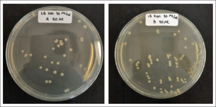
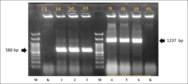
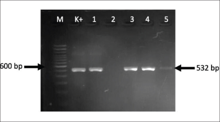
AbstractBackground: Methicillin-resistant Staphylococcus aureus (MRSA) is a major public health and veterinary concern due to the widespread dissemination of the mecA gene, which encodes penicillin-binding protein 2a (PBP2a). Although PBP2a-based diagnostic assays are widely used, antibodies developed from locally derived MRSA strains remain limited despite regional genetic variation. Aim: This study aimed to develop and characterize anti-recombinant PBP2a (anti-rPBP2a) antibodies using recombinant mecA-derived proteins from clinical MRSA isolates in Indonesia and to assess their preliminary applicability for MRSA detection using immunoblotting and latex agglutination assays. Methods: Two mecA gene fragments corresponding to amino acid positions 341–453 and 341–670 were amplified from Indonesian MRSA isolates, cloned into pET-24a(+), expressed in Escherichia coli BL21 (DE3), and purified to obtain recombinant PBP2a (rPBP2a) antigens. BALB/c mice were immunized with soluble and insoluble antigen preparations. Antibody titers were determined by an indirect enzyme-linked immunosorbent assay (ELISA), and antibody reactivity was evaluated by sodium dodecyl sulphate-polyacrylamide gel electrophoresis, western blotting (WB), dot-blot assays, and latex agglutination against MRSA and methicillin-susceptible Staphylococcus aureus (MSSA) isolates. Results: Both recombinant constructs were successfully expressed, yielding proteins of approximately 14 and 40 kDa for the mecA341–453 and mecA341–670 constructs, respectively. The elution B (EB) antigen fraction induced the strongest immune response, with ELISA optical density values of 2.4–3.5 during booster immunizations and ascites titers of 3.41–3.50. WB revealed a distinct immunoreactive band at approximately 40 kDa, consistent with the predicted molecular weight of rPBP2a. Anti-rPBP2a antibodies generated against the EB antigen fraction showed stronger and more consistent reactivity than those raised against pellet A and pellet B antigens in dot-blot assays. In latex agglutination assays, EB-derived antibodies produced visible agglutination with MRSA isolates but not with MSSA, supporting their preliminary applicability for differentiating MRSA from MSSA. Conclusion: To the best of our knowledge, this study is the first to develop and characterize anti-rPBP2a antibodies derived from Indonesian MRSA isolates. These antibodies demonstrated strong immunoreactivity and consistent recognition of rPBP2a across multiple assay formats, providing a promising foundation for the future development of rapid and affordable MRSA detection methods in veterinary and clinical settings. Keywords: Anti-rPBP2a antibody, Latex agglutination, mecA, MRSA, Recombinant antigen. IntroductionMethicillin-resistant Staphylococcus aureus (MRSA) is a globally significant zoonotic pathogen associated with extensive antimicrobial resistance, particularly to β-lactam antibiotics (World Health Organization (WHO), 2024). MRSA is commonly referred to as a “superbug” and has been linked to severe healthcare-associated infections, such as septic shock, bacteremia, and postoperative wound infections, resulting in considerable morbidity and mortality (Khan et al., 2017; Curcio et al., 2018; Nataraj and Mallappa, 2021). MRSA has been reported in companion animals, including dogs and cats (Fitranda et al., 2024; Feuer et al., 2024), as well as in livestock, such as cattle and goats (Salasia et al., 2013; Widianingrum et al., 2016). Increasing evidence of bidirectional transmission between humans and animals has further emphasized MRSA as an emerging One Health pathogen and highlighted the importance of integrated surveillance and antimicrobial stewardship (Worthing et al., 2018; Genath et al., 2024; Dewulf et al., 2025). Resistance to β-lactam antibiotics in MRSA is primarily mediated by the acquisition of the mecA gene, which is carried on SCCmec (Abebe and Birhanu, 2023; Tabassum et al., 2025). The mecA gene encodes penicillin-binding protein 2a (PBP2a), a class B transpeptidase that allows cell wall synthesis to continue in the presence of β-lactam antibiotics (Abebe and Birhanu, 2023). In addition, SCCmec often harbors other resistance determinants, contributing to multidrug resistance and limiting therapeutic options. MRSA remains a significant pathogen in clinical, veterinary, and community settings because of these characteristics. MRSA is encountered across a wide range of environments, including hospitals, veterinary clinics, farms, and community settings, reinforcing the need for integrated One Health surveillance strategies (Igrejas et al., 2018; Kourtis et al., 2019; Fitranda et al., 2023, 2024). Such approaches aim to link human, animal, and environmental data to better understand the transmission and risk factors associated with antimicrobial resistance (Murray et al., 2022; Dalton et al., 2020; Lo et al., 2022; Nigo et al., 2024). Although early detection is crucial for infection control, clinical management, and epidemiological mapping, important diagnostic challenges remain. Culture-based methods and antimicrobial susceptibility testing are time-consuming, whereas molecular techniques, such as polymerase chain reaction (PCR), require specialized equipment that may not be readily available, particularly in veterinary or resource-limited settings. Rapid diagnostic assays targeting PBP2a have increasingly been explored as complementary tools for MRSA detection, enabling faster decision-making and broader applicability in human and veterinary medicine. Several rapid diagnostic assays targeting PBP2a have been developed, including latex agglutination tests based on monoclonal antibodies, which enable the rapid identification of MRSA directly from bacterial isolates (van Griethuysen et al., 1999; Gupta et al., 2023). Moreover, antibody-based approaches targeting PBP2a have been explored as diagnostic tools and experimental immunotherapies (Saraiva et al., 2019). In experimental models, recombinant PBP2a (rPBP2a) has been explored as an immunogenic antigen capable of inducing specific antibody responses against MRSA, highlighting its potential utility for immunological applications and diagnostic antibody development (Haghighat et al., 2017). The development of anti-recombinant PBP2a (anti-rPBP2a) antibodies can enhance the performance of rapid detection methods, including latex agglutination, western blotting (WB), and dot-blot assays. In this context, the use of animal-derived MRSA isolates in Indonesia offers regionally relevant material and contributes to efforts to develop diagnostic tools that are better aligned with local epidemiological conditions. However, most available PBP2a-based diagnostic antibodies have been developed using reference laboratory strains and may not fully represent the antigenic variations in regional MRSA populations. Therefore, the use of locally derived recombinant antigens may improve antibody specificity and diagnostic performance in specific epidemiological settings. Accordingly, this study aimed to develop and characterize anti-rPBP2a antibodies using recombinant mecA-derived proteins from Indonesian MRSA isolates and to evaluate their preliminary applicability through molecular, immunological, and functional assays, including immunoblotting and latex agglutination. The findings of this study are expected to support the further development of rapid and affordable MRSA detection tools for veterinary practice and broader One Health surveillance initiatives. Materials and MethodsStudy duration and locationThis study was conducted between April 2024 and November 2025 at the Clinical Pathology Laboratory, Faculty of Veterinary Medicine, Universitas Gadjah Mada, Yogyakarta, Indonesia. Bacterial isolates and DNA extractionClinical Staphylococcus aureus isolates were obtained from the Integrated Laboratory Installation of Dr. Sardjito General Hospital, Yogyakarta, Indonesia, and from the Veterinary Clinical Pathology Laboratory, Faculty of Veterinary Medicine, Universitas Gadjah Mada, Yogyakarta, Indonesia. A total of eight clinical isolates were included in this study and originated from routine clinical specimens such as blood and wound samples. Isolates were selected based on colony morphology, Gram staining, catalase and coagulase positivity, and phenotypic methicillin resistance screening. We excluded mixed cultures or isolates with inconsistent microbiological identification from further analysis. To screen for methicillin resistance, antimicrobial susceptibility testing was performed using the oxacillin disk diffusion method according to CLSI guidelines (Clinical and Laboratory Standards Institute, 2023). Isolates showing phenotypic resistance were further confirmed by PCR detection of the mecA gene. The isolates were cultured on 5% sheep blood agar (Oxoid, Germany) at 37°C for 24 hours. Bacterial DNA was extracted using the QIAamp DNA Mini Kit (Qiagen, Germany) according to the manufacturer’s instructions. Before PCR amplification, DNA concentration and purity were assessed using spectrophotometry, and DNA integrity was confirmed by agarose gel electrophoresis. Confirmed MRSA DNA from a previous study (Fitranda et al., 2024) was included as an additional reference. Molecular detection of mecAAll isolates were confirmed to be S. aureus based on amplification of the 23S rRNA and nuc genes. Methicillin resistance was detected by PCR targeting the mecA gene using the following primers: forward 5′-AGT TCT GCA GTA CCG GAT TTG-3′ and reverse 5′-AAA ATC GAT GGT AAA GGT TGG-3′. PCR amplification was performed in a thermal cycler with an initial denaturation at 95°C for 5 minutes, followed by 35 cycles of denaturation at 95°C for 30 seconds, annealing at 55°C for 30 seconds, and extension at 72°C for 10 seconds, with a final extension at 72°C for 5 minutes. The amplified products were separated by electrophoresis on 1.5% agarose gels stained with RedSafe (Intron, Korea) and visualized under UV illumination. The primer set and amplification protocol were adapted from Fitranda et al. (2023) with minor modifications. An MRSA strain was used as the positive control, while S. aureus ATCC 25923 served as the negative control. The expected amplicon size was 532 bp. Recombinant inserts were commercially synthesized and sequence-verified before cloning. Cloning of mecA fragmentsTwo recombinant mecA constructs corresponding to the PBP2a transpeptidase region were designed from Indonesian clinical MRSA isolates. The first construct, mecA341–453 (372 bp), represented a partial transpeptidase domain fragment containing predicted immunogenic regions while allowing efficient recombinant protein expression. The second construct, mecA341–670 (1,023 bp), represented the full transpeptidase domain and was designed to preserve a broader antigenic region of native PBP2a. The PCR products were digested with NdeI and BamHI and ligated into the pET-24a(+) expression vector (Novagen, USA). Recombinant plasmids carrying mecA341–453 and mecA341–670 inserts were transformed into competent Escherichia coli BL21 (DE3) cells (Thermo Fisher Scientific, USA). Positive transformants were initially screened by colony PCR using insert-specific primers to verify the expected amplicon sizes. Recombinant construct identity and insert orientation were further confirmed by sequence verification before recombinant protein expression. The validated plasmids were subsequently used for protein expression and purification. Expression and purification of rPBP2a proteinsTransformed E. coli BL21 (DE3) cultures were grown in Luria–Bertani broth (Sigma-Aldrich, USA) supplemented with kanamycin (30 µg/ml) (Sigma-Aldrich, USA) at 37°C with shaking at 200 rpm until the optical density (OD) reached OD600 ≈ 0.6–0.8. Protein expression was induced by the addition of 1 mM isopropyl-β-D-thiogalactopyranoside (Sigma-Aldrich, USA), and the cultures were incubated at 37°C for 16 hours. Bacterial cells were harvested by centrifugation at 6,000 × g for 10 minutes at 4°C, resuspended in lysis buffer, and disrupted by sonication on ice. The lysates were centrifuged at 12,000 × g for 20 minutes at 4°C to separate the soluble and insoluble fractions. Recombinant His-tagged PBP2a proteins were purified using Ni-NTA affinity chromatography (Qiagen, Germany) according to the manufacturer’s protocol. For construct A (mecA341–453), the purified elution fraction was designated as elution A (EA), whereas the insoluble fraction recovered after cell lysis and centrifugation was designated as pellet A (PA). Similarly, for construct B (mecA341–670), the purified elution fraction was designated as elution B (EB), the soluble fraction was designated as Supernatant B (SB), and the insoluble fraction was designated as pellet B (PB). Although SB was not used for immunization or enzyme-linked immunosorbent assay (ELISA)-based antibody evaluation, it was retained for selected WB to assess the distribution of rPBP2a in the soluble protein fraction. Subsequently, EA, EB, PA, and PB were evaluated as antigen preparations for immunization and antibody characterization, whereas SB was retained only for selected immunoblot analyses. Protein expression and purity were evaluated by sodium dodecyl sulphate-polyacrylamide gel electrophoresis (SDS-PAGE), which revealed protein bands of approximately 14 kDa for the partial transpeptidase domain (mecA341–453) and 40 kDa for the larger recombinant construct (mecA341–670), consistent with their predicted molecular weights. Immunization of mice and antibody productionFemale BALB/c mice aged 6–8 weeks were used for antibody production. Mice were randomly allocated into five experimental groups according to antigen preparation: (EA; n=5), (EB; n=5), (PA; n=3), (PB; n=3), and a control group receiving Phosphate-Buffered Saline (PBS) (n=3). All procedures were performed according to the approved animal ethics protocol and animal welfare guidelines. For primary immunization (day 0), each mouse received 5 µg rPBP2a protein by intraperitoneal injection, emulsified 1:1 (v/v) with Complete Freund’s Adjuvant (CFA, Sigma-Aldrich, USA) to a final volume of 250 µl per mouse. The administered antigen volume varied according to protein concentration as follows: EA, 7.7 µl; EB, 21.6 µl; PA, 12.3 µl; and PB, 4.4 µl. Sterile PBS was added to complete the 125 µl aqueous phase before emulsification with CFA. Control mice were treated with PBS emulsified with CFA without antigen. Booster immunizations were administered at 2-week intervals (days 14, 28, and 42) using the same antigen dose (5 µg/mouse, IP) emulsified with Incomplete Freund’s Adjuvant (Sigma-Aldrich, USA). The immunization schedule and booster frequency were identical in all antigen groups. After each booster, blood samples were collected to monitor antibody responses. Small-volume blood samples were obtained from the tail vein during routine monitoring. Serum was separated and stored at –20°C until indirect ELISA and immunoblot analysis. The final serum collection was performed through the retro-orbital plexus under anesthesia following institutional procedures. The animals were monitored daily throughout the study for general condition, feed and water intake, and any signs of injection-site reactions or distress. Indirect ELISA for antibody titrationThe resulting antibody responses were measured using indirect ELISA. Microplates were coated with rPBP2a antigens prepared as (EA), (EB), (PA), and (PB). Following blocking, serial serum dilutions were added, and bound antibodies were detected using Horseradish Peroxidase (HRP)-conjugated antimouse Immunoglobulin G (IgG) (Promega, USA). The OD was recorded at 450 nm. To identify the most immunogenic antigen preparation, antibody responses were evaluated based on signal intensity, frequency of OD (3.5), and consistency across replicates. Western blottingPurified rPBP2a proteins from (EB), SB, and (PB) were separated by SDS-PAGE and transferred to Immobilon®-P polyvinylidene difluoride membrane (Merck Millipore, Germany). Membranes were probed with anti-rPBP2a antibodies generated using the (EB) antigen preparation, which exhibited the strongest and most consistent antibody responses in ELISA. Bound antibodies were detected using HRP-conjugated secondary antibodies. Detection of an immunoreactive band at approximately 40 kDa was consistent with antibody recognition of the rPBP2a protein. Dot blottingDot-blot assays were performed by spotting rPBP2a antigen preparations (EA, EB, PA, and PB) onto nitrocellulose membranes. Membranes were subsequently incubated with mouse anti-rPBP2a antibodies generated against the EB, PA, or PB immunogens (EB, PA, and PB antibody groups, respectively). PBS was used as a negative control. Detection was performed using HRP-conjugated antimouse IgG, and signal intensity was assessed qualitatively. Latex agglutination test resultsPolystyrene latex beads (Sigma-Aldrich, USA) were coated with purified anti-rPBP2a antibodies and incubated with MRSA and methicillin-susceptible Staphylococcus aureus (MSSA) bacterial suspensions. The formation of visible clumping or crystalline aggregates was interpreted as a positive agglutination reaction. MRSA isolates consistently exhibited agglutination, whereas MSSA isolates exhibited no visible reaction. Resazurin staining was used as a visualization aid to enhance contrast during the macroscopic observation of latex agglutination on a glass slide. The dye facilitates a clear differentiation between aggregated and non-aggregated latex particles. The staining was applied after mixing and does not interfere with the antigen–antibody interaction or the agglutination reaction, serving only as a visual indicator. Statistical analysisStatistical analysis was performed only for the ELISA data, as the SDS-PAGE, WB, dot blot, and latex agglutination assays were evaluated qualitatively and presented as descriptive observations. The ELISA results were analyzed using GraphPad Prism version 9.0 (GraphPad Software, USA). Antibody titers were expressed as mean ± SD from triplicate measurements. Differences between groups (mecA341–453, mecA341–670, and control) were analyzed using one-way analysis of variance followed by Tukey’s post hoc test. Repeated-measures Analysis of Variance was applied for repeated measurements across immunization time points. A p-value of < 0.05 was considered statistically significant. Ethical approvalEthical approval for the use of human-derived S. aureus isolates was obtained from the Medical and Health Research Ethics Committee, Universitas Gadjah Mada, Yogyakarta, Indonesia (Ref. No. KE/FK/0131/EC/2023). The use of mouse immunization procedures was approved by the Ethics Committee of the Faculty of Veterinary Medicine, Universitas Gadjah Mada (Approval No. 143/EC-FKH/Int./2024). All procedures were conducted under institutional and national guidelines for animal welfare and biosafety. ResultsDetection of mecA in MRSA isolatesThe initial screening of clinical S. aureus isolates for methicillin resistance was performed by amplifying the mecA gene. As shown in Fig. 1, PCR successfully produced the expected 532 bp amplicon in the MRSA positive control (Lane K⁺) and several clinical isolates (Lanes 1 and 3–5). Band intensity varied slightly among isolates, with one isolate showing a weaker but still clearly detectable amplicon, likely reflecting differences in template concentration or PCR amplification efficiency. No amplification was observed in the negative control MSSA (Lane 2), confirming the specificity of the assay. These results verified that the selected isolates carried the mecA gene and were suitable for developing rPBP2a constructs downstream. Construction and screening of recombinant mecA expression plasmidsTwo segments of mecA were amplified and cloned into the pET-24a (+) vector to generate rPBP2a fragments for antibody production: mecA341–453 (construct A) and mecA341–670 (construct B). Following the transformation into E. coli BL21 (DE3), the presence of the inserts was confirmed by PCR screening of transformants. As shown in Figure 2, construct A colonies produced a specific fragment at 586 bp, whereas construct B produced a 1,237 bp fragment, consistent with their predicted insert lengths. These results demonstrate that two recombinant plasmids encoding truncated forms of PBP2a were successfully generated.
Fig. 2. Representative colony growth of Escherichia coli BL21 (DE3) transformed with recombinant plasmids mecA341–453_pET-24a(+) (A) and mecA341–670_pET-24a(+) (B) on LB agar plates supplemented with kanamycin (50 µg/ml). Colonies observed after incubation at 37°C for 16–18 hours indicate successful bacterial transformation and kanamycin-resistant transformant selection. The recombinant insert verification was subsequently confirmed by colony PCR (Fig. 3). Verification of positive recombinant clonesThe colony PCR was performed to validate the integrity of positive transformants. Representative results are shown in Fig. 3, where construct A clones consistently displayed amplicons of 586 bp, and construct B clones displayed 1,237 bp bands. The presence of clear, single bands of the expected sizes confirmed the correct insertion and stable maintenance of the recombinant mecA fragments within the host cells, enabling subsequent protein expression.
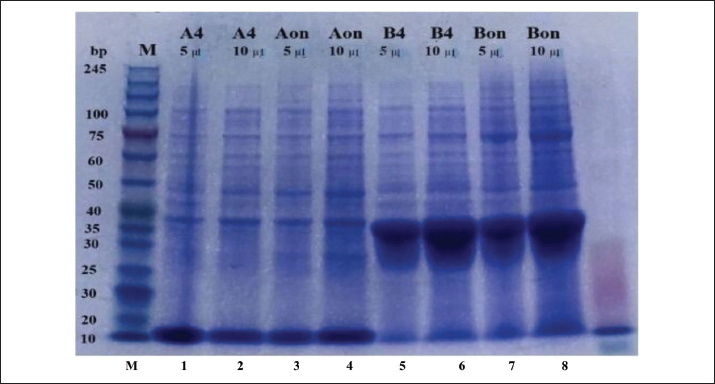
Fig. 3. Colony PCR analysis confirming positive recombinant clones. Lane M: 100 bp DNA ladder; Lane K: negative control (PCR mixture without template DNA). Lanes 1–3: construct A (mecA341–453) showing the expected amplicon at 586 bp; Lanes 4–6: construct B (mecA341–670) showing the expected amplicon at 1,237 bp, confirming the successful insertion of the recombinant plasmids. Expression and purification of rPBP2a fragmentsRecombinant protein expression was induced in E. coli BL21 (DE3) cells carrying the validated recombinant plasmids. SDS-PAGE analysis of purified protein fractions (Fig. 4) revealed distinct bands corresponding to the predicted molecular weights of the rPBP2a fragments encoded by constructs A and B. Both recombinant constructs were successfully expressed, yielding proteins of approximately 14 kDa for the mecA341–453 construct and 40 kDa for the mecA341–670 construct, consistent with the predicted molecular weights of the respective rPBP2a fragments. A pre-stained molecular weight protein marker was included in the SDS-PAGE gel to confirm the estimated molecular weights of the expressed proteins.
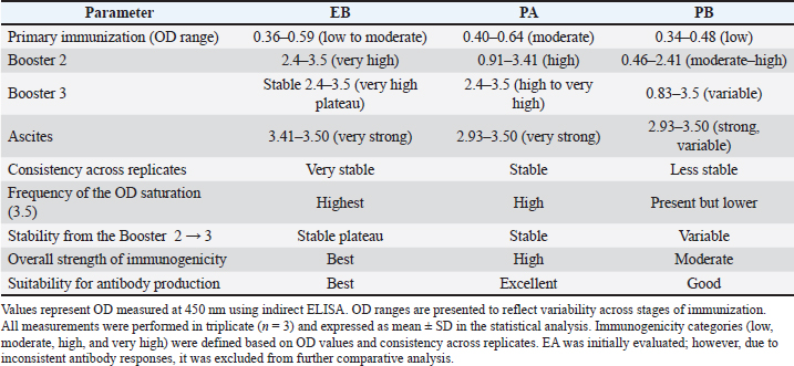
Fig. 4. SDS-PAGE analysis of purified rPBP2a proteins expressed from construct A (mecA341–453) and construct B (mecA341–670). Lane M: protein molecular weight marker; Lanes 1–4: purified recombinant proteins from construct A showing bands at approximately 14 kDa; Lanes 5–8: purified recombinant proteins from construct B showing bands at approximately 40 kDa. The observed bands correspond to the predicted molecular weights of the respective rPBP2a fragments. The recombinant proteins were recovered predominantly in the soluble fractions following Ni-NTA affinity chromatography, indicating the successful expression and purification of the rPBP2a antigens. Protein quantification revealed variable yields among the purified fractions, with 7.62 μg obtained from EA, 21.57 μg from EB, 12.25 μg from PA, and 4.37 μg from PB, resulting in an average yield of approximately 11.45 μg per fraction, which was consistent with the band intensity observed in SDS-PAGE analysis. These purified recombinant proteins were subsequently used as immunogens to develop antibodies and for further immunological assays. Antibody response following rPBP2a antigen immunizationThe antibody responses generated against the four rPBP2a antigen preparations were evaluated across all immunization stages (Table 1). ELISA measurements were performed in triplicate wells for each sample, and the results are presented as mean OD values at 450 nm ± SD. EB consistently produced the strongest and most stable IgG response among the tested antigens throughout the immunization series. During the primary immunization, EB induced a low but detectable response (OD 0.36–0.59), which increased markedly following Booster 2, reaching very high OD values ranging from 2.4 to 3.5. This strong response remained stable after Booster 3, indicating the establishment of high-affinity IgG with minimal inter-replicate variation. Table 1. Summary of antibody responses and antigen performance across stages of immunization.
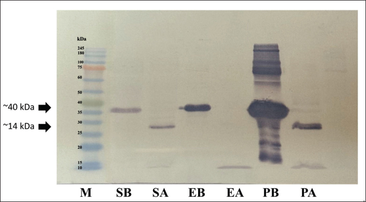
In comparison, PA generated a moderate response during the primary immunization and subsequently reached OD values of up to 3.5. However, the response exhibited greater variability than that of EB. PB, while capable of inducing moderate to high antibody levels, showed the highest variability across replicates and immunization stages, suggesting less consistent antigenic performance. Although EA was initially evaluated as an immunogen, it generated less consistent antibody responses than the other antigen preparations and was therefore not included in subsequent comparative analyses. The evaluation of ascites further supported these patterns. The antibodies derived from EB displayed the strongest and most consistent reactivity (OD 3.41–3.50), with minimal variation between replicates. PA and PB also produced strong ascites responses (OD 2.93–3.50), although both antigens, particularly PB, demonstrated broader variability. Statistical analysis was performed using one-way analysis of variance followed by Tukey’s multiple comparison test, with p < 0.05 considered statistically significant. The combined immunogenicity scoring, based on response magnitude, inter-replicate consistency, OD saturation frequency, and ascites strength, confirmed that EB was the most robust antigen, outperforming PA, PB, and EA across all evaluated parameters. Therefore, the antibodies generated by immunization with EB were selected as the primary antibodies for subsequent analyses, including WB validation, dot-blot antigen reactivity assays, and latex agglutination testing. WB confirmation of the anti-rPBP2a antibody specificityWB analysis was performed using rPBP2a preparations obtained from EB, SB, and PB antigens to further evaluate the immunoreactivity of anti-rPBP2a antibodies generated against the EB antigen fraction (anti-EB antibodies). As shown in Fig. 5, the anti-EB antibodies recognized a distinct immunoreactive band at approximately 40 kDa, which corresponds to the expected molecular weight of the rPBP2a protein used in this study. A pre-stained molecular weight marker was included in the gel to verify the estimated protein size.
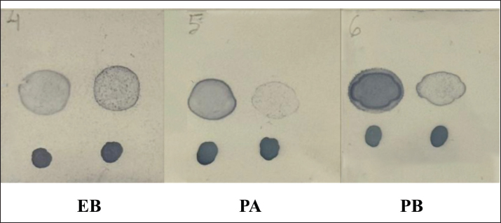
Fig. 5. WB analysis of anti-rPBP2a antibodies generated using the EB antigen preparation. A distinct immunoreactive band of approximately 40 kDa was detected in EB, and a similar band was observed in SB and PB, indicating recognition of the rPBP2a protein across soluble, insoluble, and purified protein fractions derived from construct B. Importantly, a similar ~40 kDa immunoreactive band was consistently detected in the EB, SB, and PB fractions, demonstrating that the anti-rPBP2a antibody recognized rPBP2a across different protein preparations. No additional nonspecific bands were observed at other molecular weights, indicating a high degree of antibody specificity. Because purified recombinant proteins were used as the antigen source, protein loading was normalized based on equal protein quantities determined during purification and confirmed by SDS-PAGE analysis before WB. The absence of cross-reactive bands suggests that the antibody recognizes the PBP2a antigen rather than unrelated proteins present in the preparation. These findings further support the ELISA results, which demonstrated strong antibody reactivity and affinity maturation following Booster 2 and 3 immunization. Integrated interpretation of ELISA and WB findingsThe combined ELISA and WB results confirm that immunization with the rPBP2a antigen from EB yields high-quality, high-affinity, and highly specific antibodies. The ELISA data reflect rapid titer elevation and strong booster responses, with EB outperforming all other antigen preparations in terms of strength, reproducibility, and stability. WB analysis further verified antigen specificity by demonstrating a single, clear band corresponding to PBP2a at ~40 kDa across multiple antigen fractions. Dot blotting of antibody–antigen reactivityDot-blot assays were performed using four rPBP2a antigen preparations: EA, EB, PA, and PB antigens to further assess the immunoreactivity of the anti-rPBP2a antibodies. Each antigen was probed separately with anti-EB, PA, and PB antibodies, while PBS served as the negative control. As shown in Fig. 6, the anti-EB antibody exhibited the strongest and most consistent dot signal intensity across the recombinant antigen preparations tested. This pattern was consistent with the ELISA and WB findings and supported the stronger immunoreactivity of anti-EB antibodies under the experimental conditions. In contrast, antibodies derived from PA and PB exhibited comparatively weaker and more variable dot signal intensities. No visible signal was observed in the PBS-negative control. Collectively, these findings indicate that immunization with rPBP2a antigen from EB generated an antibody preparation with consistent recognition of multiple recombinant antigen fractions and supported its selection for subsequent latex agglutination testing.
Fig. 6. Dot-blot analysis of rPBP2a antigens derived from EB, PA, and PB tested against anti-rPBP2a antibodies produced from (EB, left panel), (PA, middle panel), and (PB, right panel) produced distinct dot signals, with PBS serving as the negative control. The recombinant antibody generated from the EB produced the strongest and most consistent dot signals. Each antigen–antibody reaction was tested in triplicate, and the representative results are shown. The latex agglutination test for PBP2a functional detectionA latex agglutination test was performed using clinical MRSA and MSSA isolates to evaluate the functional performance of the anti-rPBP2a antibody developed from EB. As shown in Fig. 7, when mixed with the MRSA isolate, the EB-coated latex beads produced visible agglutination, forming coarse aggregates suggestive of rPBP2a antibody interactions. In contrast, no visible agglutination was observed when the same antibody-coated latex beads were tested against the MSSA isolate, which lacks the mecA-encoded PBP2a protein. The absence of visible clumping in MSSA confirms the high specificity of the anti-rPBP2a antibody and supports its potential for distinguishing MRSA from the tested MSSA isolate. The visible agglutination observed with MRSA was consistent with the ELISA, WB, and dot-blot findings, in which EB demonstrated the strongest and most consistent immunoreactivity among the tested antibody preparations. Collectively, these findings indicate that the recombinant antibody generated from EB showed promising performance in a latex agglutination–based assay and may have potential for future antibody-based MRSA screening. These observations were qualitative and were based on representative images from independent replicate experiments. DiscussionThis study successfully demonstrated the stepwise development of rPBP2a-targeting antibodies using mecA gene fragments derived from MRSA isolates collected in Indonesia. The molecular confirmation of methicillin resistance through mecA amplification (Fig. 1) is consistent with the well-established role of this gene as the primary determinant of β-lactam resistance in S. aureus (Idrees et al., 2023). The presence of clear mecA amplicons among the tested isolates further validated the suitability of locally circulating strains as a relevant antigen source for the development of recombinant antibodies.
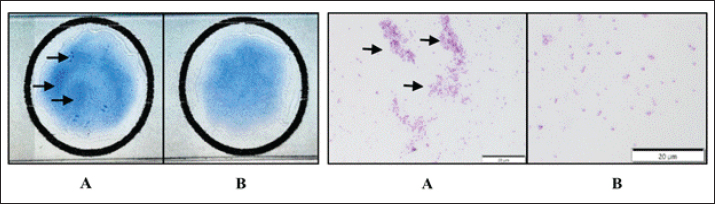
Fig. 1. PCR amplification of mecA (532 bp). Lane M: 100 bp DNA ladder; Lane K+: MRSA positive control; Lane 2: negative control (MSSA); Lanes 1, 3–5: MRSA isolates showing clear amplicons of the expected size. Two recombinant constructs, mecA341–453 and mecA341–670, were successfully cloned and verified (Figs. 2–3). These fragments encompass key regions of the PBP2a transpeptidase domain, which mediates low-affinity β-lactam binding (Fergestad et al., 2020). Previous studies have demonstrated that truncated or domain-focused PBP2a constructs retain critical immunogenic epitopes capable of inducing specific antibody responses (Haghighat et al., 2017). The development of diagnostic approaches targeting PBP2a remains highly relevant due to its central role in methicillin resistance. The mecA-encoded PBP2a exhibits a markedly reduced affinity for β-lactam antibiotics, enabling MRSA to maintain peptidoglycan cross-linking even in the presence of antimicrobial pressure (Lade and Kim, 2023). Structural studies have revealed that PBP2a possesses a unique conformational architecture, including a transpeptidase catalytic domain and regulatory regions that influence antibiotic accessibility to the active site (Ambade et al., 2023). More recent investigations have also highlighted the importance of allosteric regulatory sites in modulating PBP2a activity, providing additional opportunities for both therapeutic targeting and diagnostic development (Yao et al., 2025). The successful expression and purification of these recombinant proteins (Fig. 4) confirmed that these fragments maintained structural characteristics suitable for antigenic stimulation and antibody generation. Immunization of BALB/c mice produced differential responses, with antigens from the full transpeptidase domain construct (mecA341–670) producing the strongest and most consistent antibody responses (Table 1). The stronger immune response produced by the mecA341–670 construct suggests that the preservation of the full transpeptidase domain may improve epitope presentation and antibody recognition compared with the shorter partial-domain construct (mecA341–453). This finding supports the rationale for selecting these two recombinant fragments, as the shorter construct was intended to represent an epitope-focused region, whereas the larger construct preserved a broader antigenic structure of PBP2a. These results are consistent with previous reports indicating that larger or domain-complete PBP2a fragments expose a broader epitope repertoire and promote more robust antibody maturation (Peacock and Paterson, 2015). Interestingly, although the PB preparation also originated from the mecA341–670 construct and was capable of inducing moderate to high antibody responses, greater variability was observed among biological replicates and immunization stages. This variability may reflect differences in protein solubility, folding state, or the presence of aggregated proteins within the insoluble fraction, which could affect epitope accessibility and contribute to less consistent immune stimulation. These findings further support the use of the purified EB fraction as the preferred antigen source for subsequent antibody development. Furthermore, the sustained ELISA responses observed after Booster 3, together with the low variability among replicates, indicate that the mecA341–670 construct induced a stable humoral immune response. Although antibody affinity was not directly measured in this study, the observed pattern is consistent with progressive antibody maturation following repeated booster immunization. Comparable observations have been reported in experimental MRSA vaccine and antibody studies, where PBP2a-derived antigens induced robust immune responses in murine models (Haghighat et al., 2017; Mortazavi et al., 2020; Bagherzadeh et al., 2025). Saraiva et al. (2019) demonstrated that monoclonal antibodies targeting PBP2a can provide protective effects in MRSA infection models, further emphasizing the biological relevance of this protein as an immunogenic and diagnostically valuable target. The specificity of the recombinant antibody derived from EB was confirmed through WB analysis, which consistently detected a ~40 kDa band corresponding to the rPBP2a fragment used in this study (Fig. 5). The absence of additional nonspecific bands suggests that the antibody recognizes PBP2a epitopes without cross-reacting with unrelated proteins. Previous studies evaluating diagnostic antibodies targeting PBP2a reported similar observations (Lakhundi and Zhang, 2018). Dot-blot analysis further supported these findings by demonstrating strong and clean antigen–antibody interactions with minimal background signals (Fig. 6), indicating the high analytical specificity of the generated antibody. From a diagnostic perspective, the development of antibodies targeting PBP2a is particularly relevant because this protein is the phenotypic expression product of the mecA gene, the hallmark determinant of methicillin resistance. Molecular methods, such as PCR-based detection of mecA, remain the gold standard for MRSA confirmation due to their high sensitivity and specificity. However, PCR requires specialized equipment, trained personnel, and high operational costs, which may limit its accessibility in routine diagnostic laboratories, particularly in resource-limited settings. Therefore, alternative rapid diagnostic approaches, including commercial latex agglutination assays and immunochromatographic tests targeting PBP2a, have been developed. Latex agglutination assays based on monoclonal antibodies allow the rapid detection of PBP2a directly from bacterial cultures and are widely used in clinical microbiology laboratories. Our results demonstrated that the anti-rPBP2a antibody produced clear agglutination reactions with MRSA isolates but not with MSSA (Fig. 7). This performance pattern is comparable to that of previously reported PBP2a-based rapid diagnostic assays (Gupta et al., 2023; Bushra et al., 2024), suggesting that recombinant antibodies derived from locally circulating MRSA strains may provide an effective alternative to commercially imported diagnostic reagents.
Fig. 7. Latex agglutination test using the anti-rPBP2a antibody against MRSA (strain R6) and MSSA (strain K2). Macroscopic observations on a glass slide (left panel) with resazurin staining to improve visualization and microscopic views at 1,000× magnification (right panel). MRSA (A) showed rapid formation of coarse agglutination aggregates (arrows) within 30 s, indicating a positive interaction with PBP2a-coated latex beads. MSSA (B) showed no visible agglutination throughout the observation period, demonstrating the high specificity of the recombinant antibody for PBP2a-expressing MRSA. The assay was performed in three independent replicates, and the representative images are presented. An additional contribution of this work is its potential relevance to the One Health framework. While MRSA surveillance studies in Indonesia have predominantly focused on livestock-associated strains or human hospital isolates, limited research has investigated MRSA and MRSP in companion animals. In this study, the integration of phenotypic screening, mecA PCR confirmation, and species-specific identification provides a valuable foundation for future diagnostic and epidemiological investigations of methicillin-resistant staphylococci in veterinary contexts. The use of locally derived genetic material for antigen design may also improve the diagnostic antibody relevance and performance in regional settings. Despite these promising findings, this study has several limitations. First, the number of bacterial isolates used for antibody validation was limited, which may restrict the generalizability of the observed findings. Second, the anti-rPBP2a antibody system was primarily evaluated under laboratory conditions using cultured isolates, and broader clinical validation using a larger and more diverse collection of clinical samples has not yet been performed. Diagnostic performance parameters such as sensitivity, specificity, predictive values, and cross-reactivity with other bacterial species, were not evaluated in this study. Therefore, future studies should include expanded isolate panels and prospective clinical validation to further assess this antibody-based detection system’s diagnostic performance and practical applicability. The full transpeptidase domain construct (mecA341–670), particularly the EB fraction, produced the most stable and immunogenic recombinant antigen and generated the strongest anti-rPBP2a antibody response. EB consistently demonstrated superior immunoreactivity across ELISA, WB, dot blot, and latex agglutination assays compared with the other antigen preparations evaluated, supporting its selection as the most promising antigen source for anti-rPBP2a antibody development. These findings indicate the feasibility of developing locally derived anti-rPBP2a antibodies and provide a promising foundation for the development of rapid and affordable MRSA detection approaches in veterinary and clinical settings in Indonesia. ConclusionThis study successfully developed rPBP2a-targeting antibodies using mecA fragments derived from locally circulating MRSA isolates in Indonesia. The cloning, expression, and purification of rPBP2a constructs enabled effective immunization in BALB/c mice, with the EB antigen from the full transpeptidase domain (mecA341–670) producing the strongest and most consistent antibody response. The resulting anti-rPBP2a antibody demonstrated high specificity and reliable antigen recognition in ELISA, WB, dot blot, and latex agglutination assays. These findings indicate that rPBP2a has promising potential as a candidate reagent for the development of antibody-based MRSA detection systems. The use of indigenous MRSA isolates provides additional novelty and regional relevance, which may support the development of locally adapted diagnostic approaches. However, further studies involving a larger number of isolates and comprehensive diagnostic validation are necessary to evaluate the sensitivity, specificity, and practical applicability of this antibody system. AcknowledgmentsThe authors would like to thank the Laboratory of Clinical Pathology staff and collaborators for their technical support. The authors would also like to acknowledge Universitas Gadjah Mada for facility support and Genetika Science for technical assistance in gene synthesis and cloning. Conflict of interestThe authors declare that they have no conflicts of interest. FundingThis study was supported by the Ministry of Education, Culture, Research, and Technology of the Republic of Indonesia through Regular Fundamental Research Grant No. 067/C3/DT.05.00/P L/2025; 2473/UN1/DITLIT/Dit-Lit/PT.01.03/2025. Authors’ contributionsConceptualization, S.I.O.S. and F.A.; Funding Acquisition, S.I.O.S. and W.S.N.; Writing Original Draft Preparation, S.I.O.S., N.H., and F.A.; Methodology and Investigation, F.A., N.H, M.W., W.S.N.; Data Analysis, F.A., W.S.N.; Validation and Visualization, N.H., and M.W. The authors have reviewed and approved the published edition of this paper. Data availabilityAll data supporting the findings of this study are available within the manuscript. ReferencesAbebe, A. and Birhanu, A. 2023. Methicillin-resistant Staphylococcus aureus: molecular mechanisms underlying drug resistance development and novel strategies to combat. Infect Drug. Resist. 16, 7641–7662. Ambade, S.S., Gupta, V.K., Bhole, R.P., Khedekar, P.B. and Chikhale, R.V. 2023. A review on five- and six-membered heterocyclic compounds targeting penicillin-binding protein 2a (PBP2a) of methicillin-resistant Staphylococcus aureus. Molecules 28(20), 7008. Bagherzadeh, M., Haghighat, S. and Mahdavi, M. 2025. Killed whole-cell Staphylococcus aureus formulation in Montanide ISA266 and Alum adjuvants: different vaccine formulations varied in the vaccine’s potency and efficacy. Immunol Res. 73(1), 1–11. Bushra, R., Naqvi, S.H., Mirza, I.A., Imtiaz, A., Sajjad, R. and Gill, M.M. 2024. Evaluation of penicillin-binding protein 2a (PBP2a) latex agglutination assay for rapid identification of methicillin-resistant Staphylococcus aureus isolated from blood culture growth. Pak. Armed. Forces. Med. J. 74(6), 1604–1608. Clinical and Laboratory Standards Institute (CLSI). 2023. Performance standards for antimicrobial susceptibility testing, 33rd ed. CLSI supplement M100. Wayne, PA: CLSI. Curcio, D., Cane, A., Fernández, F. and Correa, J. 2018. Surgical site infection in elective clean and clean-contaminated surgeries in developing countries. Int. J. Infect. Dis. 34, 34–45. Dalton, K.R., Rock, C., Carroll, K.C. and Davis, M.F. 2020. One Health in hospitals: understanding the dynamics of people, animals, and the built environment to inform interventions for antimicrobial-resistant gram-positive infections. Antimicrob. Resist. Infect Control. 9(1), 78. Dewulf, S., Boyen, F., Paepe, D., Clercx, C., Tilman, N., Dewulf, J. and Boland, C. 2025. Antimicrobial resistance characterization of methicillin-resistant Staphylococcus aureus and Staphylococcus pseudintermedius isolates from clinical cases in dogs and cats in Belgium. Antibiotics 14(7), 631. Fergestad, M.E., Stamsås, G.A., Morales Angeles, D., Salehian, Z., Wasteson, Y. and Kjos, M. 2020. Penicillin-binding protein PBP2a provides variable levels of protection toward different β-lactams in Staphylococcus aureus RN4220. Microbiol. Open 9(8), 1057. Feuer, L., Frenzer, S.K., Merle, R., Leistner, R., Bäumer, W., Bethe, A., Lübke-Becker, A., Klein, B. and Bartel, A. 2024. Prevalence of MRSA in canine and feline clinical samples from one-third of veterinary practices in Germany from 2019–2021. J. Antimicrob. Chemother. 79(9), 225. Fitranda, M., Salasia, S.I.O., Sianipar, O., Dewananda, D.A., Arjana, A.Z., Aziz, F., Wasissa, M., Lestari, F.B. and Santosa, C.M. 2023. Methicillin-resistant Staphylococcus aureus isolates derived from humans and animals in Yogyakarta, Indonesia. Vet. World 16(1), 239–245. Fitranda, M., Salasia, S.I.O., Sianipar, O., Sukorini, U., Aziz, F., Wasissa, M., Lestari, F.B., Khair, R.E. and Dahesihdewi, A. 2024. Distribution of antimicrobial resistance genes of methicillin-resistant Staphylococcus aureus isolated from animals and humans in Yogyakarta, Indonesia. Int. J. One Health 10(1), 38–44. Genath, A., Hackmann, C., Denkel, L., Weber, A., Maechler, F., Kola, A., Schwarz, S., Gastmeier, P. and Leistner, R. 2024. The genetic relationship between human and pet isolates: a core genome multilocus sequence analysis of multidrug-resistant bacteria. Antimicrob. Resist. Infect Control. 13, 107. Gupta, N., Jais, M., Sharma, A. and Shrivastava, P.K. 2023. Evaluation of diagnostic accuracy of latex agglutination test and E-test for the detection of methicillin-resistant Staphylococcus aureus. Int. J. Scientific. Rep. 9(12), 398–405. Haghighat, S., Siadat, S.D., Sorkhabadi, S.M.R., Sepahi, A.A., Sadat, S.M., Yazdi, M.H. and Mahdavi, M. 2017. RPBP2a as a vaccine candidate against methicillin-resistant Staphylococcus aureus: immunogenicity and protectivity. Microbial Pathogenesis 108, 32–39. Idrees, M.M., Saeed, K., Shahid, M.A., Akhtar, M., Qammar, K., Hassan, J., Khaliq, T. and Saeed, A. 2023. Prevalence of mecA and mecC associated methicillin-resistant Staphylococcus aureus in clinical specimens, Punjab, Pakistan. Biomedicines 11(3), 878. Igrejas, G., Correia, S., Silva, V., Hébraud, M., Caniça, M., Torres, C., Gomes, C., Nogueira, F. and Poeta, P. 2018. Planning a One Health case study to evaluate methicillin-resistant Staphylococcus aureus and its economic burden in Portugal. Front. Microbiol. 9, 2964. Khan, H.A., Baig, F.K. and Mehboob, R. 2017. Nosocomial infections: epidemiology, prevention, control and surveillance. Asian. Pacific. J. Trop. Biomed. 7(5), 478–482. Kourtis, A.P., Hatfield, K., Baggs, J., Mu, Y., See, I., Epson, E. , Nadle, J., Kainer, M.A., Dumyati, G., Petit, S. and Ray, S.M. 2019. Vital signs: epidemiology and recent trends in methicillin-resistant and methicillin-susceptible Staphylococcus aureus bloodstream infections—United States. MMWR. Morbidity Mortality Weekly Rep. 68(9), 214–219. Lade, H. and Kim, J.S. 2023. Molecular determinants of β-lactam resistance in methicillin-resistant Staphylococcus aureus (MRSA): an updated review. Antibiotics 12(9), 1362. Lakhundi, S. and Zhang, K. 2018. Methicillin-resistant Staphylococcus aureus: molecular characterization, evolution, and epidemiology. Clin. Microbiol. Rev. 31(4), e00020–e00018. Lo, D.K.H., Muhlebach, M.S. and Smyth, A.R. 2022. Interventions for the eradication of meticillin-resistant Staphylococcus aureus in people with cystic fibrosis. Cochrane Database Systematic Rev. 2022(12), CD009650. Mortazavi, S.S., Haghighat, S. and Mahdavi, M. 2020. RPBP2a of methicillin-resistant Staphylococcus aureus formulation in Alum and Montanide ISA266 adjuvants induced cellular and humoral immune responses with protection in BALB/c mice. Microbial. Pathogenesis. 140, 103945. Murray, C.J., Ikuta, K.S., Sharara, F., Swetschinski, L., Robles Aguilar, G., and Gray, A. 2022. Global burden of bacterial antimicrobial resistance in 2019: a systematic analysis. Lancet 399(10325), 629–655. Nataraj, B.H. and Mallappa, R.H. 2021. Antibiotic resistance crisis: an update on antagonistic interactions between probiotics and methicillin-resistant Staphylococcus aureus (MRSA). Curr. Microbiol. 78(6), 2194–2211. Nigo, M., Rasmy, L., Mao, B., Kannadath, B.S., Xie, Z. and Zhi, D. 2024. Deep learning model for personalized prediction of positive MRSA culture using time-series electronic health records. Nature. Commun. 15, 2036. Peacock, S.J. and Paterson, G.K. 2015. Mechanisms of methicillin resistance in Staphylococcus aureus. Annu. Rev. Biochem. 84, 577–601. Salasia, S.I.O., Tato, S., Prabawati, F. and Ariyanti, D. 2013. Clonal relationship of methicillin-resistant Staphylococcus aureus in cattle and humans. Jurnal. Kedokteran. Hewan. 7(2), 125–128. Saraiva, F.B., De Araújo, A.C.C., De Araújo, A.E.V. and Senna, J.P.M. 2019. Monoclonal antibody anti-PBP2a protects mice against MRSA infections. PLoS One 14(11), 225752. Tabassum, F., Munir, A., Shakeel, F., Zia, T., Khan, M.U.Z. and Tahir, Z. 2025. Methicillin resistance in Staphylococcus aureus: a review of the mecA gene and its role in antibiotic resistance. Indus J. Bioscience Res. 3(9), 124–129. Van Griethuysen, A., Pouw, M., Van Leeuwen, N., Heck, M., Willemse, P., Buiting, A. and Kluytmans, J. 1999. Rapid slide latex agglutination test for detection of methicillin resistance in Staphylococcus aureus. J. Clin. Microbiol. 37(9), 2789–2792. Widianingrum, D.C., Windria, S. and Salasia, S.I.O. 2016. Antibiotic resistance and methicillin-resistant Staphylococcus aureus isolated from bovine, crossbred Etawa goat, and human. Asian. J. Anim. Vet. Adv. 11(2), 122–129. World Health Organization (WHO). 2024. Antimicrobial resistance. Available via https://www.who.int/europe/news-room/fact-sheets/item/antimicrobial-resistance (Accessed 19 September 2024). Worthing, K.A., Brown, J., Gerber, L., Trott, D.J., Abraham, S. and Norris, J.M. 2018. Methicillin-resistant staphylococci among veterinary personnel, personnel-owned pets, patients, and the hospital environment in two veterinary hospitals. Vet. Microbiol. 223, 79–85. Yao, S., Fang, C., Xu, B., Hu, Y., Chen, Z., Xue, X., Liu, J., Li, M. and Li, P. 2025. Designing novel nucleoside inhibitors targeting the allosteric site of PBP2a: a strategic approach to overcome resistance in MRSA. Bioorganic. Med. Chem. 122, 118133. Values represent OD measured at 450 nm using indirect ELISA. OD ranges are presented to reflect variability across stages of immunization. All measurements were performed in triplicate (n=3) and expressed as mean ± SD in the statistical analysis. Immunogenicity categories (low, moderate, high, and very high) were defined based on OD values and consistency across replicates. EA was initially evaluated; however, due to inconsistent antibody responses, it was excluded from further comparative analysis. | ||
| How to Cite this Article |
| Pubmed Style Salasia SIO, Hanifah N, Wasissa M, Aziz F, Nugroho WS. Development and characterization of anti-recombinant PBP2a antibodies for preliminary detection of methicillin-resistant Staphylococcus aureus. doi:10.5455/OVJ.2026.v16.i7.49 Web Style Salasia SIO, Hanifah N, Wasissa M, Aziz F, Nugroho WS. Development and characterization of anti-recombinant PBP2a antibodies for preliminary detection of methicillin-resistant Staphylococcus aureus. https://www.openveterinaryjournal.com/?mno=302770 [Access: July 15, 2026]. doi:10.5455/OVJ.2026.v16.i7.49 AMA (American Medical Association) Style Salasia SIO, Hanifah N, Wasissa M, Aziz F, Nugroho WS. Development and characterization of anti-recombinant PBP2a antibodies for preliminary detection of methicillin-resistant Staphylococcus aureus. doi:10.5455/OVJ.2026.v16.i7.49 Vancouver/ICMJE Style Salasia SIO, Hanifah N, Wasissa M, Aziz F, Nugroho WS. Development and characterization of anti-recombinant PBP2a antibodies for preliminary detection of methicillin-resistant Staphylococcus aureus. doi:10.5455/OVJ.2026.v16.i7.49 Harvard Style Salasia, S. I. O., Hanifah, . N., Wasissa, . M., Aziz, . F. & Nugroho, . W. S. (2026) Development and characterization of anti-recombinant PBP2a antibodies for preliminary detection of methicillin-resistant Staphylococcus aureus. doi:10.5455/OVJ.2026.v16.i7.49 Turabian Style Salasia, Siti Isrina Oktavia, Nada Hanifah, Madarina Wasissa, Fatkhanuddin Aziz, and Widagdo Sri Nugroho. 2026. Development and characterization of anti-recombinant PBP2a antibodies for preliminary detection of methicillin-resistant Staphylococcus aureus. doi:10.5455/OVJ.2026.v16.i7.49 Chicago Style Salasia, Siti Isrina Oktavia, Nada Hanifah, Madarina Wasissa, Fatkhanuddin Aziz, and Widagdo Sri Nugroho. "Development and characterization of anti-recombinant PBP2a antibodies for preliminary detection of methicillin-resistant Staphylococcus aureus." doi:10.5455/OVJ.2026.v16.i7.49 MLA (The Modern Language Association) Style Salasia, Siti Isrina Oktavia, Nada Hanifah, Madarina Wasissa, Fatkhanuddin Aziz, and Widagdo Sri Nugroho. "Development and characterization of anti-recombinant PBP2a antibodies for preliminary detection of methicillin-resistant Staphylococcus aureus." doi:10.5455/OVJ.2026.v16.i7.49 APA (American Psychological Association) Style Salasia, S. I. O., Hanifah, . N., Wasissa, . M., Aziz, . F. & Nugroho, . W. S. (2026) Development and characterization of anti-recombinant PBP2a antibodies for preliminary detection of methicillin-resistant Staphylococcus aureus. doi:10.5455/OVJ.2026.v16.i7.49 |


