





| Research Article | ||
Open Vet. J.. 2026; 16(7): 4241-4249 Open Veterinary Journal, (2026), Vol. 16(7): 4241-4249 Research Article Diagnostic value of endometrial cytology in mares with normal uterine ultrasonographyMihaela Ioana Ciuta, Mirela Alexandra Tripon*, Iancu Adrian Morar, Cristian Mihăiță Crecan and Mihai Cosmin CenariuFaculty of Veterinary Medicine, University of Agricultural Sciences and Veterinary Medicine, Cluj-Napoca, Romania *Corresponding Author: Mirela Alexandra Tripon. Faculty of Veterinary Medicine, University of Agricultural Sciences and Veterinary Medicine, Cluj-Napoca, Romania. Email: mirela.tripon [at] usamvcluj.ro Submitted: 12/12/2025 Revised: 22/05/2026 Accepted: 03/06/2026 Published: 02/07/2026 © 2026 Open Veterinary Journal
AbstractBackground: Endometritis is a major cause of subfertility in mares and may occur in both clinical and subclinical forms. Although transrectal ultrasonography is routinely used to evaluate uterine health, its sensitivity for detecting mild inflammatory changes is limited. Endometrial cytology can identify inflammatory cells directly and may reveal uterine pathology even when ultrasonographic findings appear normal. Aim: The aim of this study was to determine the incidence of positive uterine cytology in mares with normal ultrasonographic findings and to evaluate the relationship between cytological inflammation, bacteriological culture results, and reproductive outcomes. Methods: The study included 43 mares aged 4–20 years (mean ± SD: 10.6 ± 3.8 years) presented for reproductive evaluation at the Equine Clinic of the University of Agricultural Sciences and Veterinary Medicine, Cluj-Napoca. Only mares with normal uterine ultrasonographic appearance were included. Endometrial cytology and uterine bacterial culture were performed during estrus. Cytological samples were classified based on the number of polymorphonuclear neutrophils per high-power field. Results: Cytological evidence of endometrial inflammation was detected in 6 of 43 mares (14.0%), despite normal ultrasonographic findings. Bacterial culture was positive in 5 mares (11.6%). A significant association was observed between positive cytology and bacterial culture (OR=72; p=0.0005). Pregnancy rates were significantly lower in mares with positive cytology (p=0.028). Conclusion: Subclinical endometrial inflammation may occur in mares with normal ultrasonographic uterine appearance. These findings highlight the importance of incorporating endometrial cytology into routine reproductive evaluations to improve the detection of uterine inflammation and support better fertility management in breeding mares. Keywords: Endometritis, Mare, Subclinical inflammation, Ultrasonography, Uterine cytology. IntroductionReproductive performance is a key determinant of efficiency in equine breeding programs, as reduced fertility in mares can lead to important economic and management consequences for breeding operations. Reduced conception rates in mares may result from multiple reproductive disorders, including uterine inflammation, impaired uterine clearance, anatomical abnormalities of the reproductive tract, and age-related endometrial degeneration. Among these conditions, endometritis—defined as inflammation of the endometrial lining and occurring in both clinical and subclinical forms—remains one of the most frequently diagnosed causes of subfertility and infertility in mares (Christoffersen et al., 2012; Christoffersen and Troedsson, 2017; Canisso et al., 2020; Morris et al., 2020). Despite its routine use in reproductive examinations, ultrasonography has limited sensitivity for detecting subclinical endometritis, because mild inflammatory changes may occur in the absence of detectable intrauterine fluid or structural abnormalities (Christoffersen et al., 2012; Rua et al., 2018). The reported prevalence of positive cytology in mares without ultrasonographic abnormalities varies widely. Previous studies have reported that approximately 10%–20% of clinically healthy mares may present positive cytology or bacteriological culture results despite the absence of clinical or ultrasonographic abnormalities, while higher prevalences of up to 28%–30% have been reported in breeding populations evaluated before insemination (Christoffersen et al., 2012; Rasmussen et al., 2015; Diel De Amorim et al., 2016). Similar findings have been reported in breeding populations across Europe and North America, highlighting the global importance of uterine inflammatory disease as a limiting factor in equine reproductive efficiency. Endometritis in mares may result from a variety of infectious and non-infectious causes. Bacterial infections represent the most common etiology and are frequently associated with organisms such as Streptococcus equi subsp. zooepidemicus, Escherichia coli, Klebsiella pneumoniae, and Pseudomonas aeruginosa (El Atafy et al., 2018). Fungal infections, although less common, may also occur, particularly in mares subjected to repeated intrauterine antibiotic treatments or poor perineal conformation. In addition to infectious causes, several non-infectious factors may predispose mares to persistent uterine inflammation, including impaired uterine clearance, cervical dysfunction, anatomical abnormalities of the reproductive tract, and age-related degenerative endometrial changes (Troedsson, 2011). Diagnosis of uterine inflammation in mares typically relies on a combination of transrectal ultrasonography, endometrial cytology, and bacteriological culture. Ultrasonography is widely used in clinical practice to evaluate uterine fluid accumulation, endometrial edema, and structural abnormalities. However, its sensitivity for detecting mild inflammatory changes or early stages of endometritis is limited (Rua et al., 2018). On the contrary, endometrial cytology allows direct evaluation of inflammatory cells within the uterine lumen and can detect polymorphonuclear neutrophils (PMNs) infiltration even when ultrasonographic findings appear normal. Previous studies have shown that mares with apparently normal ultrasonographic uterine findings may still present positive cytological results, indicating the presence of subclinical endometritis (Diel De Amorim et al., 2016; Rua et al., 2018). Such cases are particularly relevant in clinical practice because mares are often considered reproductively healthy based solely on ultrasonographic examination. As a result, inflammatory changes may remain undiagnosed when cytology is not routinely performed during reproductive evaluation. Therefore, the objective of the present study was to determine the incidence of positive uterine cytology in mares with normal ultrasonographic findings and to evaluate the diagnostic value of cytology for detecting subclinical uterine inflammation. Materials and MethodsStudy populationThe study was conducted between March 2024 and July 2025 at the Equine Clinic of the Faculty of Veterinary Medicine, University of Agricultural Sciences and Veterinary Medicine, Cluj-Napoca, Romania. All procedures were performed as part of routine clinical reproductive examinations, and written informed consent was obtained from the owners prior to sample collection. A total of 43 mares were included in the study, ranging in age from 4 to 20 years (mean ± SD: 10.6 ± 3.8 years). The study population consisted of 24 warmblood mares, 7 Arabians, and 12 draft-type mares. Based on reproductive history, 26 mares were classified as barren, 11 as maiden, and 6 as postpartum. Only mares with a normal uterine appearance on ultrasonographic examination were included. Inclusion criteria required the absence of pathological intrauterine fluid accumulation, excessive endometrial edema, or uterine wall thickening during ultrasonographic evaluation. Small amounts of intrauterine fluid occasionally observed during estrus were considered physiological and were not regarded as evidence of uterine pathology. Management conditions and breeding protocols were not standardized across all mares and may represent a potential source of variability. Ultrasonographic examinationTransrectal ultrasonography was performed using a 5-MHz linear transducer connected to a portable veterinary ultrasound system (MyLab Omega VET, Esaote S.p.A., Genoa, Italy). The uterus was examined systematically to evaluate endometrial echotexture and identify structural abnormalities (Kidd et al., 2022). Particular attention was given to the presence of intrauterine fluid accumulation, focal endometrial thickening, and the degree of endometrial edema. Uterine horn diameter was assessed in transverse section at the mid-horn region. In adult mares, uterine horn diameter during estrus typically ranges between approximately 3 and 5 cm (McKinnon et al., 2011). In all mares included in the study, both uterine horns appeared symmetrical and within normal size limits, with no evidence of focal thickening or abnormal echogenic structures. During estrus, the endometrium displayed the characteristic physiological edema pattern with distinct radial folds (“cartwheel” appearance). Small amounts of intrauterine fluid occasionally observed during estrus were considered physiological and were not classified as pathological findings (Kidd et al., 2022 ). Only mares without clinically significant intrauterine fluid accumulation, uterine wall thickening, or abnormal echogenic structures were included in the study. Estrus was confirmed by the presence of at least one follicle ≥35 mm in diameter together with a uterine edema score of grade 2 (McKinnon et al., 2011; Kidd et al., 2022). To minimize variability related to operator technique, all ultrasonographic examinations were performed by the same experienced clinician throughout the study period. Uterine culture samplingUterine culture samples were collected during estrus, when cervical relaxation facilitates transcervical sampling. Prior to sampling, the perineal region was thoroughly cleaned and disinfected. A sterile double-guarded swab was introduced transcervically into the uterine lumen under aseptic conditions. Once positioned in the uterine body, the swab was briefly exposed to the endometrial surface and then retracted into its protective sheath before removal (Kozdrowski et al., 2015). Samples were immediately inoculated onto aerobic culture media, including blood agar and MacConkey agar, and incubated at 37°C for 24–48 hours. Anaerobic cultures were not performed because obligate anaerobic uterine infections are considered uncommon in mares (Ferrer and Palomares, 2018). When clinically indicated, fungal culture media were used to evaluate the presence of mycotic pathogens. Bacterial growth was recorded based on colony morphology, hemolysis patterns, and Gram-staining characteristics. Endometrial cytology sampling and evaluationEndometrial cytology samples were obtained using a sterile double-guarded cytology brush (Minitüb GmbH, Tiefenbach, Germany) designed for collection of uterine epithelial cells in mares. Smears were air-dried, stained using the Diff-Quik® staining method (Sysmex Corporation, Kobe, Japan), and examined under light microscopy at 400× magnification using a light microscope (Olympus CX23, Olympus Corporation, Tokyo, Japan), following previously described cytological protocols for the diagnosis of endometritis in mares (Overbeck et al., 2011; Kozdrowski et al., 2015). Cytological evaluation was performed by a single experienced observer who was blinded to bacteriological culture results and the reproductive history of the mares. For each smear, at least five non-overlapping high-power fields (HPF; 400×) were randomly selected and examined, and the mean number of PMNs per field was recorded. Samples were classified as negative when ≤1 PMN per 5 HPF was observed, suspicious when 2–4 PMNs per 5 HPF were present, and positive when >5 PMNs per 5 HPF were detected, according to previously published criteria (Kozdrowski et al., 2015). Although sampling during estrus may be associated with mild physiological infiltration of PMNs due to estrogen-mediated immune responses, this stage was selected because cervical relaxation allows safer and more reliable transcervical sampling. In the present study, cytological samples from mares classified as negative showed ≤1 PMN per 5 HPF, indicating that no evidence of physiological estrus-associated PMN infiltration was observed in the study population. Therapeutic managementMares diagnosed with positive cytology and/or positive bacterial culture received treatment according to the underlying clinical diagnosis. Inflammatory (non-infectious) endometritisIn mares with cytological evidence of inflammation and negative bacterial culture, treatment consisted of uterine lavage combined with ecbolic agents and anti-inflammatory therapy (Köhne et al., 2020). Oxytocin was administered to stimulate uterine contractions and promote uterine clearance. Uterine lavages using sterile saline or diluted povidone–iodine solution were performed at 24–48 hours. A non-steroidal anti-inflammatory agent, flunixin meglumine, was administered for 3–5 days, depending on clinical response. Ultrasonographic monitoring was used to assess treatment efficacy (Morris et al., 2020). Infectious endometritisMares with confirmed infectious endometritis based on positive culture results were treated according to antimicrobial susceptibility testing. Initial management included repeated uterine lavage with sterile saline or diluted povidone–iodine solution until clear effluent was obtained (Köhne et al., 2020). Oxytocin or prostaglandin F2α was administered to enhance uterine evacuation. Intrauterine antibiotics were subsequently infused under aseptic conditions using an insemination catheter. The choice of antibiotic was guided by the antibiogram and commonly included gentamicin, amikacin, enrofloxacin, penicillin, or ceftiofur. Aminoglycosides were buffered with sodium bicarbonate to reduce endometrial irritation. Systemic antimicrobial therapy was administered when indicated (Morris et al., 2020). Chronic degenerative endometritisChronic degenerative endometritis, characterized by irreversible endometrial fibrosis and degeneration, was managed conservatively, as no curative treatment is currently available (Köhne et al., 2020). Affected mares were excluded from breeding programs. Preventive strategies focused on early diagnosis and treatment of inflammatory and infectious endometritis. Mares diagnosed and treated for endometritis during the study period were not bred in the same reproductive season and were therefore excluded from pregnancy outcome analysis. Statistical analysisData analysis included both descriptive and inferential statistics. The proportion of mares with positive cytology was expressed as a percentage, and 95% confidence intervals (CI) were calculated using the Wilson method for binomial proportions. Associations between cytological findings and bacteriological results, as well as between cytology and pregnancy outcome, were assessed using Fisher’s exact test due to small expected cell counts. Odds ratios (OR) with corresponding 95% CIs were calculated to estimate the strength of associations. Statistical significance was set at p < 0.05. Agreement between cytological and bacteriological findings was evaluated using Cohen’s kappa (κ) coefficient. Interpretation of κ values followed the criteria proposed by Landis and Koch (1977). Pregnancy rates were compared between cytology-positive and cytology-negative mares using Fisher’s exact test, and ORs were calculated to estimate the likelihood of pregnancy. Statistical analyses were performed using GraphPad Prism version 10 (GraphPad Software, San Diego, USA). Ethical approvalAll procedures performed in this study were part of routine clinical reproductive examinations carried out at the Equine Clinic of the University of Agricultural Sciences and Veterinary Medicine, Cluj-Napoca. No experimental procedures were performed on the animals. Written informed consent was obtained from the owners prior to sample collection. All procedures complied with institutional guidelines for veterinary clinical practice and animal welfare. ResultsOverall findingsA small proportion of mares (6/43; 14.0%) showed cytological evidence of uterine inflammation despite a normal ultrasonographic appearance. The diagnostic and reproductive outcomes of the 43 mares included in the study are summarized in Table 1. Most mares (33/43; 76.7%) had negative uterine cytology, whereas six mares (14.0%) showed clear cytological evidence of inflammation. Four additional samples (9.3%) were classified as suspicious based on intermediate PMN counts. Bacteriological culture was positive in five mares (11.6%), while the remaining 38 samples yielded no bacterial growth. Among the positive cultures, three isolates were identified as Streptococcus equi subsp. zooepidemicus and two as E. coli. Table 1. Distribution of cytological findings, uterine culture results, and pregnancy outcomes in mares with normal ultrasonographic uterine appearance (original).
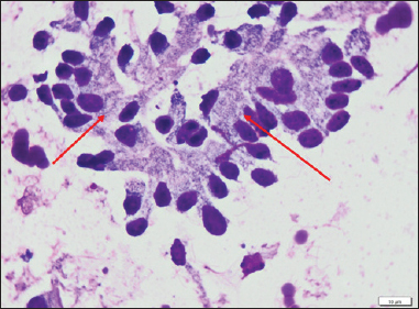
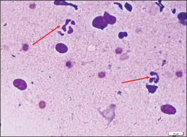
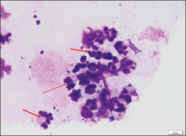
Among the cytologically negative samples, PMNs were absent or rare (≤1 PMN/ 5 HPF) (Fig. 1). Samples classified as suspicious contained low numbers of PMNs (2–4 PMNs/ 5 HPF) (Fig. 2). On the contrary, six samples showed marked PMN infiltration (≥5 PMNs/ 5 HPF), consistent with cytological evidence of endometritis (Fig. 3). Culture and cytology results were concordant in four mares, all of which tested positive using both diagnostic methods.
Fig. 1. Endometrial cytology smear from a mare with negative cytological findings. The smear was air-dried, stained using the Diff-Quik® staining method (Sysmex Corporation, Kobe, Japan), and examined under light microscopy at 400× magnification. Endometrial epithelial cells with normal morphology are visible, and no PMNs are present in the evaluated field. The red arrow indicates endometrial epithelial cells.
Fig. 2. Endometrial cytology smear classified as suspicious. The smear was air-dried, stained using the Diff-Quik® staining method (Sysmex Corporation, Kobe, Japan), and examined under light microscopy at 400× magnification. Endometrial epithelial cells are visible together with a small number of PMNs (2–4 PMNs per 5 HPF), consistent with intermediate cytological findings. Red arrows indicate PMNs.
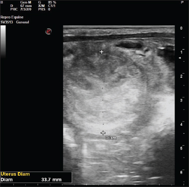
Fig. 3. Endometrial cytology smear from a mare with positive cytological findings consistent with endometritis. The smear was air-dried, stained using the Diff-Quik® staining method (Sysmex Corporation, Kobe, Japan), and examined under light microscopy at 400× magnification using a light microscope (Olympus CX23, Olympus Corporation, Tokyo, Japan). Numerous PMNs (>5 PMNs per 5 HPF) are present, indicating cytological evidence of uterine inflammation. Red arrows indicate PMNs. Pregnancy occurred in 26 of 33 mares (78.78%) with negative cytology, whereas none of the mares with positive cytology conceived during the study period (0/6). Agreement between cytology and culture was substantial, with a Cohen’s kappa coefficient of 0.68, indicating moderate to substantial concordance between the two diagnostic methods. However, it should be noted that the presence of bacterial growth in uterine culture does not necessarily indicate pathogenic infection. Some isolates may represent environmental contamination, transient colonization, or bacteria with limited pathogenic significance. Therefore, bacteriological results should always be interpreted together with cytological findings and clinical examination. Pregnancy outcomes were available for mares that underwent breeding during the study period. Approximately 70% of mares with negative cytology achieved pregnancy. On the contrary, conception rates were markedly lower in mares with cytological evidence of inflammation. Overall, the majority of mares with normal ultrasonographic findings exhibited negative cytology and culture results. A representative ultrasonographic image of a normal uterus observed in the study population is shown in Figure 4. However, a relevant minority showed inflammatory or infectious changes detectable only through cytological or microbiological examination.
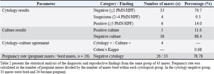
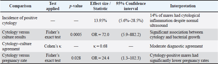
Fig. 4. Normal ultrasonographic appearance of the uterus in a mare during estrus. Transrectal ultrasonographic image showing a homogeneous uterine echotexture with physiological endometrial edema and no evidence of intrauterine fluid accumulation. The uterine diameter was measured in transverse section at the mid-portion of the left uterine horn (33.7 mm). CorrelationsStatistical associations between diagnostic findings and reproductive outcomes are presented in Table 2. Cytological evidence of uterine inflammation was identified in approximately 14% of mares with normal ultrasonographic appearance. The calculated 95% CI (5.6%–28.5%) reflects variability related to sample size but confirms the presence of subclinical inflammation within this population. Table 2. Associations between cytology, bacteriological culture, and pregnancy outcome in mares with normal uterine ultrasonography (original).
A strong association was observed between positive cytology and positive bacterial culture. Mares with cytological inflammation were significantly more likely to have a positive culture result (OR=72), indicating that microscopic PMN infiltration is a reliable indicator of underlying bacterial involvement. Agreement between cytology and culture was further supported by a kappa value of 0.68, consistent with moderate to substantial agreement. While the two methods were concordant in most cases, each test identified a small number of cases not detected by the other. A statistically significant relationship was also observed between cytology and fertility outcome (p < 0.05). Mares with positive cytology were 24.4 times more likely to fail to conceive compared with mares with normal cytology, demonstrating that even subclinical uterine inflammation may adversely affect reproductive performance. DiscussionThe present study demonstrates that cytological evidence of endometrial inflammation can be detected in mares with a normal ultrasonographic uterine appearance. In this population, approximately 14% of mares showed cytological evidence of uterine inflammation despite the absence of ultrasonographic abnormalities, indicating that subclinical endometritis may remain undetected when ultrasonography is used as the sole diagnostic tool. These findings highlight the importance of cytological evaluation even in mares that appear reproductively normal on ultrasonographic examination. Similar bacteriological findings have recently been reported in mares using culture-based diagnostic approaches (Carvalho et al., 2025). Furthermore, uterine lavage and inflammatory markers have been investigated as adjunctive diagnostic tools for subclinical endometritis (Sikora et al., 2016). Subclinical endometrial inflammation has also been described in other species. In dairy cattle, cytological detection of uterine inflammation has been widely used to diagnose subclinical endometritis and has been associated with reduced conception rates and impaired reproductive performance. Similar inflammatory conditions have been reported in women and small animals, where chronic endometrial inflammation may negatively affect implantation and fertility (Moreno et al., 2018; Praderio et al., 2019). Although diagnostic criteria vary between species, these findings support the broader concept that mild or subclinical uterine inflammation may significantly influence reproductive success. This prevalence is consistent with previous reports indicating that 10%–20% of clinically normal mares may present positive cytology or culture results in the absence of ultrasonographic changes (Christoffersen et al., 2012; Diel De Amorim et al., 2016; El Atafy et al., 2018). Such mares often lack overt clinical signs, yet subclinical inflammation has been shown to negatively affect fertility (Canisso et al., 2020). It is important to recognize that the presence of a small number of polymorphonuclear neutrophils (1–2 PMNs/ 5 HPF) in the uterine lumen during estrus may represent a physiological response rather than pathological inflammation. Estrogen-induced cervical relaxation, increased exposure of the uterus to the vaginal environment, and transient immune activation can lead to mild PMN migration into the uterine lumen (Katila and Ferreira-Dias, 2022). Previous studies have reported low PMN counts in cytological samples (Walter et al., 2012; Kozdrowski et al., 2015; Teixeira-Soares et al., 2022). Consequently, intermediate PMN counts (2–4 PMNs/ 5 HPF) should be interpreted with caution, as they may represent subclinical endometritis (Overbeck et al., 2011). In the present study, cytology proved to be a sensitive method for detecting uterine inflammation. The substantial agreement observed between cytology and bacterial culture (κ=0.68) supports their complementary diagnostic roles. Similar levels of agreement have been reported previously (Christoffersen et al., 2012; Diel De Amorim et al., 2016). Cytology may reveal PMN infiltration even in the absence of detectable bacterial growth, possibly due to transient infections, low bacterial loads, or non-infectious inflammatory processes. The marked reduction in pregnancy rates among cytology-positive mares highlights the clinical importance of identifying subclinical uterine inflammation. In the present study, mares diagnosed with endometritis were treated and were not bred during the same reproductive season and therefore treatment did not influence the pregnancy outcomes reported in this study. Mares with positive cytology were significantly less likely to conceive than cytology-negative mares, a finding consistent with earlier studies linking increased PMN counts to reduced fertility (Overbeck et al., 2011; Moreno et al., 2018). Persistent endometrial inflammation may impair uterine clearance mechanisms, disrupt sperm transport, and compromise early embryonic development (Troedsson, 2011; Morris et al., 2020). These findings confirm that a normal ultrasonographic appearance does not necessarily reflect a healthy uterine environment. Comprehensive reproductive evaluation should therefore include cytological assessment, particularly in mares presented for unexplained infertility. The combined use of ultrasonography, cytology, and microbial culture remains the most reliable approach for diagnosing uterine pathology in mares (Rasmussen et al., 2015; Christoffersen and Troedsson, 2017). Interpretation of combined diagnostic results allows differentiation between non-infectious and infectious forms of endometritis. Positive cytology with negative culture findings suggests inflammatory endometritis, which is often reversible, especially in younger or maiden mares. Concurrent positivity of cytology and culture confirms infectious endometritis requiring targeted antimicrobial therapy. Negative results for both tests are consistent with a healthy uterine environment, whereas isolated positive cultures may reflect early infection or contamination. From a clinical perspective, these findings support the use of a comprehensive diagnostic approach in mares with reduced fertility, even when ultrasonographic examination appears normal. Routine inclusion of endometrial cytology may help identify mares with subclinical uterine inflammation that could benefit from early therapeutic intervention and closer reproductive monitoring. Future studies involving larger populations of mares, combined diagnostic approaches including cytology, bacteriology, and endometrial biopsy, and evaluation at different stages of the estrous cycle may further clarify the relationship between subclinical inflammation and reproductive performance. LimitationsThis study has several limitations that should be considered when interpreting the results. First, the sample size was relatively modest (43 mares), and the study population was heterogeneous, which may have reduced statistical power and limited the possibility of detailed subgroup analyses. Second, only mares with a normal ultrasonographic uterine appearance were included, which prevented direct comparison with mares presenting ultrasonographic abnormalities suggestive of uterine pathology. An additional limitation is the absence of histopathological evaluation through endometrial biopsy. Cytological evidence of uterine inflammation does not always correspond to histopathological endometritis, and the lack of biopsy evaluation may therefore lead to underestimation or overestimation of the true prevalence of uterine pathology. Third, microbiological evaluation relied on routine aerobic bacterial culture, which may fail to detect fastidious microorganisms or non-bacterial causes of uterine inflammation. In addition, several potential confounding factors—including the precise stage of the estrous cycle beyond estrus confirmation, previous reproductive treatments, management conditions, and detailed reproductive history—were not assessed in a standardized manner. These factors may have influenced both cytological findings and fertility outcomes. Furthermore, cytological evaluation was performed by a single observer, which may introduce observer-related bias despite the use of standardized evaluation criteria. Future studies should involve larger and more homogeneous populations of mares and incorporate expanded diagnostic approaches, including improved microbiological techniques, molecular diagnostics, and histopathological evaluation through endometrial biopsy. Prospective longitudinal studies following mares throughout the breeding season would also help to better clarify the relationship between subclinical uterine inflammation, treatment strategies, and reproductive performance. ConclusionThe present study suggests that uterine inflammation may be present in a notable proportion of mares despite a normal ultrasonographic uterine appearance. These findings indicate that subclinical endometritis can remain undetected when ultrasonography is used as the sole diagnostic method. The routine inclusion of endometrial cytology in reproductive examinations, particularly in mares with unexplained infertility or reduced reproductive performance, may improve the detection of subclinical uterine pathology and support more effective fertility management in breeding mares. AcknowledgmentsThe authors thank the staff of the Equine Clinic of the Faculty of Veterinary Medicine for their assistance with clinical examinations and sample collection throughout the study period. Conflict of interestThe authors declare no conflict of interest. FundingThis research did not receive any specific grant from funding agencies in the public, commercial, or not-for-profit sectors. Authors’ contributionsMihaela Ioana Ciuta and Mihai Cosmin Cenariu conceived the study and drafted the manuscript. Mirela Alexandra Tripon designed the methodology, supervised the research, and critically revised the manuscript. Cristian Mihăiță Crecan and Iancu Adrian Morar were involved in data collection and contributed to data analysis and interpretation. All authors reviewed and approved the final version of the manuscript. Data availabilityThe datasets generated and analyzed during the current study are not publicly available due to institutional policies and the clinical nature of the data. Relevant data supporting the findings are available from the corresponding author, Mirela Alexandra Tripon, upon reasonable request and subject to appropriate confidentiality agreements. ReferencesCanisso, I.F., Segabinazzi, L.G.T.M. and Fedorka, C.E. 2020. Persistent breeding-induced endometritis in mares—A multifaceted challenge: from clinical aspects to immunopathogenesis and pathobiology. Int. J. Mol. Sci. 21(4), 1432. Carvalho, I.B., Branco, S., Laranjo, M., Queiroga, M.C. and Bettencourt, E. 2025. Characteristics of the mare-uterine-culture-based bacterial composition using practical clinical evaluation methods. Pathogens 14(4), 357. Christoffersen, M. and Troedsson, M. 2017. Inflammation and fertility in the mare. Reprod. Domest. Anim. 52(S3), 14–20. Christoffersen, M., Woodward, E.M., Bojesen, A.M., Petersen, M.R., Squires, E.L. and LeBlanc, M.M. 2012. Diagnostic markers of subclinical endometritis in mares. Theriogenology 78(9), 1929–1941. Diel De Amorim, M., Gartley, C.J., Foster, R.A., Hill, A., Scholtz, E.L., Hayes, A. and Chenier, T.S. 2016. Comparison of clinical signs, endometrial culture, endometrial cytology, uterine low-volume lavage, and uterine biopsy and combinations in the diagnosis of equine endometritis. J. Equine Vet. Sci. 44, 54–61. El Atafy, W., Montaser, A. and El-Sheikh, H. 2018. Diagnosis of endometritis using ultrasound and low–volume uterine flush in arabian mares. Mansoura Vet. Med. J. 19(1), 371–381. Ferrer, M.S. and Palomares, R. 2018. Aerobic uterine isolates and antimicrobial susceptibility in mares with post‐partum metritis. Equine Vet. J. 50(2), 202–207. H A Morris, L., M Mccue, P. and Aurich, C. 2020. Equine endometritis: a review of challenges and new approaches. Reproduction 160(5), R95–R110. Katila, T. and Ferreira-Dias, G. 2022. Evolution of the concepts of endometrosis, post breeding endometritis, and susceptibility of mares. Animals 12(6), 779. Kidd, J.A., Lu, K.G. and Frazer, M.L. 2022. Atlas of equine ultrasonography. 2nd ed. Hoboken, NJ: Wiley-Blackwell. Köhne, M., Kuhlmann, M., Tönißen, A., Martinsson, G. and Sieme, H. 2020. Diagnostic and treatment practices of equine endometritis—a questionnaire. Front. Vet. Sci. 7, 547. Kozdrowski, R., Sikora, M., Buczkowska, J., Nowak, M., Raś, A. and Dzięcioł, M. 2015. Effects of cycle stage and sampling procedure on interpretation of endometrial cytology in mares. Anim. Reprod. Sci. 154, 56–62. Landis, J.R. and Koch, G.G. 1977. The measurement of observer agreement for categorical data. Biometrics 33(1), 159–174. McKinnon, A.O., Squires, E.L., Vaala, W.E. and Varner, D.D. 2011. Equine reproduction. Ames, IA: Wiley-Blackwell. Moreno, I., Cicinelli, E., Garcia-Grau, I., Gonzalez-Monfort, M., Bau, D., Vilella, F., De Ziegler, D., Resta, L., Valbuena, D. and Simon, C. 2018. The diagnosis of chronic endometritis in infertile asymptomatic women: a comparative study of histology, microbial cultures, hysteroscopy, and molecular microbiology. Am. J. Obstetrics Gynecol. 218(6), 602.e1–602.e16. Overbeck, W., Witte, T.S. and Heuwieser, W. 2011. Comparison of different diagnostic methods to identify endometritis in mares. Tierarztl. Prax. 39(1), 23–30. Praderio, R.G., García Mitacek, M.C., Núñez Favre, R., Rearte, R., De La Sota, R.L. and Stornelli, M.A. 2019. Uterine endometrial cytology, biopsy, bacteriology, and serum C-reactive protein in clinically healthy diestrus bitches. Theriogenology 131, 153–161. Rasmussen, C.D., Petersen, M.R., Bojesen, A.M., Pedersen, H.G., Lehn-Jensen, H. and Christoffersen, M. 2015. Equine infectious endometritis—clinical and subclinical cases. J. Equine. Vet. Sci. 35(2), 95–104. Rua, M.A.S., Quirino, C.R., Ribeiro, R.B., Carvalho, E.C.Q., Bernadino, M.D.L.A., Bartholazzi Junior, A., Cipagalta, L.F. and Barreto, M.A.P. 2018. Diagnostic methods to detect uterus illnesses in mares. Theriogenology 114, 285–292. Sikora, M., Król, J., Nowak, M., Stefaniak, T., Aubertsson, G. and Kozdrowski, R. 2016. The usefulness of uterine lavage and acute phase protein levels as a diagnostic tool for subclinical endometritis in Icelandic mares. Acta Vet. Scand. 58, 50. Teixeira-Soares, C.M., Viana, A.G.D.A., Ribeiro, I.M., Silva, K.D.P., Sancler-Silva, Y.F.R. and Machado-Neves, M. 2022. Comparison between gynecological examination methods and sample collection techniques for the diagnosis of endometritis in subfertile mares. J. Equine Vet. Sci. 119, 104147. Troedsson, M.H.T. 2011. Uterine clearance and resistance to persistent endometritis in the mare. Theriogenology 76(9), 1071–1079. Walter, J., Neuberg, K.P., Failing, K. and Wehrend, A. 2012. Cytological diagnosis of endometritis in the mare: investigations of sampling techniques and relation to bacteriological results. Anim. Reprod. Sci. 132(3-4), 178–186. | ||
| How to Cite this Article |
| Pubmed Style Ciuta MI, Tripon MA, Morar IA, Crecan CM, Cenariu MC. Diagnostic value of endometrial cytology in mares with normal uterine ultrasonography. Open Vet. J.. 2026; 16(7): 4241-4249. doi:10.5455/OVJ.2026.v16.i7.11 Web Style Ciuta MI, Tripon MA, Morar IA, Crecan CM, Cenariu MC. Diagnostic value of endometrial cytology in mares with normal uterine ultrasonography. https://www.openveterinaryjournal.com/?mno=302996 [Access: June 30, 2026]. doi:10.5455/OVJ.2026.v16.i7.11 AMA (American Medical Association) Style Ciuta MI, Tripon MA, Morar IA, Crecan CM, Cenariu MC. Diagnostic value of endometrial cytology in mares with normal uterine ultrasonography. Open Vet. J.. 2026; 16(7): 4241-4249. doi:10.5455/OVJ.2026.v16.i7.11 Vancouver/ICMJE Style Ciuta MI, Tripon MA, Morar IA, Crecan CM, Cenariu MC. Diagnostic value of endometrial cytology in mares with normal uterine ultrasonography. Open Vet. J.. (2026), [cited June 30, 2026]; 16(7): 4241-4249. doi:10.5455/OVJ.2026.v16.i7.11 Harvard Style Ciuta, M. I., Tripon, . M. A., Morar, . I. A., Crecan, . C. M. & Cenariu, . M. C. (2026) Diagnostic value of endometrial cytology in mares with normal uterine ultrasonography. Open Vet. J., 16 (7), 4241-4249. doi:10.5455/OVJ.2026.v16.i7.11 Turabian Style Ciuta, Mihaela Ioana, Mirela Alexandra Tripon, Iancu Adrian Morar, Cristian Mihăiță Crecan, and Mihai Cosmin Cenariu. 2026. Diagnostic value of endometrial cytology in mares with normal uterine ultrasonography. Open Veterinary Journal, 16 (7), 4241-4249. doi:10.5455/OVJ.2026.v16.i7.11 Chicago Style Ciuta, Mihaela Ioana, Mirela Alexandra Tripon, Iancu Adrian Morar, Cristian Mihăiță Crecan, and Mihai Cosmin Cenariu. "Diagnostic value of endometrial cytology in mares with normal uterine ultrasonography." Open Veterinary Journal 16 (2026), 4241-4249. doi:10.5455/OVJ.2026.v16.i7.11 MLA (The Modern Language Association) Style Ciuta, Mihaela Ioana, Mirela Alexandra Tripon, Iancu Adrian Morar, Cristian Mihăiță Crecan, and Mihai Cosmin Cenariu. "Diagnostic value of endometrial cytology in mares with normal uterine ultrasonography." Open Veterinary Journal 16.7 (2026), 4241-4249. Print. doi:10.5455/OVJ.2026.v16.i7.11 APA (American Psychological Association) Style Ciuta, M. I., Tripon, . M. A., Morar, . I. A., Crecan, . C. M. & Cenariu, . M. C. (2026) Diagnostic value of endometrial cytology in mares with normal uterine ultrasonography. Open Veterinary Journal, 16 (7), 4241-4249. doi:10.5455/OVJ.2026.v16.i7.11 |


