





| Case Report | ||
Open Vet. J.. 2026; 16(7): 4295-4304 Open Veterinary Journal, (2026), Vol. 16(7): 4295-4304 Case Report Concomitant occurrence of canine mast cell tumor and cutaneous melanoma in a geriatric dogPalagan Senopati Sewoyo1*, Willy Moris Nainggolan2 and I Nyoman Mantik Astawa31Laboratory of Veterinary Pathology, Faculty of Veterinary Medicine, Udayana University, Denpasar, Indonesia 2BVC Animal Hospital, Denpasar, Indonesia 3Laboratory of Veterinary Immunology, Faculty of Veterinary Medicine, Udayana University, Denpasar, Indonesia *Corresponding Author: Palagan Senopati Sewoyo. Laboratory of Veterinary Pathology, Faculty of Veterinary Medicine, Udayana University, Denpasar, Indonesia. Email: senopati.sewoyo [at] unud.ac.id Submitted: 14/12/2025 Revised: 14/04/2026 Accepted: 23/04/2026 Published: 02/07/2026 © 2026 Open Veterinary Journal
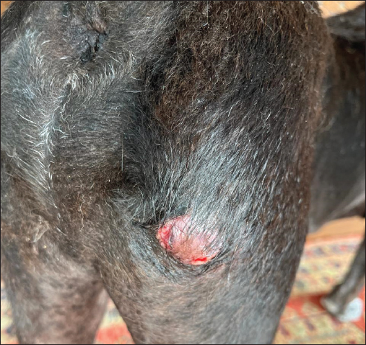
AbstractBackground: Skin tumors frequently occur in dogs, which can arise from various layers of the skin. This case reported the concomitant occurrence of mast cell tumor (MCT) and cutaneous melanoma in dogs. Case Description: A 14-year-old male local dog weighing 23 kg was presented to the Bali Veterinary Clinic Animal Hospital, Indonesia, with a 1-month history of a lump on the right hind limb. Clinical examination revealed a 3-cm mass in the right femoral region and three smaller nodules, each measuring 0.5–1 cm in diameter, in the ventral abdominal area. Hematology and blood biochemistry analyses showed that all parameters were within normal limits, except for a slight increase in alanine aminotransferase. Fine-needle aspiration cytology (FNAC) of the right hind limb mass showed a population of mast cells, while FNAC of the ventral abdominal nodules yielded inconclusive results because the cytological features were non-specific and did not support a definitive tumor diagnosis. Histopathological examination confirmed that the right hind limb mass was MCT, whereas the ventral abdominal masses were cutaneous melanomas. Immunohistochemical staining was performed against CD117 and cyclooxygenase-2 (COX-2). Histopathologically confirmed MCT showed CD117 focal localization in the cytoplasm and low COX-2 expression, suggesting a favorable prognosis. Surgical excision was performed with a lateral margin of 2 cm. The dog received amoxicillin as an antibiotic and tolfenamic acid as an anti-inflammatory agent postoperatively. During the recovery period, carprofen and cefadroxil were prescribed as analgesics and antibiotics, respectively. Conclusion: The dog fully recovered within 14 days. A 1-year follow-up revealed no evidence of tumor recurrence. This case report describes the successful surgical treatment of a concomitant MCT and cutaneous melanoma. Keywords: Cytology, Histopathology, Immunohistochemistry, Skin tumors. IntroductionThe skin is the largest organ in the body, with a complex structure that functions as a physical barrier, protecting against mechanical damage, pathogens, toxic substances, and allergens (Ibrahim et al., 2021; Shah et al., 2024). Due to its constant exposure to external factors, the skin is prone to tumor development (Winaya et al., 2024). It contains a large population of cells that continuously renew throughout the cell cycle, making it particularly susceptible to neoplastic transformation (Melnikova and Ananthaswamy, 2005; Hassan et al., 2022). Tumors can arise from various skin layers, such as the epidermis, dermis, subcutaneous tissue, and adnexal structures (Brønden et al., 2010). Skin tumors are more frequently detected than other types of tumors because they are easily visible, facilitating early examination and diagnosis. Several retrospective epidemiological studies have investigated skin tumors in dogs. Martins et al. (2022) reported that among 1,185 cases of canine skin tumors, 62.9% were benign and the remainder were malignant. The evaluation of skin tumors in dogs typically involves a combination of clinical examination, cytology, histopathology, and imaging techniques. Fine-needle aspiration cytology (FNAC) is widely used as an initial diagnostic tool due to its minimally invasive nature and rapid results; however, in certain cases, it may yield inconclusive findings (Yumuşak and Kutsal, 2016). Histopathology remains the gold standard for tumor diagnosis, providing detailed insights into tissue architecture and cellular morphology (Šimkus et al., 2016; Fragoso-Garcia et al., 2023). Mastocytomas, commonly known as mast cell tumors (MCTs), are hematopoietic neoplasms composed of mast cells (de Nardi et al., 2022). These tumors exhibit highly variable biological behavior, ranging from benign to malignant. Epidemiological studies across different countries indicate that MCTs are among the most frequently diagnosed skin tumors in dogs, with some studies identifying them as the most common type (Brønden et al., 2010; Martins et al., 2022). Melanocytic tumors are relatively common in dogs, with approximately 70% being malignant and the remainder benign. Melanomas are most frequently found in the oral cavity (62%), followed by cutaneous locations (27%), digital (6%), and subungual sites (4%) (Gillard et al., 2014). In general, cutaneous melanomas tend to be benign, whereas oral melanomas are often malignant. Digital and subungual melanomas exhibit a high recurrence rate (Stevenson et al., 2023; Polton et al., 2024). Cutaneous melanoma is a melanocytic neoplasm originating from skin melanocytes (Smedley et al., 2022). Although most cases are considered benign, some can exhibit aggressive behavior (Hritcu et al., 2023). Middle-aged and older dogs are generally at a higher risk of developing melanoma than younger dogs, especially heavily pigmented breeds (Polton et al., 2024). This case report describes a case of neoplastic synchronism involving MCT and cutaneous melanoma in a geriatric dog, detailing the clinical presentation, clinicopathological findings, immunohistochemical characteristics, and management approach. Case DetailsA 14-year-old male local dog was brought by his owner to Bali Veterinary Clinic (BVC) Animal Hospital, Bali, Indonesia, with a complaint of a lump on the right hind limb that had been present for 1 month. The owner had applied an antiseptic treatment, but the lump showed no improvement. The dog weighed 23 kg and had a rectal temperature of 38.5°C, with a capillary refill time under 2 seconds. The heart rate, pulse, and respiratory rate were within normal limits. A firm, 3-cm mass was identified in the right femoral region, with alopecia in the surrounding area and an ulcerated surface (Fig. 1). Three small, darkly pigmented lumps, approximately 0.5–1 cm in diameter, were observed in the ventral abdomen.
Fig. 1. The macroscopic characteristics of the tumor. A reddish mass on the right hind limb. Hematology and blood biochemistry analyses were performed using blood samples collected from the saphenous vein. Hematological parameters were assessed using a Veterinary Hematology Analyzer (CC-3200Vet, Shenzhen Licare Biomedical Technology Co., Ltd., Shenzhen, China), while blood biochemistry was analyzed using a Veterinary Chemistry Analyzer (SMT-120VP, Chengdu Seamaty Technology Co., Ltd., China). Cytological examination of the tumor mass was performed using the negative pressure fine-needle aspiration technique with a 3 ml syringe as described by Sewoyo and Nainggolan (2023). The obtained samples were stained using the Diff-Quik (Diff-Quik Staining Kit, PT. Mora Anugerah Berkat, Indonesia) and examined under a microscope at 1,000× magnification. An incisional biopsy was performed by taking the center of the lesion under general anesthesia. The biopsy site was then sutured with a monofilament nylon suture. The collected tissue was fixed in 10% formalin for histological processing and stained with Hematoxylin-Eosin. Two veterinary pathologists subsequently examined the prepared histological slides for diagnostic confirmation. Paraffin-embedded tumor samples were sectioned and subjected to immunohistochemical staining using the immuno-enzyme polymer method. The primary antibodies used in this study for examinations were cyclooxygenase-2 (COX-2) and c-Kit/CD117 (Table 1). Endogenous peroxidase was blocked with H2O2 0.3%. The secondary antibody was goat anti-mouse-rabbit conjugated with horseradish peroxidase (N-Histofine Simple Stain MAX PO (MULTI), Nichirei Biosciences Inc., Japan). Chromogen diaminobenzidine (Histofine DAB-2V, Nichirei Biosciences Inc., Japan) was added after the introduction of the secondary antibody. The slides were counterstained with Mayer hematoxylin (Merck, Germany). The expression of COX-2 was evaluated using a semiquantitative scoring system (Gregório et al., 2017). Table 1. Primary antibodies used in this study.
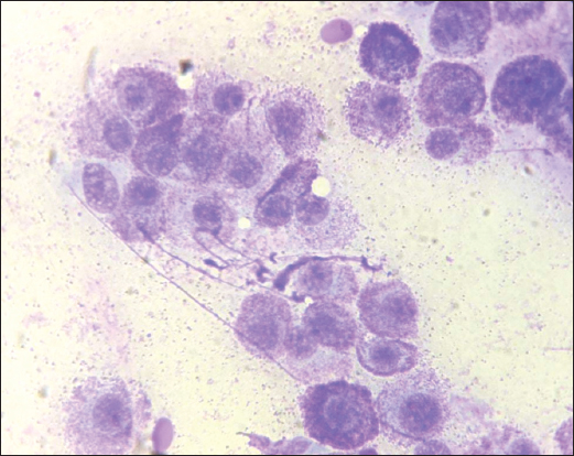
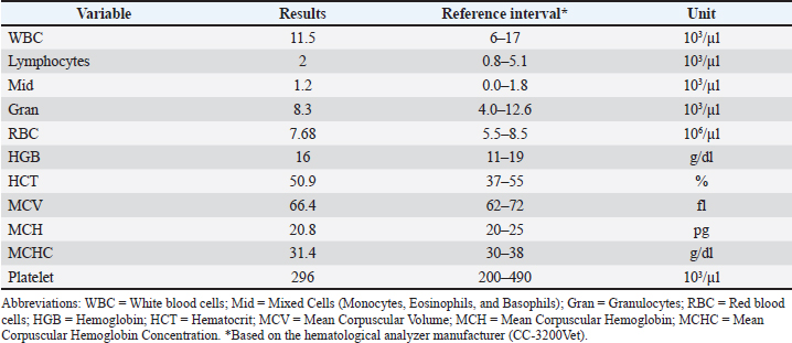
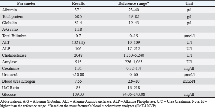
The complete blood count (CBC) showed no abnormalities (Table 2), whereas blood biochemistry revealed a slight increase in alanine aminotransferase (ALT) levels (Table 3). The mass in the right femoral region exhibited a population of mast cells of varying sizes (Fig. 1). Some mast cells displayed distinct granulation, whereas others lacked clearly visible granules, indicating an MCT (Fig. 2). Cytological examination of the mass in the ventral abdomen was also conducted; however, the results were inconclusive because the cytological features were non-specific and did not support a definitive tumor diagnosis. No cytological evidence, such as melanin pigment deposition, suggestive of a melanocytic tumor, was observed. Therefore, a histopathological examination was performed for further confirmation.
Fig. 2. FNAC of the right hind limb mass showing several mast cells with moderate anisocytosis and anisokaryosis. Some cells are well-granulated, whereas others exhibit scant cytoplasmic granules (Diff-Quik, 1,000×). Table 2. CBC results.
Table 3. Blood biochemistry test results.
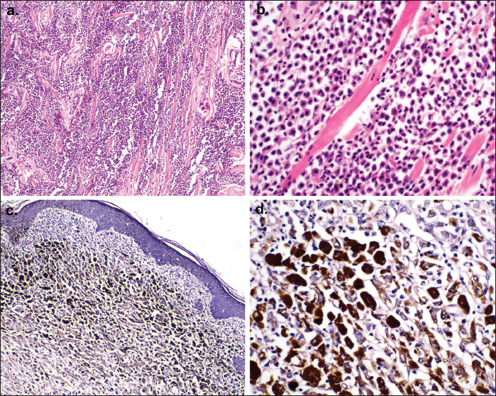
Histopathological examination of the tumor mass in the right femoral region revealed a dense population of well-differentiated mast cells arranged in groups (Fig. 3a). The mast cells exhibited round to oval nuclei. Mitoses ranged from 2 to 4 per high-power field (HPF). Additionally, eosinophil infiltration was observed among the mast cell population, and abundant collagen was evident as brightly eosinophilic bands running between the neoplastic cells (Fig. 3b). Based on the Kiupel grading system, the MCT was classified as low grade, whereas it was categorized as Grade II according to the Patnaik grading system (Patnaik et al., 1984; Kiupel et al., 2011). Histopathological evaluation of the ventral abdominal mass revealed brown melanin pigmentation within the dermis with moderate inflammatory cell infiltration (Fig. 3c). Round to polygonal epithelioid cells and melanophages were also observed (Fig. 3d).
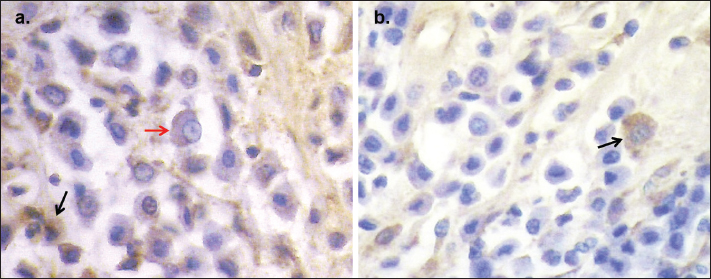
Fig. 3. Histopathological tumor mass retrieved from the hind limb (a, b) and ventral area of the abdomen (c, d). (a) Photomicrograph of low-grade MCT: The tumor consists of densely packed neoplastic cells arranged in groups, with the presence of collagen coursing among neoplastic cells. The area of scattered inflammatory infiltrates is observed. (b) Neoplastic mast cells exhibit moderate pleomorphism, with round to oval nuclei and variably distinct nucleoli. Moderate mitotic figures are present. Scattered eosinophils are observed. (c) Photomicrograph of cutaneous melanoma: Areas of pigmentation due to melanin deposits are visible on the dermis, giving a brown appearance. Large nests of neoplastic cells infiltrate the dermis. (d) Several round to polygonal pigmented epithelioid cells are found along with melanophages. Mild inflammation in the dermis was also observed (Hematoxylin-Eosin, 100× & 400×). Immunohistochemical staining for CD117 and COX-2 was performed on MCT. Neoplastic mast cells demonstrated focal cytoplasmic immunoreactivity for CD117 (Fig. 4a). COX-2 was expressed in the cytoplasm, and the staining intensity was generally weak (Fig. 4b). Immunoreactivity scoring revealed that the MCT mass exhibited weak COX-2 expression. Based on these findings, the prognosis was considered good. The selected treatment for this case was surgical excision. Based on hematology and blood biochemistry results (Tables 2 and 3), the animal was deemed fit for surgery. Prior to surgery, the animal was positioned in left lateral recumbency. Preanesthetic medication included atropine sulfate (V-Tropin, Agrovet, Peru) at 0.027 mg/kg body weight (BW) administered subcutaneously (SC) and xylazine (Xyla, Interchemie, The Netherlands) at 1.7 mg/kg BW administered intramuscularly (IM). Anesthesia was induced and maintained with propofol (Nupovel, Novell Pharmaceutical Laboratories, Indonesia) at 1.3 mg/kg BW, administered slowly intravenously.
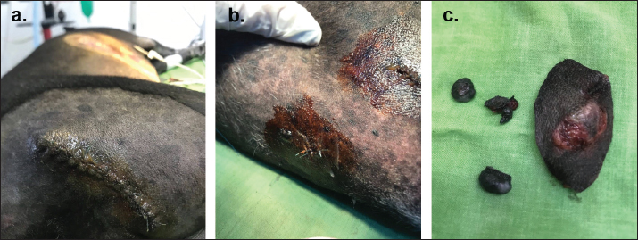
Fig. 4. Immunohistochemistry evaluation of MCT against anti-CD117 and anti-COX-2 (a) CD117: Neoplastic mast cells showed immunoreactivity for CD117 with focal cytoplasmic localization (black arrow), while the perimembranous staining was decreased (red arrow). (b) COX-2: COX-2 expressed weakly in cytoplasm (arrow). Based on scoring, the COX-2 expression was considered weak immunoreactivity (DAB and hematoxylin counterstain, 1,000×). The tumor mass was excised with a lateral margin of 2 cm. The postoperative wound was closed using nylon 3–0 with a simple interrupted suture pattern (Fig. 5). Postoperatively, the dog received amoxicillin (Betamox, Norbrook, UK) at 15 mg/kg BW IM and tolfenamic acid (Tolfedine, Vetoquinol, France) at 4 mg/kg BW SC. For postoperative recovery, carprofen (Rimadyl, Zoetis, US) at 2.2 mg/kg BW PO b.i.d. for 5 days and cefadroxil (Cefadroxil, PT. Dankos Farma, Indonesia) at 22 mg/kg BW PO b.i.d. for 14 days were prescribed. The dog recovered well within 14 days, and a 1-year follow-up examination confirmed no tumor recurrence.
Fig. 5. Postoperative appearance of surgical sites and excised tumors. (a) The surgical site on the hind limb. (b) Surgical site on the ventral abdomen. (c) Excised tumor masses after surgical removal. DiscussionOlder dogs are generally prone to developing skin tumors, with several studies indicating a significant increase in tumor incidence with age. Dogs older than 7 years have been reported to develop skin tumors more frequently (Kim et al., 2009; Hassan et al., 2022; García et al., 2019). Certain tumor types, such as MCT, also exhibit age predisposition (Martins et al., 2021). FNAC and histopathology are commonly used diagnostic methods for skin tumors. FNAC has a sensitivity of 89.3% and specificity of 97.9% in diagnosing skin tumors (Ghisleni et al., 2006). However, histopathology remains the gold standard due to its high accuracy and ability to provide detailed tissue architecture (Fournier et al., 2018). FNAC is still highly valuable as it is rapid and minimally invasive. MCTs are classified as cutaneous round cell tumors (RCTs), and some RCTs share similar morphological characteristics, making differentiation challenging (Rissi and Oliveira, 2022). Macroscopically, they appear as raised, firm masses and are ulcerated in 30% of cases (de Nardi et al., 2022). In this case, the MCT mass appeared to be slightly ulcerated and exhibited an area of alopecia in the tumor region. The secondary clinical signs of MCT, known as Darier’s sign, occur in half of the affected dogs and include delayed wound healing, coagulation abnormalities, hypotension, and circulatory collapse (Welle et al., 2008). However, these signs were not observed in this case. Biochemistry analysis showed a slight increase in ALT in the dog. According to Hall and German (2016), a less than twofold increase in ALT levels is usually considered clinically insignificant in dogs. In cases where diagnosis is challenging, immunohistochemical markers such as c-Kit/CD117 can aid in diagnosis (Cruz et al., 2020). In normal mast cells, CD117 is localized on the cell surface. In neoplastic mast cells, the CD117 localization is usually found in the cytoplasm, with three patterns described. The first pattern is perimembrane localization, the same as on normal mast cells. The second pattern is stippled to focal localization, with a decrease in perimembrane localization. The last pattern is diffuse cytoplasmic localization (Kiupel and Camus, 2019). In addition to diagnostic purposes, CD117 is also used as a prognostic indicator, as there is a significant association between cytoplasmic localization and decreased overall survival in dogs with cutaneous MCTs. The first and second patterns are most commonly found in low-grade (Kiupel) and grade 1 and 2 (Patnaik) MCTs, while the third pattern is usually found in high-grade (Kiupel) or grade 3 (Patnaik) MCTs (Giantin et al., 2012). In this case, CD117 was focally distributed in the cytoplasm and was histopathologically classified as low grade (Kiupel). COX-2 is a commonly used prognostic marker in veterinary oncology due to its association with tumor proliferation, angiogenesis, invasion, and metastasis. In MCT, high COX-2 expression is correlated with elevated Ki67 levels, increased microvascular density, and a high mitotic index, all of which indicate a more aggressive phenotype (Gregório et al., 2017). Furthermore, intratumoral collagen composition can predict mortality and survival in MCT-positive dogs. Daniel et al. (2019) showed that collagen levels were lower in high-grade MCT cases than in low-grade cases. A low collagen composition was also associated with an increased risk of death compared with MCT with a high collagen composition. In this case, histopathological evaluation revealed abundant collagen fibers among the tumor cells and low COX-2 expression, along with a low-grade category (Kiupel), suggesting a favorable prognosis. This was supported by the absence of tumor recurrence 1 year after surgery and the stable condition of the dog during routine health checks. Cutaneous melanoma, a melanocytic tumor, is typically a raised, pigmented mass (Prouteau and André, 2019). In this case, histopathological findings classified the tumor as a benign melanoma (melanocytoma). Malignant melanoma is more likely to ulcerate and display asymmetry, whereas melanocytoma tends to be symmetrical (Resende et al., 2015). Histopathologically, malignant melanoma exhibits a mitotic rate of >3 per 10 HPF. Melanocytoma is characterized by various cellular morphologies, including epithelioid, spindle, balloon, and signet-ring cells (Smedley et al., 2011). In this case, the tumor was classified as epithelioid melanocytoma. The primary treatment for MCT is surgical excision with lateral margins of 2–3 cm and a deep margin extending to at least one fascial plane (Selmic and Ruple, 2020). Recently, an alternative therapy using tigilanol tiglate has been reported, offering a less invasive option, particularly for tumors in surgically challenging locations (Verbrugghe et al., 2023). Similarly, the primary treatment for cutaneous melanoma is surgical excision, with recommended margins of 2–3 cm laterally and a deep subfascial resection (Fonseca-Alves et al., 2021; Polton et al., 2024). The concomitant occurrence of MCT and melanoma has been reported in several studies. Scott et al. (2017) reported a collision between a grade II MCT and melanoma in a cutaneous location, as well as a collision between a grade III MCT and melanoma in the eyelid. Rebhun and Thamm (2010) reported that several distinct tumor types were overrepresented in dogs with multiple tumors, including thyroid carcinoma, MCT, and malignant melanoma. This study states that no breed-related factor plays a role, as nearly 30% of cases involved mixed-breed dogs, despite evidence that mutations in the MET proto-oncogene in Rottweiler dogs convincingly increase the potential for the development of multiple tumors. In humans, an increased incidence of melanoma has been observed in patients with systemic mastocytosis (Vojvodic et al., 2019). Both conditions share molecular signaling pathways, including c-Kit, SCF, STAT3, and MITF (Testa and U, 2008; Vojvodic et al., 2019). A registry-based study in humans also showed an increased risk of developing melanoma in patients with mastocytosis (Bergström et al., 2025). AcknowledgmentsThe completion of this article was made possible through the support of BVC Animal Hospital, the Veterinary Pathology Laboratory, Faculty of Veterinary Medicine, Udayana University, and the Ministry of Higher Education, Science, and Technology of the Republic of Indonesia. This publication is the result of a collaboration between the Faculty of Veterinary Medicine, Udayana University and BVC Animal Hospital based on Collaborative Agreement No. B/15/UN14.2.9/HM.01.01/2022. Conflict of interestNo competing interest is declared by all authors. FundingNone. Author’s contributionsP.S.S. drafted and wrote the original manuscript, processed histological slides, and analyzed histopathological and immunohistochemical images. W.M.N. is the clinician who performed the clinical examination, fine-needle aspiration cytology and analysis, hematology and biochemical analyses, surgery, and postoperative management of the animal. INMA performed immunohistochemical staining and reviewed and validated the manuscript. Data availabilityAll data were provided in the manuscript. ReferencesBergström, A., Hägglund, H., Berglund, A., Nilsson, G. and Lambe, M. 2025. Evaluating melanoma risk in adult mastocytosis: potential impact of detection bias – a registry-based study (Sweden). Adv. Dermatol. Venereol. 18, 105. Brønden, L.B., Eriksen, T. and Kristensen, A.T. 2010. Mast cell tumours and other skin neoplasia in Danish dogs—data from the Danish Veterinary Cancer Registry. Acta. Vet. Scand. 52(1), 6. Cruz, V.S., Borges, J.C.A., Nepomuceno, L.L., Gonçalves, P.A.M., Prado, Y.C.L., Bianchi, C., Fioravanti, M.C.S. and Araújo, E.G. 2020. Histological classification and expression of markers of canine mast cell tumors. Vet. World 13(8), 1627–1634. Daniel, J., Barra, C.N., Pulz, L.H., Kleeb, S.R., Xavier, J.G., Catão‐Dias, J.L., Nishiya, A.T., Fukumasu, H. and Strefezzi, R.F. 2019. Intratumoral collagen index predicts mortality and survival in canine cutaneous mast cell tumours. Vet. Dermatol. 30(2), 162–e48. De Nardi, A.B., Dos Santos Horta, R., Fonseca-Alves, C.E., De Paiva, F.N., Linhares, L.C.M., Firmo, B.F., Ruiz Sueiro, F.A., De Oliveira, K.D., Lourenço, S.V., De Francisco Strefezzi, R., Brunner, C.H.M., Rangel, M.M.M., Jark, P.C., Castro, J.L.C., Ubukata, R., Batschinski, K., Sobral, R.A., Da Cruz, N.O., Nishiya, A.T., Fernandes, S.C., Dos Santos Cunha, S.C., Gerardi, D.G., Challoub, G.S.G., Biondi, L.R., Laufer-Amorim, R., De Oliveira Paes, P.R., Lavalle, G.E., Huppes, R.R., Grandi, F., De Carvalho Vasconcellos, C.H., Dos Anjos, D.S., Luzo, A.C.M., Matera, J.M., Vozdova, M. and Dagli, M.L.Z. 2022. Diagnosis, prognosis and treatment of canine cutaneous and subcutaneous mast cell tumors. Cells 11(4), 618. Fonseca-Alves, C.E., Ferreira, E., De Oliveira Massoco, C., Strauss, B.E., Fávaro, W.J., Durán, N., Oyafuso Da Cruz, N., Dos Santos Cunha, S.C., Castro, J.L.C., Rangel, M.M.M., Brunner, C.H.M., Tellado, M., Dos Anjos, D.S., Fernandes, S.C., Barbosa De Nardi, A., Biondi, L.R. and Dagli, M.L.Z. 2021. Current status of canine melanoma diagnosis and therapy: report from a colloquium on canine melanoma organized by ABROVET (Brazilian Association of Veterinary Oncology). Front. Vet. Sci. 8, 707025. Fournier, Q., Cazzini, P., Bavcar, S., Pecceu, E., Ballber, C. and Elders, R. 2018. Investigation of the utility of lymph node fine-needle aspiration cytology for the staging of malignant solid tumors in dogs. Vet. Clin. Pathol. 47(3), 489–500. Fragoso-Garcia, M., Wilm, F., Bertram, C.A., Merz, S., Schmidt, A., Donovan, T., Fuchs-Baumgartinger, A., Bartel, A., Marzahl, C., Diehl, L., Puget, C., Maier, A., Aubreville, M., Breininger, K. and Klopfleisch, R. 2023. Automated diagnosis of 7 canine skin tumors using machine learning on H&E-stained whole slide images. Vet. Pathol. 60(6), 865–875. García, E., Alpízar, A., Fajardo, R., Córdova, D., Pérez, L. and Martínez, S. 2019. Epidemiology of tumors in dogs in the capital of the state of Mexico from 2002–2016. Arq. Bras. Med. Vet. Zootec. 71(4), 1085–1092. Ghisleni, G., Roccabianca, P., Ceruti, R., Stefanello, D., Bertazzolo, W., Bonfanti, U. and Caniatti, M. 2006. Correlation between fine-needle aspiration cytology and histopathology in the evaluation of cutaneous and subcutaneous masses from dogs and cats. Vet. Clin. Pathol. 35(1), 24–30. Giantin, M., Vascellari, M., Morello, E.M., Capello, K., Vercelli, A., Granato, A., Lopparelli, R.M., Nassuato, C., Carminato, A., Martano, M., Mutinelli, F. and Dacasto, M. 2012. C-KIT messenger RNA and protein expression and mutations in canine cutaneous mast cell tumors: correlations with post-surgical prognosis. J. Vet. Diagn. Invest. 24(1), 116–126. Gillard, M., Cadieu, E., De Brito, C., Abadie, J., Vergier, B., Devauchelle, P., Degorce, F., Dréano, S., Primot, A., Dorso, L., Lagadic, M., Galibert, F., Hédan, B., Galibert, M.D. and André, C. 2014. Naturally occurring melanomas in dogs as models for non-UV pathways of human melanomas. Pigment Cell Melanoma Res. 27(1), 90–102. Gregório, H., Raposo, T., Queiroga, F.L., Pires, I., Pena, L. and Prada, J. 2017. High COX-2 expression in canine mast cell tumours is associated with proliferation, angiogenesis and decreased overall survival. Vet. Comp. Oncol. 15(4), 1382–1392. Hall, E.J. and German, A.J. 2016. Laboratory evaluation of hepatic disease. In BSAVA manual of canine and feline clinical pathology, 3rd. Eds. Villiers, E. and Ristic, J Hassan, B.B., Al-mokaddem, A.K., Abdelrahman, H.A., Samir, A. and Mousa, M.R. 2022. Cutaneous tumors in dogs: a retrospective epidemiological and histological study of 112 cases. Adv. Anim. Vet. Sci. 10(1), 170–184. Hritcu, O.M., Bocaneti Daraban, F., Bacusca, F.D. and Pasca, A.S. 2023. Unusual canine cutaneous melanoma presenting parietal bone metastasis: a case report. Vet. Sci. 10(4), 282. Ibrahim, A.E., Bagherani, N., Smoller, B., Bagherani, N. and Reyes-Barron, C. 2021. Functions of the skin. In Atlas of dermatology, dermatopathology and venereology. Eds., Smoller, B. and Bagherani, N. Cham, Switzerland: Springer, pp: 133–43. Kim, Y.H., Ahn, N.K., Roh, I.S., Yoon, B.Y. and Han, J.H. 2009. Retrospective investigation of canine skin and mammary tumors in Korea. J. Vet. Clin. 26(6), 556–562. Kiupel, M. and Camus, M. 2019. Diagnosis and prognosis of canine cutaneous mast cell tumors. Vet. Clin. North. Am. Small. Anim. Pract. 49(5), 819–836. Kiupel, M., Webster, J.D., Bailey, K.L., Best, S., Delay, J., Detrisac, C.J., Fitzgerald, S.D., Gamble, D., Ginn, P.E., Goldschmidt, M.H., Hendrick, M.J., Howerth, E.W., Janovitz, E.B., Langohr, I., Lenz, S.D., Lipscomb, T.P., Miller, M.A., Misdorp, W., Moroff, S., Mullaney, T.P., Neyens, I., O’Toole, D., Ramos-Vara, J., Scase, T.J., Schulman, F.Y., Sledge, D., Smedley, R.C., Smith, K., W. Snyder, P., Southorn, E., Stedman, N.L., Steficek, B.A., Stromberg, P.C., Valli, V.E., Weisbrode, S.E., Yager, J., Heller, J. and Miller, R. 2011. Proposal of a 2-tier histologic grading system for canine cutaneous mast cell tumors to more accurately predict biological behavior. Vet. Pathol. 48(1), 147–155. Martins, A., Faria, F., Mesquita, J., Rtner, F. and Amorim, I. 2021. Analysis of risk factors for canine mast cell tumors based on the Kiupel and Patnaik grading system among dogs with skin tumors. Open Vet. J. 11(4), 619–634. Martins, A.L., Canadas-Sousa, A., Mesquita, J.R., Dias-Pereira, P., Amorim, I. and Gärtner, F. 2022. Retrospective study of canine cutaneous tumors submitted to a diagnostic pathology laboratory in Northern Portugal (2014–2020). Canine Med. Genet. 9(1), 2. Melnikova, V.O. and Ananthaswamy, H.N. 2005. Cellular and molecular events leading to the development of skin cancer. Mutat. Res. Fundam. Mol. Mech. Mutagen. 571(1–2), 91–106. Patnaik, A.K., Ehler, W.J. and Macewen, E.G. 1984. Canine cutaneous mast cell tumor: morphologic grading and survival time in 83 dogs. Vet. Pathol. 21(5), 469–474. Polton, G., Borrego, J.F., Clemente-Vicario, F., Clifford, C.A., Jagielski, D., Kessler, M., Kobayashi, T., Lanore, D., Queiroga, F.L., Rowe, A.T., Vajdovich, P. and Bergman, P.J. 2024. Melanoma of the dog and cat: consensus and guidelines. Front. Vet. Sci. 11, 1359426. Prouteau, A. and André, C. 2019. Canine melanomas as models for human melanomas: clinical, histological, and genetic comparison. Genes 10, 501. Rebhun, R.B. and Thamm, D.H. 2010. Multiple distinct malignancies in dogs: 53 cases. J. Am. Anim. Hosp. Assoc. 46, 20–30. Resende, L., Moreira, J., Prada, J., Queiroga, F.L. and Pires, I. 2015. Current insights into canine cutaneous melanocytic tumours diagnosis. In Melanoma-current clinical management and future therapeutics. Ed. Murph, M. Rijeka, Croatia: IntechOpen. Rissi, D.R. and Oliveira, F.N. 2022. Review of diagnostic histologic features of cutaneous round cell neoplasms in dogs. J. Vet. Diagn. Investig. 34(5), 769–779. Scott, J.E., Liptak, J.M. and Powers, B.E. 2017. Malignant collision tumors in two dogs. J. Am. Anim. Hosp. Assoc. 251, 941–945. Selmic, L.E. and Ruple, A. 2020. A systematic review of surgical margins utilized for removal of cutaneous mast cell tumors in dogs. BMC. Vet. Res. 16(1), 5. Sewoyo, P.S. and Nainggolan, W.M. 2023. Sebaceous adenoma case in a golden retriever dog. J. Appl. Vet. Sci. Technol. 4(2), 122–126. Shah, J., McKnight, G. and Hargest, R. 2024. Physiology of the skin. Surgery (Oxf) 42(11), 788–792. Šimkus, D., Petkevičius, S., Pridotkas, G., Zorgevica-Pockeviča, L., Maskaliovas, V., Šimkienė, V. and Pockevičius, A. 2016. Histological and immunohistochemical practical studies of canine cutaneous tumors. Med. Weter. 72(9), 571–579. Smedley, R.C., Sebastian, K. and Kiupel, M. 2022. Diagnosis and prognosis of canine melanocytic neoplasms. Vet. Sci. 9(4), 175. Smedley, R.C., Spangler, W.L., Esplin, D.G., Kitchell, B.E., Bergman, P.J., Ho, H.Y., Bergin, I.L. and Kiupel, M. 2011. Prognostic markers for canine melanocytic neoplasms: a comparative review of the literature and goals for future investigation. Vet. Pathol. 48, 54–72. Stevenson, V.B., Klahn, S., Leroith, T. and Huckle, W.R. 2023. Canine melanoma: a review of diagnostics and comparative mechanisms of disease and immunotolerance in the era of the immunotherapies. Front. Vet. Sci. 9, 1046636. Testa, U. 2008. Kit mutations in cancer and their treatment with protein kinase inhibitors. Drugs. Future. 33(2), 161–174. Verbrugghe, A., De Vos, S., Krupa, A., Vandenabeele, S. and De Rooster, H. 2023. The use of tigilanol tiglate (Stelfonta®) for the treatment of canine mast cell tumors. Vlaam. Diergeneeskd. Tijdschr. 92(2), 51–57. Vojvodic, A., Vlaskovic-Jovicevic, T., Vojvodic, P., Vojvodic, J., Goldust, M., Peric-Hajzler, Z., Matovic, D., Sijan, G., Stepic, N., Wollina, U., Fioranelli, M., Tirant, M., Nguyen, V.T. and Lotti, T. 2019. Melanoma and mastocytosis. Open Access Maced. J. Med. Sci. 7(18), 3050. Welle, M.M., Bley, C.R., Howard, J. and Rüfenacht, S. 2008. Canine mast cell tumours: a review of the pathogenesis, clinical features, pathology and treatment. Vet. Dermatol. 19(6), 321–339. Winaya, I.B.O., Adi, A.A.A.M., Sudimartini, L.M., Merdana, I.M., Sudipa, P.H., Pemayun, I.G.A.G.P. and Sewoyo, P.S. 2024. Sebaceous adenoma in a geriatric poodle dog: a case report. J. Medik. Vet. 7(2), 413–419. Yumuşak, N. and Kutsal, O. 2016. A comparative study between fine needle aspiration biopsy (FNAB) findings and histopathology in the evaluation of canine skin and skin adnexal tumors. Ankara. Univ. Vet. Fak. Derg. 63(4), 393–400. | ||

| How to Cite this Article |
| Pubmed Style Sewoyo PS, Nainggolan WM, Astawa INM. Concomitant occurrence of canine mast cell tumor and cutaneous melanoma in a geriatric dog. Open Vet. J.. 2026; 16(7): 4295-4304. doi:10.5455/OVJ.2026.v16.i7.17 Web Style Sewoyo PS, Nainggolan WM, Astawa INM. Concomitant occurrence of canine mast cell tumor and cutaneous melanoma in a geriatric dog. https://www.openveterinaryjournal.com/?mno=303195 [Access: June 30, 2026]. doi:10.5455/OVJ.2026.v16.i7.17 AMA (American Medical Association) Style Sewoyo PS, Nainggolan WM, Astawa INM. Concomitant occurrence of canine mast cell tumor and cutaneous melanoma in a geriatric dog. Open Vet. J.. 2026; 16(7): 4295-4304. doi:10.5455/OVJ.2026.v16.i7.17 Vancouver/ICMJE Style Sewoyo PS, Nainggolan WM, Astawa INM. Concomitant occurrence of canine mast cell tumor and cutaneous melanoma in a geriatric dog. Open Vet. J.. (2026), [cited June 30, 2026]; 16(7): 4295-4304. doi:10.5455/OVJ.2026.v16.i7.17 Harvard Style Sewoyo, P. S., Nainggolan, . W. M. & Astawa, . I. N. M. (2026) Concomitant occurrence of canine mast cell tumor and cutaneous melanoma in a geriatric dog. Open Vet. J., 16 (7), 4295-4304. doi:10.5455/OVJ.2026.v16.i7.17 Turabian Style Sewoyo, Palagan Senopati, Willy Moris Nainggolan, and I Nyoman Mantik Astawa. 2026. Concomitant occurrence of canine mast cell tumor and cutaneous melanoma in a geriatric dog. Open Veterinary Journal, 16 (7), 4295-4304. doi:10.5455/OVJ.2026.v16.i7.17 Chicago Style Sewoyo, Palagan Senopati, Willy Moris Nainggolan, and I Nyoman Mantik Astawa. "Concomitant occurrence of canine mast cell tumor and cutaneous melanoma in a geriatric dog." Open Veterinary Journal 16 (2026), 4295-4304. doi:10.5455/OVJ.2026.v16.i7.17 MLA (The Modern Language Association) Style Sewoyo, Palagan Senopati, Willy Moris Nainggolan, and I Nyoman Mantik Astawa. "Concomitant occurrence of canine mast cell tumor and cutaneous melanoma in a geriatric dog." Open Veterinary Journal 16.7 (2026), 4295-4304. Print. doi:10.5455/OVJ.2026.v16.i7.17 APA (American Psychological Association) Style Sewoyo, P. S., Nainggolan, . W. M. & Astawa, . I. N. M. (2026) Concomitant occurrence of canine mast cell tumor and cutaneous melanoma in a geriatric dog. Open Veterinary Journal, 16 (7), 4295-4304. doi:10.5455/OVJ.2026.v16.i7.17 |


