





| Research Article | ||
Open Vet. J.. 2026; 16(7): 4731-4745
Open Veterinary Journal, (2026), Vol. 16(7): 4731-4745 Research Article Differential responsiveness of the midbrain dopaminergic system to acute and chronic lead intoxications and the neuroprotective potential of curcumin I in rat: A possible involvement of astrogliaHind Benammi1,2*, Kamal Smimih3, Ahmed Draoui3,4, Bilal El-Mansoury2, Hicham Chatoui1,2,3, Morad Belkouri5, Faissal Aziz6, Omar El Hiba2 and Halima Gamrani71Higher Institute of Nursing Professions and Health Techniques, Ministry of Health, Marrakesh, Morocco 2Nutritional Physiopathologies, Neuroscience and Toxicology Team, Laboratory of Anthropogenic, Biotechnology and Health, Faculty of Sciences, Chouaib Doukkali, University, El Jadida, Morocco 3Biological Engineering Laboratory, Faculty of Sciences and Techniques (FST), Sultan Moulay Slimane University, Beni Mellal, Morocco 4Graduate School of Education and Training, Beni Mellal, Morocco 5Mohammed VI University Hospital Center, Marrakesh, Morocco 6Faculty of Sciences Semlalia, Laboratory of Water Sciences, Microbial Biotechnologies and Natural Resources Sustainability, Cadi Ayyad University, Marrakesh, Morocco 7Laboratory of Clinical, Experimental and Environmental Neurosciences, Cadi Ayyad University, Marrakesh, Morocco *Corresponding Author: Hind Benammi. Higher Institute of Nursing Professions and Health Techniques, Ministry of Health, Marrakesh, Morocco. Email: hbenammi85 [at] gmail.com Submitted: 21/12/2025 Revised: XX/XX/XX Accepted: 11/06/2026 Published: XX/XX/XX © 2025 Open Veterinary Journal
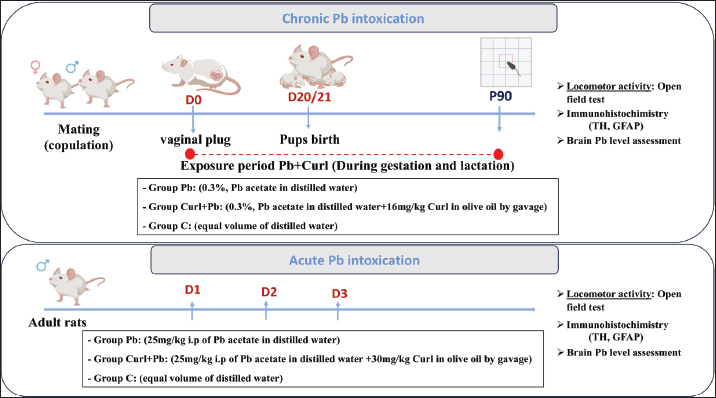
AbstractBackground: Lead (Pb) is a powerful heavy metal that was associated with various neurological diseases, including saturnism. Previous studies have reported its marked neurotoxicity, involving several neurotransmitter systems and their related neurobehavioral functions. Aim: This study aims to investigate the neurotoxicological impacts of acute and chronic Pb exposure on the dopaminergic (DAergic) innervation and astroglial reactivity, along with the subsequent locomotor outcomes. Methods: The acute model involved daily i.p. injections of Pb acetate [25 mg/kg body weight (B.W.)] for 3 days, while chronic exposure was achieved by supplementing maternal drinking water with 0.3% Pb acetate over 82 days (gestation through lactation). Curcumin I (Cur I) was administered by oral gavage at a dose of 30-mg/kg B.W. in the acute model, and 16-mg/kg B.W. in the chronic one to assess its neuroprotective potential. Using immunohistochemistry, we monitored tyrosine hydroxylase (TH) levels within the substantia nigra pars compacta (SNpc) and glial fibrillary acidic protein (GFAP) expression in the frontal cortex, while locomotor activity was assessed via the open-field test. Results: The results demonstrated that both acute and chronic exposures to Pb significantly increased cerebral Pb concentrations. Behaviorally, acute exposure-induced locomotor hyperactivity is contrary to the chronic Pb exposure. At the cellular level, acute Pb exposure was associated with increased TH immunoreactivity within the SNpc, while chronic Pb exposure elicited a significant decline in TH expression in the same region. Furthermore, GFAP-immunoreactivity in the frontal cortex was elevated across both intoxication models. Notably, these neurochemical and behavioral alterations were significantly attenuated by curcumin administration, underscoring its neuroprotective potential against Pb-induced toxicity. Conclusion: Our findings demonstrated that Pb exposure alters the DAergic and astroglial systems, leading to significant locomotor disturbances. These impairments were significantly restored by Cur I, leading to the consideration of this molecule as a potential natural neuroprotective agent against Pb-induced neurotoxicity. Keywords: Dopaminergic system, Intoxication, Lead, Neurotoxicity, Rat. IntroductionLead (Pb) is a heavy metal that can pass through the placenta and the blood-brain barrier (BBB) and has several negative impacts on human health, including neurological, haematological, gastrointestinal, reproductive, and immunological functions (Hu et al., 2006; Plusquellec et al., 2007; Wani et al., 2015; Ramírez Ortega et al., 2021; Aliche et al., 2025). Several experimental and epidemiological studies have suggested that the neurotoxic effects of Pb are mediated by interference with neurotransmission systems and/or astroglia (Leret et al., 2002; Mitra et al., 2017; Parithathvi et al., 2024). Due to its ease of extraction and versatile chemical properties, Pb has been utilized extensively across the globe for centuries. Thus, due to its wide availability throughout the environment, Pb remains a major public health and veterinary concern, affecting both humans and domestic and wildlife animals through contaminated water, soil, air, food (mainly plants growing on soils rich in Pb), and industrial pollutants (Bischoff et al., 2010; Tchounwou et al., 2012; Assi et al., 2016). Also, exposure to Pb can be through hair dyes, pigments, and other cosmetic products, Pb mines, fertilisers, pesticides, and Gasoline Pb (Al-Saleh et al., 2009; Ullah et al., 2023; Aslam et al., 2025; Laoye et al., 2025). Notably, Pb is a ubiquitous environmental toxin that can cause numerous acute and chronic deleterious effects. Chronic low-level exposure is particularly problematic, as both humans and animals may serve as sentinels of environmental contamination (Assi et al., 2016). Therefore, understanding the differential effects of acute and chronic Pb exposure on neural and glial systems is essential for elucidating its impact on brain function and for developing effective preventive and therapeutic strategies in both clinical and veterinary toxicology. The mechanism of Pb toxicity involves oxidative damage, which has been widely reported as one of the major mechanisms underlying Pb-induced neurotoxicity. Under the influence of Pb, oxidative stress is initiated via two primary pathways. The first involves the generation of reactive oxygen species (ROS), including hydroperoxides, singlet oxygen, and hydrogen peroxide (Liu et al., 2017). The second is due to the reduction of systemic antioxidant enzymes such as glutathione-S-transferase (GST), glutathione (GSH), glutathione peroxidase (GPx), catalase, and superoxide dismutase (SOD) (Flora et al., 2012; Vacchi-Suzzi et al., 2018), leading to neuronal death. Several experimental and epidemiological studies have shown the involvement of heavy metals, including Pb, in neurological alterations and neurodegenerative processes associated with disorders such as saturnism, Parkinson's disease (PD), or Alzheimer's disease (AD) (De Lau and Breteler, 2006; Abbaoui et al., 2016; Xu et al., 2018). PD, which affects 0.3% of the global population (Tysnes and Storstein, 2017; Draoui et al., 2020), is the most prevalent neurodegenerative disease after AD (Duty and Jenner, 2011; Jagmag et al., 2016). PD is characterised mainly by its primary cardinal symptoms, including akinesia, bradykinesia, stiffness, and resting tremor (Duty and Jenner, 2011; De Virgilio et al., 2016). Neurologically, PD is characterised by progressive, severe, and irreversible degeneration of the dopaminergic (DAergic) neurons within the substantia nigra pars compacta (SNpc), leading to the onset of motor dysfunctions (Lang and Obeso, 2004). Currently, the primary treatment strategy for PD relies on symptomatic pharmacological treatments aiming at restoring DAergic neurotransmission (Huot and Fox, 2011). Among these treatments, levodopa (L-DOPA), a metabolic precursor of dopamine (DA), remains the standard of care and is widely used to relieve the motor symptoms associated with PD (Connolly and Lang, 2014; Poewe et al., 2017). However, long-term use of L-DOPA is often associated with several complications, including motor fluctuations and dyskinesias, which limit its long-term effectiveness (Ahlskog and Muenter, 2001; Olanow et al., 2009). Consequently, there has been a growing focus on developing complementary or alternative therapeutic strategies with potential neuroprotective properties capable of slowing neurodegeneration or reducing neuronal damage. In general, the best remedy against heavy metal poisoning is by chelation or by the powerful antioxidant activity, which gives molecules endowed with these properties a very great importance. Several plants have a therapeutic interest because of their richness in bioactive molecules, and are used against several pathologies. Among the plants that have attracted great interest is the Curcuma longa L., given the richness of these curcumin rhizomes, which have been reported to exhibit several biological activities, including anti-inflammatory, anticancer, antimutagenic, antibacterial, as well as a powerful antioxidant (Esatbeyoglu et al., 2012; Hewlings and Kalman, 2017). Curcumin is a molecule able to cross the BBB (Garcia‐Alloza et al., 2007), which allows it to act at the central level against neurodegeneration. Indeed, several experimental and epidemiological studies have shown the beneficial effect of curcumin on the damage of free radicals. It is, therefore, considered a molecule with potential neuroprotective effects (B. Mythri and M. Srinivas Bharath, 2012; Esatbeyoglu et al., 2012). The purpose of the present investigation is to assess, in rats, the neurotoxicological effects of acute and chronic Pb exposures on the DAergic and astroglial systems, as well as locomotor performance, and to evaluate the potential protective effects of curcumin I (Cur I). Materials and MethodsChemicalsPb (II), acetate trihydrate (Panreac Quimica S.L.U. (Barcelona, Spain)]. Cur I, 95% (total curcuminoid content), (Alfa Aser, Johnson Mattey Compagny Karlsruhe, Germany, Lot: 10143876). AnimalsThe study was conducted exclusively on adult Wistar rats aged 3 months from the central animal care facilities of the Faculty of Sciences of Cadi Ayyad University of Marrakech (UCAM), Morocco. The rats used for all manipulations had an average weight of between 200 and 300 g. They were housed at a constant ambient temperature (25°C) on a 12-hour light/dark cycle, with free access to water and food for several generations in Plexiglas cages. All experiments were conducted in accordance with the guidelines of the Moroccan Ethics Committee of the Moroccan Society for Ethics and Animal Research. Intoxication and drug treatmentAcute Pb intoxication and treatment with Cur IExperimental protocol and the doses of Pb and Cur I were based on our previous study (Benammi et al., 2017). Indeed, adult male rats aged 3 months were divided into three groups: Group I (C): control rats (n=8, m=225 ± 37.03 g) injected with physiological saline buffer (0.9% NaCl i.p.) for 3 days consecutively. Group II (Pb): (n=8, m=230 ± 28.12 g) injected i.p with Pb (Pb acetate dissolved in distilled water) at a dose of 25-mg/kg body weight (B.W.) daily for 3 consecutive days. Group III (Cur I + Pb): (n=8, m=232 ± 22.84 g) received Cur I (dissolved in olive oil) at a dose of 30-mg/kg B.W. by oral gavage daily for 3 days. Rats also received i.p injections of Pb at a dose of 25-mg/kg B.W 2 hours after the last dose of Cur I (Fig. 1).
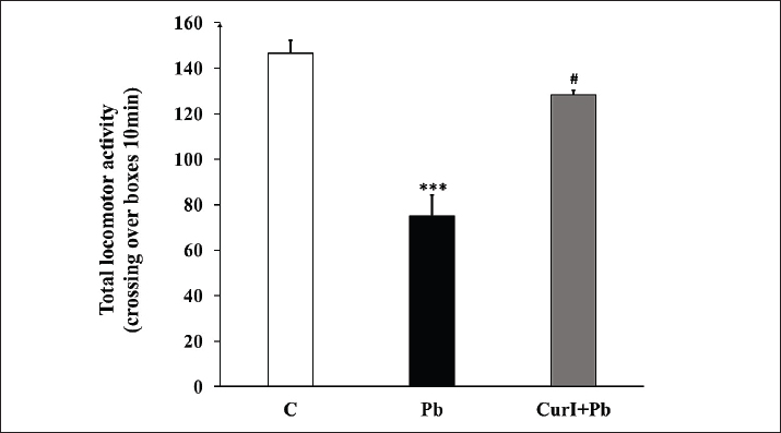
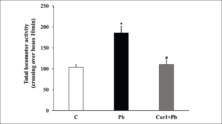
Fig. 1. Experimental design and timeline schedule of the experiments. Chronic Pb intoxication and treatment with Cur IWistar female rats weighing 250–300 g (n=18) were mated, and sperm-positive vaginal smears were taken to indicate the first day of gestation. The female rats were divided into three groups with every three females and one male to increase the gestation probability: Group I (C): control pregnant dams and their offspring (pups) received only distilled water and were provided with ad libitum access to food. Group II (Pb): Pb-treated dams, wherein drinking water was replaced by Pb acetate solution prepared in distilled water (0.3%) during the gestational stage (from positive plugs verification to the adult stage of their pups). Group III (Cur I + Pb): dams were treated as group Pb with Cur I given concomitantly by gavage at a dose of 16 mg/kg (dissolved in olive oil). For the next experiments, a total of 72 male offspring rats (aged 3 months) from the three groups were included (Fig. 1). For both acute and chronic treatments with Cur I, we have used low doses of Cur I below the human toxicity range of 6 g/day orally for 4–7 weeks as documented previously (Soleimani et al., 2018) Pb assaysFor central Pb quantification, five adult rats from each group were used (C, Pb, and Cur I + Pb) were anesthetised using urethane (1%, i.p.), then, brains were gently dissected, and 100 mg of the fresh tissue was used for Pb quantification and dissolved in 2 ml of ultra-pure nitric acid (Franzblau et al., 1988), for flame absorption spectrophotometry coupled to a graphite furnace. Locomotor activitySpontaneous locomotor activity was assessed using the open-field test. The apparatus consisted of a square black wooden enclosure (100 × 100 × 50 cm) (Seibenhener and Wooten, 2015; Smimih et al., 2026). The test was performed by placing the rats individually and simultaneously in the center of the open field (thoroughly cleaned), then filming the animals as they freely explored the open field for 5 minutes using a camera placed at the top of the apparatus. The videos are then reviewed, and the number of squares crossed is counted for each animal (Smimih et al., 2023). Immunohistochemistry (IHC)The IHC technique used was previously described (Benammi et al., 2014; El Hiba et al., 2016; Draoui et al., 2019; El Khiat et al., 2019). Briefly, the rat brains were gently dissected (from the three groups) following deep anaesthesia using sodium pentobarbital through the intraperitoneal route, followed by intracardiac perfusion with paraformaldehyde (4%) solution in phosphate buffer (PBS, 0.1 M, pH 7.4). Then, brains were post-fixed in the same fixative for at least 24 hours. The organs are then dehydrated through different baths of graded ethanol concentrations. Later, brains are included in polyethylene glycol. Finally, frontal sections through the midbrain are performed throughout the SNpc (bregma −5.3 mm) and the frontal cortex (Bregma −3.8 mm) according to the stereotaxic coordinates (Paxinos and Watson, 2004). For immunolabelling, the DAergic neurons as well as astroglia, we used monoclonal anti-sera [anti-tyrosine hydroxylase (TH) for DAergic neurons 1:1,000, Santa Cruz, mouse monoclonal CA, USA; catalogue No. SC-25269] (El Hiba et al., 2013) and anti-glial fibrillary acid protein (GFAP) for cortical astroglia (DAKO, monoclonal mouse anti-human GFAP, 1:1,000, lot#00072911, ref#M0761) (El Hiba et al., 2013). Thus, slices were either incubated with anti-TH or anti-GFAP solution containing PBS (0.1 M, pH 7.4), Triton (0.3%), and bovine serum albumin (BSA; 1%) overnight. Then, washed with PBS (0.1 M, pH 7.4) containing BSA (1%) three times and finally incubated with rabbit anti-immunoglobulin (secondary antibody, 1/1,000) for 2 hours at ambient temperature. Later, slices were, following three washes, incubated for 90 minutes in PBS-Triton (0.3%) solution with Avidin-Biotin peroxidise complex (Vector; 1/500). To visualize the positive immunolabelling, slices were placed finally in 3,3′-diaminobenzidine solution (3-3-diaminobenzidine, 0.03%, Sigma-Aldrich, Oakville, Canada) prepared in 0.05 M Tris buffer, (pH 7.5) with H2 O2 (0.01%). Antibodies specificity testsFor both anti-TH and anti-GFAP antibodies, preliminary essays were performed to test their specificities before application in our experiments. Thus, slices from the rat midbrain (for TH) and cortex (for GFAP) were subjected to the above-described protocol for IHC, but with the use of either the pre-immune sera or omitting the primary antibodies (as negative controls). These tests showed that both primary antibodies used displayed specific labeling. Immunolabelling quantificationA total of five brains from each group were used, with five sections chosen from each brain through the frontal cortex and the SNpc and subjected to quantification of immunolabelling according to the protocol published by Vilaplana and Lavialle, 1999. Briefly, the images were digitised and saved using a Zeiss-Axioskop 40 microscope connected to a Canon digital camera. The images were scanned at 512 by 512 pixels with a colour depth of eight bits in grayscale and were saved in Tagged Image File Format format. The analysis and measurement of the images were performed using Adobe Photoshop version 6.0 software. After converting each image to binary mode, the percentages of black pixels were determined using the histogram function in Adobe Photoshop. This percentage is related to the TH- or GFAP-immunopositive regions in the SNpc and frontal cortex, respectively. Statistical analysisData are presented as mean ± SEM, and differences between groups were assessed using one-way analysis of variance (ANOVA), followed by Tukey's post hoc analysis using Sigma plot V12.5 software. All numerical differences in the data were considered significantly different at a probability level of p < 0.05. For all data provided, we have checked the normality using the Shapiro–Wilk test before proceeding to the ANOVA test. Ethical approvalThe experimental protocol was reviewed and approved by the Moroccan Ethics Committee of the Moroccan Society for Ethics and Animal Research (MECAR-MoSEAR, Ref. ISPITS-M-05/2024) ResultsLocomotor performanceUsing the open-field test to screen the effect of both Pb and Cur I on the locomotor performance, our data demonstrated that rats exhibited a significant (p=0.0220) increase (87%) in the number of crossed boxes following acute Pb exposure (Fig. 2) as compared to controls. While following chronic Pb exposure, we observed the opposite effect, with a 48% loss of the total number of crossed boxes (Fig. 3, p=0.0003) as compared to controls. Whereas, treatment with Cur I restores all these abnormalities significantly, with 42% of reduced number of crossed boxes compared to the acute Pb-treated group (Fig. 2, p=0.0200) and 73% increase compared to the chronic Pb-treated group (Fig. 3, p=0.0246).
Fig. 3. Effect of chronic Pb exposur e and Cur I treatment on locomotor activity in rats on the open-field test. Data were analysed for variance of ANOVA, followed by Tukey's post hoc analysis. Data are shown as group mean values ±S.E.M. A p-value < 0.05 was considered as statistically significant. *** p < 0.001 versus C. # p < 0.05 Pb versus Cur I + Pb. C: control, Pb: chronic Pb-treated rats, Cur I + Pb: chronic Pb-treated rats + Cur I. n=8.
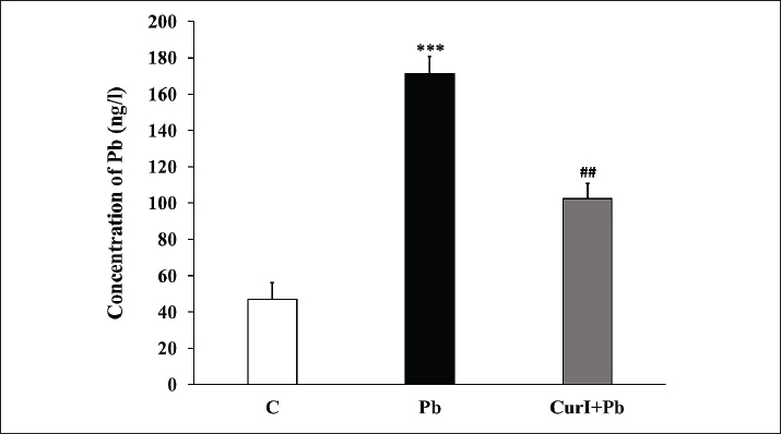
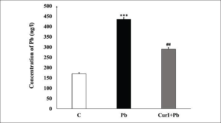
Fig. 2. Effect of acute Pb exposure and Cur I treatment on locomotor activity in rats on the open-field test. Data were analysed for variance of ANOVA, followed by Tukey's post hoc analysis. Data are shown as group mean values ±S.E.M. A p value <0.05 was considered as statistically significant, * p < 0.05 versus C. # p < 0.05 Pb versus Cur I + Pb. C: control, Pb: acute Pb-treated rats, Cur I + Pb: acute Pb-treated rats + Cur I. n=8. Pb assaysOur data showed that acute and chronic exposures to Pb induced a significant increase in brain Pb levels reaching, respectively, 145% (p=0.0006) and 254% (p=0.0003) (Fig. 4). Whereas, Cur I restored significantly such increase with 35% (p=0.0056) and 41% (p=0.0020) reduction of Pb levels compared to acute and chronic Pb group, respectively (Figs. 4 and 5).
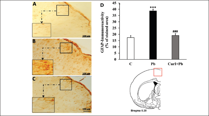
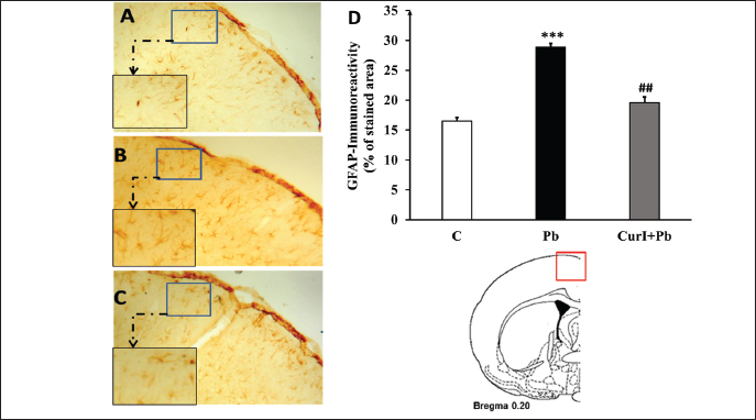
Fig. 4. Quantification of brain Pb levels in control, acute Pb-intoxicated, and Cur I + Pb-treated rats. Data were analysed for variance of ANOVA, followed by Tukey's post hoc analysis. Data are shown as group mean values ±S.E.M. A p-value <0.05 was considered as statistically significant. ***p < 0.001 versus C, ## p < 0.01 Pb versus Cur I + Pb, C: control, Pb: acute Pb-treated rats, Cur I + Pb: acute Pb-treated rats + Cur I. n=5. Effect of acute and chronic Pb and Cur I exposure on GFAP-immunoreactivity in frontal cortexFollowing both acute and chronic Pb exposures, our data showed a significant rise of GFAP-immunoreactive labelling on astrocytes within the frontal cortex (Figs. 6 and 7), reaching 122% (p=0.0005, Fig. 5D) and 70% (p=0.0001, Fig. 7D), respectively, compared to controls. However, with Cur I treatment, GFAP-immunoreactivity was significantly restored, with 58% (p=0.0005) and 28% (p=0.0023) of reduction compared to acute and Pb groups, respectively.
Fig. 6. Light micrographs showing GFAP-immunoreactivity in coronal sections of the cerebral cortex in control (A), acute Pb (B), and acute Cur I + Pb (C) groups immunolabelled with anti-GFAP antibody. D: immunolabelling quantification of the cortical GFAP-immunoreactivity. Data were analysed for variance of ANOVA, followed by Tukey's post hoc analysis. Data are shown as group mean values ±S.E.M. A p-value < 0.05 was considered as statistically significant. ***p < 0.001 versus C. ### p < 0.001 Pb versus Cur I + Pb. n=5.
Fig. 5. Quantification of brain Pb levels in control, chronic Pb-intoxicated, and Cur I + Pb-treated rats. Data were analysed for variance of ANOVA, followed by Tukey's post hoc analysis. Data are shown as group mean values ±S.E.M. A p-value < 0.05 was considered as statistically significant. ***p < 0.001 versus C, ## p < 0.01 Pb versus Cur I + Pb. C: control, Pb: chronic Pb-treated rats, Cur I + Pb: chronic Pb-treated rats + Cur I. n=5.
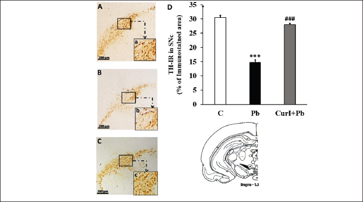
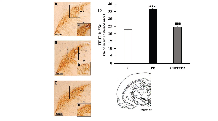
Fig. 7. Light micrographs illustrating GFAP-immunoreactivity in coronal sections of the cerebral cortex in control (A): chronic Pb (B), and chronic Cur I + Pb (C) groups immunolabelled with anti-GFAP antiserum. D: immunolabelling quantification of the cortical GFAP-immunoreactivity. Data were analysed for variance of ANOVA, followed by Tukey's post hoc analysis. Data are shown as group mean values ±S.E.M. A p-value < 0.05 was considered as statistically significant. ***p < 0.001 versus C. ## p < 0.01 Pb versus Cur I + Pb. n=5 Effect of acute and chronic Pb and Cur I exposure on TH-immunoreactivity in SNpcFollowing acute Pb exposure, TH-immunoreactivity within the SNpc nucleus (Fig. 8B) significantly (p=0.0001, Fig. 8D) increases up to 64% compared to the control level (Fig. 8A). On the contrary, following chronic Pb exposure, TH-immunoreactivity was significantly reduced (50%) in the whole nucleus (p=0.0003, Fig. 9B, D) compared to control (Fig. 9A). While, treatment with Cur I restored these abnormal TH-immunoreactivities, with 21% decrease (p=0.0001, Fig. 8C, D), compared to the acute Pb-treated group, and 86% (p=0.0007, Fig. 9C, D) increase compared to the chronic Pb-treated group.
Fig. 9. Light micrographs of frontal sections through SNpc in control (A): chronic Pb (B), and chronic Cur I + Pb (C) groups immunolabelled with anti-TH antiserum. D: immunolabelling quantification of the TH-immunoreactivity. Data were analysed for variance of ANOVA, followed by Tukey's post hoc analysis. Data are shown as group mean values ±S.E.M. A p-value < 0.05 was considered as statistically significant. ***p < 0.001 versus C. ### p < 0.001 Pb vs Cur I + Pb. n=5.
Fig. 8. Light micrographs of frontal sections through SNpc in control (A): acute Pb (B), and acute Cur I + Pb (C) groups immunolabelled with anti-TH antiserum. D: immunolabelling quantification of the TH-immunoreactivity. Data were analysed for variance of ANOVA, followed by Tukey's post hoc analysis. Data are shown as group mean values ±S.E.M. A p-value < 0.05 was considered as statistically significant. ***p < 0.001 versus C. ### p < 0.001 Pb versus Cur I + Pb. n=5. DiscussionDespite numerous investigations on the neural and glial responses to Pb exposure, many studies lack consistency regarding the duration of exposure, dosage levels, animal models, and developmental stages. The current research provides a comprehensive comparison of glial and neural responses to both acute and chronic Pb intoxications. Notably, the data reveal a profound differential neurotoxic impact on the DAergic system depending on the route and duration of administration. While acute Pb exposure resulted in significantly elevated TH levels within the SNpc, chronic intoxication caused a marked decline. These neurochemical changes were directly correlated with locomotor hyperactivity and hypoactivity, respectively. Pb is recognised as one of the potent neurotoxic heavy metals able to cross the blood-brain and placental blood milk barriers and then targets different brain neuronal systems, including the DAergic, serotoninergic, GABAergic, adrenergic, cholinergic, and glutamatergic (Leret et al., 2002; Ramírez Ortega et al., 2021; Virgolini and Aschner, 2021). An earlier neurochemical study has shown that DAergic neurons in rats are sensitive to the actions of inorganic Pb (Takeuchi et al., 2021). In fact, some authors have noted a diminished radiolabelled DA turnover in the nucleus accumbens (NAc) and frontal cortex following chronic developmental exposure to Pb (Wang et al., 2020). While an increased apparent DA synthesis was noticeable in striatum and NAc after systemically applied α-amphetamine (DA agonist), this was greater in Pb-exposed animals (Sabbar et al., 2018). These data lead to sustaining a clear dysfunction of the regulatory mechanisms in DAergic synapses; thus, support of such a view is provided by some data showing that Pb influences the y-aminobutyric acid activity in striatal and NAc efferent pathways, which appeared to be mediated via DAergic projections to these two regions (Carretero et al., 2024; Parithathvi et al., 2025). This indicates that the Pb neurotoxicity on DA neurons is mainly presynaptic and largely related to the disrupted regulation of DA production (Pyatha et al., 2023). Of importance also, acute in vivo exposure of rats to 10-mg/kg trimethyl-Pb has been reported to increase DA uptake into striatal synaptosomes (Komulainen et al., 1983). Even in vitro, using homogenates of whole mouse brain, tri-n-butyl Pb promotes the release of several neurotransmitters, in particular DA, with 100–1,000-fold more than Pb acetate (Bondy et al., 1979). Previous findings seem to support the involvement of Pb in modulating DAergic neurotransmission. Hence, triethyl Pb enhances the responsiveness of DAergic mechanisms, which participate in the modulation of motor behavior. Thus, rats treated with Tetraethyllead chloride (7.88 mg/kg) for 1 week before testing exhibited an increased response to the motor stimulant effects of DA agonists; D-amphetamine and apomorphine (Walsh et al., 1986; Sabbar et al., 2018). The observed increase in TH levels following acute Pb exposure may reflect an early compensatory response of DAergic neurons to Pb-induced neurotoxic effects (Dey et al., 2025). Acute Pb insult is known to induce oxidative stress and disrupt DA homeostasis (Akinyemi et al., 2019), which may trigger an upregulation of TH expression to maintain DA synthesis and neuronal function (Vecchio et al., 2021). This adaptive response could represent a transient neuroprotective mechanism aimed at maintaining DA synthesis. However, sustained exposure may overwhelm these compensatory mechanisms, ultimately leading to DAergic dysfunction, as observed under chronic conditions (Ma et al., 1999; Noureddine et al., 2005; Sanders et al., 2009). Otherwise, Nik Niklowitz and Yeager (1973) postulated that organo-Pb toxicity is dependent upon an alteration in the disposition of endogenous metals (Niklowitz and Yeager, 1973). Indeed, acute administration of lethal doses of tetraethyl Pb to rabbits resulted in increased Pb levels in the brain, accompanied by a concomitant reduction in iron, copper, and zinc concentrations in the hippocampus, frontal cortex, and cerebellum. These changes are relevant because iron and copper act as essential metalloprotein cofactors involved in the synthesis and metabolism of catecholamines (Morgan and O’Dell, 1977). Pb may also interact with the DA metabolism in different brain regions differentially, thus, when administered in a low dose (0.2%) in drinking water to juvenile rats, from the post-natal stage, it resulted in a drop of Monoamine oxidase activity within cerebellum, hippocampus, and cerebral cortex in young (1-month-old) as well as adult (3-month-old) rats, with more vulnerability of young rats. Also, an increased synaptosomal DA with a low dose of Pb was noted. Surprisingly, the opposite effect was noted in a high dose (1%), leading to suppose controverted Pb influences DA neurons depending on the level of exposure, which could result from the increased gastrointestinal absorption of Pb in young rats compared with adults (Ziegler et al., 1978) and then impacts on the cerebral Pb bioavailability (Devi et al., 2005). At low doses, Pb's effect on DA corroborates previous observation noting an enhancement of TH activity and reduced DA reuptake leading to an elevated central DA tonus (Silbergeld and Goldberg, 1975).To delineate the underlying mechanisms by which Pb may control TH activity, some authors have studied the effect of Pb on the presynaptic DA auto-receptors participating in the control of TH activity and then DA metabolism. Indeed, Pb decreases the responsiveness of the presynaptic DAergic neurons to DA, known to modulate TH activity, by inhibiting it when activated (Lasley and Lane, 1988). It is well-established that the main consequence of the DAergic impaired neurotransmission is an irregular locomotor behavior. Astroglia has long been considered the main supportive cells for neuronal survival and even function through different vital roles, including providing metabolic substrates, energy, neurotransmission metabolism, and neuronal defence and protection (Pannasch et al., 2014; Smimih et al., 2024). The relationships between neurons and astrocytes are very complex. The pathways that link the activity of these two groups of cells could be similarly affected in pathological conditions such as Pb toxicity. Disturbances in this bidirectional communication can lead to neuronal death depending on the dose and duration of the exhibition (Benammi et al., 2017). Astrocytes respond to brain cellular damage and support the brain's neuroprotective function against oxidative stress and harmful substances (Sofroniew, 2009; Sofroniew and Vinters, 2010). Numerous studies have demonstrated the toxic effect of Pb on astrocytes. In fact, long-term exposure to high levels of Pb leads to astrocyte death in vitro. Our immunohistochemical study of brain tissue showed astroglial involvement in different brain regions in both acute and chronic models. Indeed, in our rats poisoned (acute and chronic) at the cortical level, we note a hyper-reactivity of the GFAP immunoreactive astrocytes. Several studies are consistent with our results. Indeed, previous reports showed increased GFAP levels in the cortex, cerebellum, and corpus callosum in Pb-exposed rats (Benlahcen et al., 2009). While others showed a gliosis in the frontal cortex after chronic exposure to Pb at a dose of 5 g/l (Sansar et al., 2011). These astroglial reactions make it possible to postulate that these cells constitute a reservoir for the accumulation and sequestration of this metal, thus reducing its bioavailability (Lindahl, 1999). This accumulation contributes to the protection of neurons that are more sensitive. Late, the Pb stored in the glial cells is released continuously, which contributes to the neurotoxicity, especially of the surrounding ns or the astroglia in particular, with i.) initiating oxidative stress by the genesis of ROS, ii.) via weakening of the anti-oxidant defensive enzymes, including GST, GSH, GPx, and SOD, resulting in neuronal damage and loss (Ercal et al., 1996a,b; Lai et al., 2002; Jankovic, 2005; De Lau and Breteler, 2006; Bokara et al., 2008; Liu et al., 2017). The present study provided yet another proof of the powerful preventive property of Cur I against the toxicity of Pb. Previous studies have shown that Cur I administered alone does not induce significant changes in control animals (Benammi et al., 2014; Benammi et al., 2016). Indeed, treatment with Cur I, in our rats poisoned with Pb (acute and chronic), has shown the restoration of TH levels in SNpc with a marked improvement in the locomotor behavior. The neuroprotective effect of Cur I may be explained, at least in part, by its ability to reduce Pb accumulation within the brain (Shukla et al., 2003; Saleh et al., 2018). Cur I possesses metal-chelating properties and can interact directly with Pb ions to form stable complexes, thereby reducing their bioavailability and tissue retention (Daniel et al., 2004; Abubakar et al., 2019). In addition, by limiting oxidative stress and preserving cellular homeostasis, Cur I may reduce Pb-induced neuronal and astroglial damage, which could further contribute to lowering Pb accumulation in neural tissues (Benammi et al., 2017; Saleh et al., 2018; Abubakar et al., 2019). Such a hypothesis is supported by our chemical measurements of the central Pb level, showing a significant reduction of Pb level in both the acute and chronic Pb-intoxicated rats in the presence of Cur I. The significant decrease in brain Pb concentrations observed in Cur I-treated animals supports the hypothesis that Cur I facilitates Pb clearance or limits its cerebral retention. Other studies demonstrate an affinity between Cur I and Pb that together form a complex (Shukla et al., 2003; Daniel et al., 2004). This chelating power also makes it possible to explain the maintenance of the normal levels of GFAP despite exposure to Pb. Preventing this, gliosis is generally indicative of a neuroprotective response reducing interstitial Pb levels by its sequestration, which qualifies this cellular component (astroglia) as a first defence barrier against the neurotoxicity of heavy metals (Lindahl, 1999). Such a regulatory effect of Cur I seems to be supported by other studies showing, in other models, that the administration of Cur I reduces the rate of astrocytic proliferation in the hippocampus and the frontal cortex in mice exposed to Okadaic acid for 7 days (Rajasekar et al., 2013). In Alzheimer's models, Cur I reduced the expression of mRNA and protein levels of GFAP in the thalamic, cortical, and hippocampal regions, as well as the entorhinal cortex, piriform cortex, and amygdala (Lim et al., 2001). Another study shows that a relatively low dose of Cur I significantly reduces the inflammatory cytokine IL-1 and GFAP, also leading to a reduction in oxidative damage and decreasing the total amount of plaque, soluble amyloid, and insoluble amyloid (Lim et al., 2001). In summary, our study compares glial and neuronal responses to acute and chronic Pb exposure, revealing distinct neurotoxic effects on the DAergic system. Acute exposure increased TH levels in the SNpc and enhanced locomotor activity, whereas chronic exposure led to reduced TH levels and impaired behavioral performance. The present findings have important implications for environmental Pb exposure in both humans and animals. The differential effects observed between acute and chronic exposures on DAergic and astroglial systems may help explain the variability in clinical manifestations, ranging from transient hyperactivity to progressive neurological dysfunction. While the observed neuroprotective effects of Cur I suggest its potential as a complementary therapeutic or preventive strategy. However, further studies are required to validate its efficacy, to optimize dosing, and to assess its translational applicability in both human medicine and veterinary practice. Although this study provides further evidence on Pb-induced neurotoxicity and the potential restorative effect of curcumin, it has some limitations. First, the present study does not include other complementary neurochemical measurements (DA, DOPAC, DAT, or VMAT2 levels), which restricts the interpretation of increased TH expression as a direct indicator of enhanced DAergic function. Further studies integrating detailed neurochemical analyses are required to confirm such findings. Also, despite the promising pharmacological properties of Cur I, the clinical translation of Cur I remains limited, mainly due to its poor bioavailability, low solubility, rapid metabolism, and systemic elimination, which require implementing other strategies such as nanoparticle-based delivery systems and the use of bioavailability enhancers. ConclusionOur study revealed a remarkable differential responsiveness of the midbrain SNpc DAergic neurons to acute and chronic Pb exposures, with increased TH-immunoreactivity for the acute form and the opposite response in the chronic one. While the cortical astroglia seems to conserve the same reactivity for both intoxications. The central neuronal and glial abnormalities were associated with severe locomotor disturbances, with either increased or decreased locomotor performances. Otherwise, concomitant Cur administration with Pb appears to restore both cellular and neurobehavioral abnormalities, leading to considering this molecule as one of the potent neuroprotective agents recommended for treating heavy metal neurotoxicity. AcknowledgmentsThe authors express their deep thanks to all those who participated in the success of this work, particularly Pr Rahmoun Miloud for English revision. Conflicts of interestThe authors declare no conflict of interest. FundingThis research was funded by the FSS-UCA for research units funding (annual budget). Authors’ contributionsConceptualization: Omar Elhiba, Halima Gamrani; methodology, Omar Elhiba, Halima Gamrani; software, Hind Benammi, Ahmed Draoui, Kamal Smimih, Bilal El-Mansoury, and Hicham Chatoui; validation, Omar Elhiba and Halima Gamrani; formal analysis, Hind Benammi. and Ahmed Draoui; investigation, Ahmed Draoui, Kamal Smimih; resources, Halima Gamrani; data curation, Ahmed Draoui, Kamal Smimih, Faissal Aziz, Morad Belkouri; writing original draft preparation, Hind Benammi; writing review and editing, Kamal Smimih and Omar Elhiba; visualization, Ahmed Draoui; supervision, Omar Elhiba and Halima Gamrani; project administration, Omar Elhiba and Halima Gamrani; funding acquisition, Ahmed Draoui and Kamal Smimih. All authors have read and agreed to the published version of the manuscript. Data availabilityAll data are available in the manuscript. ReferencesAbbaoui, A., El Hiba, O. and Gamrani, H. 2016. Copper poisoning induces neurobehavioral features of Parkinson’s disease in rat: alters dopaminergic system and locomotor performance. Parkinsonism. Relat. Disord. 22, e188. Abubakar, K., Muhammad Mailafiya, M., Danmaigoro, A., Musa Chiroma, S., Abdul Rahim, E.B. and Abu Bakar [at] Zakaria, M.Z. 2019. Curcumin attenuates lead-induced cerebellar toxicity in rats via chelating activity and inhibition of oxidative stress. Biomolecules 9, 453. Ahlskog, J.E. and Muenter, M.D. 2001. Frequency of levodopa‐related dyskinesias and motor fluctuations as estimated from the cumulative literature. Mov. Disord. Off. J. Mov. Disord. Soc. 16, 448–458. Akinyemi, A.J., Miah, M.R., Ijomone, O.M., Tsatsakis, A., Soares, F.A.A., Tinkov, A.A., Skalny, A.V., Venkataramani, V. and Aschner, M. 2019. Lead (Pb) exposure induces dopaminergic neurotoxicity in Caenorhabditis elegans: involvement of the dopamine transporter. Toxicol. Rep. 6, 833–840. Aliche, K.A., Ikewuchi, C. and Diorgu, F.C. 2025. The impact of maternal lead exposure on pregnancy outcomes: a systematic review. IPS. J. Public. Heal. 5, 226–236. Al-Saleh, I., Al-Enazi, S. and Shinwari, N. 2009. Assessment of lead in cosmetic products. Regul. Toxicol. Pharmacol. 54, 105–113; doi:10.1016/j.yrtph.2009.02.005 Aslam , I., Younas, S., Fatima, W., Arshad, A. and Naz, A. 2025. Impact of environmental pollution on food safety and human health. Glob. Res. J. Nat. Sci. Technol. 03(3), 592–607. Assi, M.A., Hezmee, M.N.M., Haron, A.W., Sabri, M.Y. and Rajion, M.A. 2016. The detrimental effects of lead on human and animal health. Vet. World 9, 660. Benammi, H., El Hiba, O. and Gamrani, H. 2016. Evidence of a subcommissural organ involvement in the brain response to lead exposure and a modulatory potential of curcumin. Neuroreport 27, 264–271. Benammi, H., El Hiba, O., Romane, A. and Gamrani, H. 2014. A blunted anxiolytic like effect of curcumin against acute lead induced anxiety in rat: involvement of serotonin. Acta. Histochem. 116, 920–925. Benammi, H., Erazi, H., El Hiba, O., Vinay, L., Bras, H., Viemari, J.-C. and Gamrani, H. 2017. Disturbed sensorimotor and electrophysiological patterns in lead intoxicated rats during development are restored by curcumin. PLoS One 12, 172715. Benlahcen , K., Sansar, W., Belhabri, L. and Slimani, M. 2009. Lead in water: neurotoxicity and stressful effect on wistar rat. Glob. J. Environ. Res. 3, 52–60. Bischoff, K., Priest, H. and Mount-Long, A. 2010. Animals as sentinels for human lead exposure: a case report. J. Med. Toxicol. 6, 185–189. Bokara, K.K., Brown, E., Mccormick, R., Yallapragada, P.R., Rajanna, S. and Bettaiya, R. 2008. Lead-induced increase in antioxidant enzymes and lipid peroxidation products in developing rat brain. BioMetals 21, 9–16; doi:10.1007/s10534-007-9088-5 Bondy, S.C., Anderson, C.L., Harrington, M.E. and Prasad, K.N. 1979. The effects of organic and inorganic lead and mercury on neurotransmitter high-affinity transport and release mechanisms. Environ. Res. 19, 102–111; doi:10.1016/0013-9351(79)90038-0 Carretero, V.J., Liccardi, N., Tejedor, M.A., De Pascual, R., Campano, J.H. and Hernández-Guijo, J.M. 2024. Lead exerts a depression of neurotransmitter release through a blockade of voltage dependent calcium channels in chromaffin cells. Toxicology 505, 153809. Connolly, B.S. and Lang, A.E. 2014. Pharmacological treatment of Parkinson disease: a review. JAMA 311, 1670–1683. Daniel, S., Limson, J.L., Dairam, A., Watkins, G.M. and Daya, S. 2004. Through metal binding, curcumin protects against lead- and cadmium-induced lipid peroxidation in rat brain homogenates and against lead-induced tissue damage in rat brain. J. Inorg. Biochem. 98, 266–275; doi:10.1016/j.jinorgbio.2003.10.014 De Lau, L.M.L. and Breteler, M.M.B. 2006. Epidemiology of Parkinson’s disease. Lancet Neurol. 5, 525–535. De Virgilio, A., Greco, A., Fabbrini, G., Inghilleri, M., Rizzo, M.I., Gallo, A., Conte, M., Rosato, C., Ciniglio Appiani, M. and De Vincentiis, M. 2016. Parkinson’s disease: autoimmunity and neuroinflammation. Autoimmun. Rev; doi:10.1016/j.autrev.2016.07.022 Devi, C.B., Reddy, G.H., Prasanthi, R.P.J., Chetty, C.S. and Reddy, G.R. 2005. Developmental lead exposure alters mitochondrial monoamine oxidase and synaptosomal catecholamine levels in rat brain. Int. J. Dev. Neurosci. 23, 375–381; doi:10.1016/j.ijdevneu.2004.11.003 Dey, K.K., Kamila, S., Das, T. and Chattopadhyay, A. 2025. Chronic exposure to lead causes neurotoxicity by generating oxidative stress and inducing DNA damages in zebrafish brain: involvement of Nrf2-Keap1 regulation and DNA repair pathways. Biol. Trace. Elem. Res. 203, 4863–4875. Draoui, A., El Hiba, O., Aimrane, A., El Khiat, A. and Gamrani, H. 2020. Parkinson’s disease: from bench to bedside. Rev. Neurol. (Paris). 176, 543–559. Draoui, A., Zidour, M., Tounsi, A. and Adim, B. 2019. Static and dynamic behavior of nanotubes-reinforced sandwich plates using (FSDT). J. Nano. Res. 57, 117–135. Duty, S. and Jenner, P. 2011. Animal models of Parkinson’s disease: a source of novel treatments and clues to the cause of the disease. Br. J. Pharmacol. 164, 1357–1391; doi:10.1111/j.1476-5381.2011.01426.x El Hiba, O., Gamrani, H., Chatoui, H. and Ahboucha, S. 2013. Loss of tyrosine hydroxylase expression within the nigro-striato-cortical pathways in the cirrhotic rat: the possible restorative effect of the neurosteroid dehydroepiandrosterone sulfate. Acta. Histochem. 115, 637–645; doi:10.1016/j.acthis.2013.01.006 El Khiat, A., Tamegart, L., Draoui, A., El Fari, R., Sellami, S., Rais, H., El Hiba, O. and Gamrani, H. 2019. Kinetic deterioration of short memory in rat with acute hepatic encephalopathy: involvement of astroglial and neuronal dysfunctions. Behav. Brain. Res; doi:10.1016/j.bbr.2019.03.046 Ercal, N., Treeratphan, P., Hammond, T., Matthews, R., Grannemann, N. and Spitz, D. 1996. In vivo indices of oxidative stress in lead. Free Radic. Biol. Med. 21, 157–161. Esatbeyoglu, T., Huebbe, P., Ernst, I.M.A., Chin, D., Wagner, A.E. and Rimbach, G. 2012. Curcumin-from molecule to biological function. Angew. Chem. Int. Ed. 51, 5308–5332; doi:10.1002/anie.201107724 Flora, G., Gupta, D. and Tiwari, A. 2012. Toxicity of lead: a review with recent updates. Interdiscip. Toxicol. 5, 47. Franzblau, A., Rosenstock, L. and Eaton, D.L. 1988. Use of inductively coupled plasma-atomic emission spectroscopy (ICP-AES) in screening for trace metal exposures in an industrial population. Environ. Res. 46, 15–24; doi:10.1016/S0013-9351(88)80055-0 Garcia‐Alloza, M., Borrelli, L.A., Rozkalne, A., Hyman, B.T. and Bacskai, B.J. 2007. Curcumin labels amyloid pathology in vivo, disrupts existing plaques, and partially restores distorted neurites in an Alzheimer mouse model. J. Neurochem. 102, 1095–1104. Hewlings, S. and Kalman, D. 2017. Curcumin: a review of its effects on human health. Foods 6, 92. Hiba, O.E., Elgot, A., Ahboucha, S. and Gamrani, H. 2016. Differential regional responsiveness of astroglia in mild hepatic encephalopathy: an immunohistochemical approach in bile duct ligated rat. Acta. Histochem. 118, 338–346. Hu, H., Téllez-Rojo, M.M., Bellinger, D., Smith, D., Ettinger, A.S., Lamadrid-Figueroa, H., Schwartz, J., Schnaas, L., Mercado-García, A. and Hernández-Avila, M. 2006. Fetal lead exposure at each stage of pregnancy as a predictor of infant mental development. Environ. Health Perspect. 114, 1730–1735. Huot, P. and Fox, S.H. 2011. Nondopaminergic treatments for Parkinson’s disease. Neurodegenerative. Dis. Manage. 1, 491–512. Jagmag, S.A., Tripathi, N., Shukla, S.D., Maiti, S. and Khurana, S. 2016. Evaluation of models of Parkinson’s disease. Front. Neurosci; doi:10.3389/fnins.2015.00503 Jankovic, J. 2005. Searching for a relationship between manganese and welding and Parkinson’s disease. Neurology 64, 2021–2028. Komulainen, H., Pietarinen, R. and Tuomisto, J. 1983. Increase in dopamine uptake in rat striatal synaptosomes after an acute in vivo administration of organic and inorganic lead. Acta. Pharmacol. Toxicol. (Copenh). 52, 381–389. Lai, B.C., Marion, S.A., Teschke, K. and Tsui, J.K. 2002. Occupational and environmental risk factors for Parkinson’s disease. Parkinsonism Relat. Disord. 8, 397–309. Lang, A.E. and Obeso, J.A. 2004. Challenges in Parkinson’s disease: restoration of the nigrostriatal dopamine system is not enough. Lancet. Neurol. 3, 309–316; doi:10.1016/S1474-4422(04)00740-9 Laoye, B., Olagbemide, P., Ogunnusi, T. and Akpor, O. 2025. Heavy metal contamination: sources, health impacts, and sustainable mitigation strategies with insights from Nigerian case studies. F1000Research 14, 134. Lasley, S.M. and Lane, J.D. 1988. Diminished regulation of mesolimbic dopaminergic activity in rat after chronic inorganic lead exposure. Toxicol. Appl. Pharmacol. 95, 474–483; doi:10.1016/0041-008X(88)90365-1 Leret, M.L., Garcia-Uceda, F. and Antonio, M.T. 2002. Effects of maternal lead administration on monoaminergic, GABAergic and glutamatergic systems. Brain Res. Bull. 58, 469–473; doi:10.1016/S0361-9230(02)00819-5 Lim, G.P., Chu, T., Yang, F., Beech, W., Frautschy, S.A. and Cole, G.M. 2001. The curry spice curcumin reduces oxidative damage and amyloid pathology in an Alzheimer transgenic mouse. J. Neurosci. 21, 8370–8377; doi:10.1523/JNEUROSCI.21-21-08370.2001 Lindahl, L. 1999. Differential ability of astroglia and neuronal cells to accumulate lead: dependence on cell type and on degree of differentiation. Toxicol. Sci. 50, 236–243; doi:10.1093/toxsci/50.2.236 Liu, B., Zhang, H., Tan, X., Yang, D., Lv, Z. and Jiang, H. 2017. GSPE reduces lead-induced oxidative stress by activating the Nrf2 pathway and suppressing miR153 and GSK-3 β in rat kidney. Oncotarget 8, 42226–42237. Ma, T., Chen, H.H. and Ho, I.K. 1999. Effects of chronic lead (Pb) exposure on neurobehavioral function and dopaminergic neurotransmitter receptors in rats. Toxicol. Lett. 105, 111–121. Mitra, P., Sharma, S., Purohit, P. and Sharma, P. 2017. Clinical and molecular aspects of lead toxicity: an update. Crit. Rev. Clin. Lab. Sci. 54, 506–528. Morgan, R.F. and O’Dell, B.L. 1977. Effect of copper deficiency on the concentrations of catecholamines and related enzyme activities in the rat brain. J. Neurochem. 28, 207–213; doi:10.1111/j.1471-4159.1977.tb07728.x Mythri, R.B. and Srinivas Bharath, M.M. 2012. Curcumin: a potential neuroprotective agent in Parkinson’s disease. Curr. Pharm. Des. 18, 91–99. Niklowitz, W.J. and Yeager, D.W. 1973. Interference of Pb with essential brain tissue Cu, Fe, and Zn as main determinant in experimental tetraethyllead encephalopathy. Life Sci. 13, 897–905. Noureddine, D., Miloud, S. and Abdelkader, A. 2005. Effect of lead exposure on dopaminergic transmission in the rat brain. Toxicology 207, 363–368. Olanow, C.W., Stern, M.B. and Sethi, K. 2009. The scientific and clinical basis for the treatment of Parkinson disease (2009). Neurology 72, S1–S136. Pannasch, U., Freche, D., Dallérac, G., Ghézali, G., Escartin, C., Ezan, P., Cohen-Salmon, M., Benchenane, K., Abudara, V., Dufour, A., Lübke, J.H.R., Déglon, N., Knott, G., Holcman, D. and Rouach, N. 2014. Connexin 30 sets synaptic strength by controlling astroglial synapse invasion. Nat. Neurosci. 17, 549–558; doi:10.1038/nn.3662 Parithathvi, A., Choudhari, N. and Dsouza, H.S. 2024. Prenatal and early life lead exposure induced neurotoxicity. Hum. Exp. Toxicol. 43, 09603271241285523. Parithathvi, A., Harshitha, P., Mumbrekar, K.D. and Dsouza, H.S. 2025. Systematic review on neurotoxic implications of lead-induced gene expression alterations in the etiology of Alzheimer’s disease. Cell. Mol. Neurobiol. 45, 1–16. Paxinos, G. and Watson, C. 2004. The rat brain in stereotaxic coordinates-the new coronal set. Boston: Elsevier. Plusquellec, P., Muckle, G., Dewailly, E., Ayotte, P., Jacobson, S.W. and Jacobson, J.L. 2007. The relation of low-level prenatal lead exposure to behavioral indicators of attention in Inuit infants in Arctic Quebec. Neurotoxicol. Teratol. 29, 527–537. Poewe, W., Seppi, K., Tanner, C.M., Halliday, G.M., Brundin, P., Volkmann, J., Schrag, A.E. and Lang, A.E. 2017. Parkinson disease. Nat. Rev. Dis. Prim. 3, 17013. Pyatha, S., Kim, H., Lee, D. and Kim, K. 2023. Co-exposure to lead, mercury, and cadmium induces neurobehavioral impairments in mice by interfering with dopaminergic and serotonergic neurotransmission in the striatum. Front. Public Heal. 11, 1265864. Rajasekar, N., Dwivedi, S., Tota, S.K., Kamat, P.K., Hanif, K., Nath, C. and Shukla, R. 2013. Neuroprotective effect of curcumin on okadaic acid induced memory impairment in mice. Eur. J. Pharmacol. 715, 381–394; doi:10.1016/j.ejphar.2013.04.033 Ramírez Ortega, D., González Esquivel, D.F., Blanco Ayala, T., Pineda, B., Gómez Manzo, S., Marcial Quino, J., Carrillo Mora, P. and Pérez De La Cruz, V. 2021. Cognitive impairment induced by lead exposure during lifespan: mechanisms of lead neurotoxicity. Toxics 9, 23. Sabbar, M., Delaville, C., De Deurwaerdère, P., Lakhdar-Ghazal, N. and Benazzouz, A. 2018. Lead-induced atypical Parkinsonism in rats: behavioral, electrophysiological, and neurochemical evidence for a role of noradrenaline depletion. Front. Neurosci. 12, 173. Saleh, H.A., S. Abd El-aziz, G., N. Mustafa, H., El-Fark, M., Mansour Tashkandi, J., Hassan Alzahrani, A., Mal, A., Aborass, M. and Halim Deifalla, A. 2018. Beneficial effects of curcumin in maternal and fetal oxidative stress and brain damage induced by gestational lead administration. Biomed. Pharmacol. J. 11, 871–887. Sanders, T., Liu, Y., Buchner, V. and Tchounwou, P.B. 2009. Neurotoxic effects and biomarkers of lead exposure: a review. Rev. Environ. Health 24, 15. Sansar, W., Ahboucha, S. and Gamrani, H. 2011. Chronic lead intoxication affects glial and neural systems and induces hypoactivity in adult rat. Acta Histochem. 113, 601–607; doi:10.1016/j.acthis.2010.06.005 Seibenhener, M.L. and Wooten, M.C. 2015. Use of the open field maze to measure locomotor and anxiety-like behavior in mice. J. Vis. Exp. JoVE. 96, 52434. Shukla, P.K., Khanna, V.K., Khan, M.Y. and Srimal, R.C. 2003. Protective effect of curcumin against lead neurotoxicity in rat. Hum. Exp. Toxicol. 22, 653–658; doi:10.1191/0960327103ht411oa Silbergeld, E.K. and Goldberg, A.M. 1975. Pharmacological and neurochemical investigations of lead-induced hyperactivity. Neuropharmacology 14, 431–444; doi:10.1016/0028-3908(75)90026-X Smimih, K., El-Mansoury, B., Azzouhri, C., Ait Hamdan, Y., Saad, F.E. and Mohamed, M. 2024. Glial cells in the mature central nervous system: classification and functions. In Physiology and function of glial cells in health and disease. Bilal El-Mansoury, Omar El Hiba, and Arumugam Radhakrishnan Jayakumar. Hershey, PA:IGI Global Scientific Publishing, pp: 1–9. Smimih, K., El-Mansoury, B., Marghich, M., Azzouhri, C., Zouhairi, N., Agnaou, M., Guennouni, M., Fdil, N., Bitar, A. and Abulmeaty, M.M.A. 2026. Prenatal alcohol exposure induces anxiety and depressive-like behaviors with deficits in growth and food intake in mice. Front. Cell. Dev. Biol. 14, 1742806. Smimih, K., El-Mansoury, B., Saad, F.E., Khanouchi, M., El Amine, S., Aimrane, A., Zouhairi, N., Ferssiwi, A., Bitar, A., Merzouki, M. and El Hiba, O. 2023. Sensory motor function disturbances in mice prenatally exposed to low dose of ethanol: a neurobehavioral study in postnatal and adult stages. Neurol. Int. 15, 580–594. Sofroniew, M.V. 2009. Molecular dissection of reactive astrogliosis and glial scar formation. Trends Neurosci. 32, 638–647; doi:10.1016/j.tins.2009.08.002 Sofroniew, M.V. and Vinters, H.V. 2010. Astrocytes: biology and pathology. Acta Neuropathol. 119, 7–35; doi:10.1007/s00401-009-0619-8 Soleimani, V., Sahebkar, A. and Hosseinzadeh, H. 2018. Turmeric (Curcuma longa) and its major constituent (curcumin) as nontoxic and safe substances. Phyther. Res. 32, 985–995. Takeuchi, H., Taki, Y., Nouchi, R., Yokoyama, R., Kotozaki, Y., Nakagawa, S., Sekiguchi, A., Iizuka, K., Hanawa, S., Araki, T., Miyauchi, C.M., Sakaki, K., Nozawa, T., Ikeda, S., Yokota, S., Daniele, M., Sassa, Y. and Kawashima, R. 2021. Lead exposure is associated with functional and microstructural changes in the healthy human brain. Commun. Biol. 4, 912. Tchounwou, P.B., Yedjou, C.G., Patlolla, A.K. and Sutton, D.J. 2012. Heavy metal toxicity and the environment. Mol. Clin. Environ. Toxicol. 3, 133–164. Tysnes, O.B. and Storstein, A. 2017. Epidemiology of Parkinson’s disease. J. Neural Transm; doi:10.1007/s00702-017-1686-y Ullah, Z., Rashid, A., Ghani, J., Talib, M.A., Shahab, A. and Lun, L. 2023. Arsenic contamination, water toxicity, source apportionment, and potential health risk in groundwater of Jhelum Basin, Punjab, Pakistan. Biol. Trace. Elem. Res. 201, 514–524. Vacchi-Suzzi, C., Viens, L., Harrington, J.M., Levine, K., Karimi, R. and Meliker, J.R. 2018. Low levels of lead and glutathione markers of redox status in human blood. Environ. Geochem. Health. 40, 1175–1185. Vecchio, L.M., Sullivan, P., Dunn, A.R., Bermejo, M.K., Fu, R., Masoud, S.T., Gregersen, E., Urs, N.M., Nazari, R. and Jensen, P.H. 2021. Enhanced tyrosine hydroxylase activity induces oxidative stress, causes accumulation of autotoxic catecholamine metabolites, and augments amphetamine effects in vivo. J. Neurochem. 158, 960–979. Vilaplana, J. and Lavialle, M. 1999. A method to quantify glial fibrillary acidic protein immunoreactivity on the suprachiasmatic nucleus. J. Neurosci. Methods 88, 181–187. Virgolini, M.B. and Aschner, M. 2021. Molecular mechanisms of lead neurotoxicity. In Advances in neurotoxicology. Michael Aschner and Lucio G. Costa. Cambridge, MA: Elsevier, pp: 159–213. Walsh, T.J., Schulz, D.W., Tilson, H. and Dehaven, D.L. 1986. Acute exposure to triethyl lead enhances the behavioral effects of dopaminergic agonists: involvement of brain dopamine in organolead neurotoxicity. Brain. Res. 363, 222–229; doi:10.1016/0006-8993(86)91007-3 Wang, T., Zhang, J. and Xu, Y. 2020. Epigenetic basis of lead-induced neurological disorders. Int. J. Environ. Res. Public Health 17, 4878. Wani, A.L., Ara, A. and Usmani, J.A. 2015. Lead toxicity: a review. Interdiscipl. Toxicol. 8, 55. Xu, L., Zhang, W., Liu, X., Zhang, C., Wang, P. and Zhao, X. 2018. Circulatory levels of toxic metals (aluminum, cadmium, mercury, lead) in patients with Alzheimer’s disease: a quantitative meta-analysis and systematic review. J. Alzheimer’s. Dis. 62, 361–372. Ziegler, E.E., Edwards, B.B., Jensen, R.L., Mahaffey, K.R. and Fomon, S.J. 1978. Absorption and retention of lead by infants. Pediatr. Res. 12, 29–34. | ||
| How to Cite this Article |
| Pubmed Style Benammi H, Smimih K, Draoui A, Belkouri M, Aziz F, Hiba OE, Benammi HGH, Smimih K, Draoui A, El-mansoury B, Chatoui H, Belkouri M, Aziz F, Hiba OE, Gamrani H. Differential responsiveness of the midbrain dopaminergic system to acute and chronic lead intoxications and the neuroprotective potential of curcumin I in rat: A possible involvement of astroglia. doi:10.5455/OVJ.2026.v16.i7.53 Web Style Benammi H, Smimih K, Draoui A, Belkouri M, Aziz F, Hiba OE, Benammi HGH, Smimih K, Draoui A, El-mansoury B, Chatoui H, Belkouri M, Aziz F, Hiba OE, Gamrani H. Differential responsiveness of the midbrain dopaminergic system to acute and chronic lead intoxications and the neuroprotective potential of curcumin I in rat: A possible involvement of astroglia. https://www.openveterinaryjournal.com/?mno=303961 [Access: July 15, 2026]. doi:10.5455/OVJ.2026.v16.i7.53 AMA (American Medical Association) Style Benammi H, Smimih K, Draoui A, Belkouri M, Aziz F, Hiba OE, Benammi HGH, Smimih K, Draoui A, El-mansoury B, Chatoui H, Belkouri M, Aziz F, Hiba OE, Gamrani H. Differential responsiveness of the midbrain dopaminergic system to acute and chronic lead intoxications and the neuroprotective potential of curcumin I in rat: A possible involvement of astroglia. doi:10.5455/OVJ.2026.v16.i7.53 Vancouver/ICMJE Style Benammi H, Smimih K, Draoui A, Belkouri M, Aziz F, Hiba OE, Benammi HGH, Smimih K, Draoui A, El-mansoury B, Chatoui H, Belkouri M, Aziz F, Hiba OE, Gamrani H. Differential responsiveness of the midbrain dopaminergic system to acute and chronic lead intoxications and the neuroprotective potential of curcumin I in rat: A possible involvement of astroglia. doi:10.5455/OVJ.2026.v16.i7.53 Harvard Style Benammi, H., Smimih, . K., Draoui, . A., Belkouri, . M., Aziz, . F., Hiba, . O. E., Benammi, . H. G. H., Smimih, . K., Draoui, . A., El-mansoury, . B., Chatoui, . H., Belkouri, . M., Aziz, . F., Hiba, . O. E. & Gamrani, . H. (2026) Differential responsiveness of the midbrain dopaminergic system to acute and chronic lead intoxications and the neuroprotective potential of curcumin I in rat: A possible involvement of astroglia. doi:10.5455/OVJ.2026.v16.i7.53 Turabian Style Benammi, Hind, Kamal Smimih, Ahmed Draoui, Morad Belkouri, Faissal Aziz, Omar El Hiba, Halima Gamrani Hind Benammi, Kamal Smimih, Ahmed Draoui, Bilal El-mansoury, Hicham Chatoui, Morad Belkouri, Faissal Aziz, Omar El Hiba, and Halima Gamrani. 2026. Differential responsiveness of the midbrain dopaminergic system to acute and chronic lead intoxications and the neuroprotective potential of curcumin I in rat: A possible involvement of astroglia. doi:10.5455/OVJ.2026.v16.i7.53 Chicago Style Benammi, Hind, Kamal Smimih, Ahmed Draoui, Morad Belkouri, Faissal Aziz, Omar El Hiba, Halima Gamrani Hind Benammi, Kamal Smimih, Ahmed Draoui, Bilal El-mansoury, Hicham Chatoui, Morad Belkouri, Faissal Aziz, Omar El Hiba, and Halima Gamrani. "Differential responsiveness of the midbrain dopaminergic system to acute and chronic lead intoxications and the neuroprotective potential of curcumin I in rat: A possible involvement of astroglia." doi:10.5455/OVJ.2026.v16.i7.53 MLA (The Modern Language Association) Style Benammi, Hind, Kamal Smimih, Ahmed Draoui, Morad Belkouri, Faissal Aziz, Omar El Hiba, Halima Gamrani Hind Benammi, Kamal Smimih, Ahmed Draoui, Bilal El-mansoury, Hicham Chatoui, Morad Belkouri, Faissal Aziz, Omar El Hiba, and Halima Gamrani. "Differential responsiveness of the midbrain dopaminergic system to acute and chronic lead intoxications and the neuroprotective potential of curcumin I in rat: A possible involvement of astroglia." doi:10.5455/OVJ.2026.v16.i7.53 APA (American Psychological Association) Style Benammi, H., Smimih, . K., Draoui, . A., Belkouri, . M., Aziz, . F., Hiba, . O. E., Benammi, . H. G. H., Smimih, . K., Draoui, . A., El-mansoury, . B., Chatoui, . H., Belkouri, . M., Aziz, . F., Hiba, . O. E. & Gamrani, . H. (2026) Differential responsiveness of the midbrain dopaminergic system to acute and chronic lead intoxications and the neuroprotective potential of curcumin I in rat: A possible involvement of astroglia. doi:10.5455/OVJ.2026.v16.i7.53 |


