Open Veterinary Journal, (2026), Vol. 16(7): 4710-4723
Research Article
10.5455/OVJ.2026.v16.i7.51
Optimization of dextran sodium sulfate concentration for inducing necrotizing enterocolitis in BALB/c mice: An in vivo experimental study
Eko Sulistijono1,2, Krisni Subandiyah1, Loeki Enggar Fitri3*, Husnul Khotimah4 and
Annisatul Hakimah5
1Department of Pediatric, Faculty of Medicine, Universitas Brawijaya, Dr Saiful Anwar General Hospital, Malang, Indonesia
2Doctoral Study Program in Medical Science, Faculty of Medicine, Universitas Brawijaya, Malang, Indonesia
3Department of Clinical Parasitology, Faculty of Medicine, Universitas Brawijaya, Malang, Indonesia
4Department of Pharmacology, Faculty of Medicine, Universitas Brawijaya, Malang, Indonesia
5Department of Nutrition, Faculty of Psychology and Health, UIN Walisongo, Semarang, Indonesia
*Corresponding Author: Loeki Enggar Fitri. Department of Clinical Parasitology, Faculty of Medicine, Universitas Brawijaya, Malang, Indonesia. Email: lukief [at] ub.ac.id
Submitted: 01/01/2026 Revised: 29/05/2026 Accepted: 11/06/2026 Published: 20/07/2026
© 2025 Open Veterinary Journal
This is an Open Access article distributed under the terms of the Creative Commons Attribution-Non Commercial-No Derivatives License (http://creativecommons.org/licenses/by-nc-nd/4.0/), which permits non-commercial re-use, distribution, and reproduction in any medium, provided the original work is properly cited, and is not altered, transformed, or built upon in any way.
Abstract
Background: Necrotizing enterocolitis (NEC) is a life-threatening intestinal inflammatory disorder characterized by the breakdown of the epithelial barrier and bacterial translocation, resulting in systemic inflammation. Developing an ideal animal model is essential for gaining a deeper insight into its pathomechanism. Dextran sodium sulfate (DSS) has been widely used to induce NEC-like intestinal injury; however, the precise concentration of DSS that consistently reproduces NEC features remains uncertain.
Aim: This study aimed to determine the optimal DSS concentration for inducing NEC-like inflammation in the ileal tissue of BALB/c mice by evaluating the disease activity index (DAI), NEC histological scores, and proinflammatory cytokine expression.
Methods: A true in vivo experimental design with a randomized post-test-only controlled group was conducted using 30 male BALB/c mice (weight, 20–25 g; age, 8–10 weeks). The mice were divided into five groups (n=6): a control group receiving water and treatment groups receiving 3%, 3.5%, 4%, and 4.5% DSS in their drinking water for a duration of 9 days. The clinical parameters, including DAI scores, were recorded. Ileal tissue was collected for NEC histological score, nuclear factor kappa B (NF-кB), interleukin-1 beta (IL-1β), and tumor necrosis factor alpha (TNF-α) expression. Data were analyzed using one-way ANOVA (p ≤ 0.05).
Results: Mice treated with 4% DSS showed significantly elevated DAI and NEC histological scores and increased NF-кB, IL-1β, and TNF-α expression compared with controls. The 4.5% DSS groups exhibited more severe inflammation and higher mortality, whereas the 3%–3.5% DSS group induced only mild to moderate lesions.
Conclusion: A DSS concentration of 4% effectively induces consistent NEC-like intestinal injury in BALB/c mice, characterized by higher DAI and NEC histological scores, as well as proinflammatory cytokine upregulation. This concentration provides an optimal model for future mechanistic and therapeutic studies of NEC.
Keywords: IL-1β, Necrotizing enterocolitis, NEC histological score, NF-кB, TNF-α.
Introduction
Necrotizing enterocolitis (NEC) is a life-threatening inflammatory bowel disease that remains one of the leading causes of neonatal morbidity and mortality worldwide (Alqahtani, 2024; Kefale et al., 2025). Globally, NEC affects approximately 1 in every 1,000 live births, with up to 10% of cases occurring in preterm infants younger than 1 month (Xiong et al., 2020). Local data from Dr Saiful Anwar Hospital in Malang, Indonesia, reported 82 NEC cases among 1,278 births in 2020. A meta-analysis by Alsaied et al. (2020) also revealed that seven out of every 100 very low birth weight infants admitted to neonatal intensive care units developed NEC, emphasizing its significant clinical impact (Alsaied et al., 2020).
Clinically, NEC presents with abdominal distension, bloody stools, intestinal perforation, apnea, peritonitis, sepsis, and shock. The disease most frequently involves the terminal ileum and the proximal portion of the colon, which are highly susceptible to ischemic hypoperfusion, although lesions can occur throughout the gastrointestinal tract (Glasser, 2021). Diagnosis is generally based on a combination of systemic inflammatory signs, abdominal distension, and radiologic findings consistent with intestinal inflammation.
The pathogenesis of NEC in humans is a complex interplay of several factors, primarily affecting premature infants. These include intestinal immaturity, enteral formula feeding, microbial dysbiosis, and hypoxia-ischemia or hypothermia episodes (Cho et al., 2016; Alganabi et al., 2019; Moak et al., 2023). Disruption of the intestinal epithelial barrier facilitates bacterial translocation, triggering an excessive innate immune response (Shu et al., 2023). Several signaling pathways are involved in NEC pathogenesis at the molecular level, including the lipopolysaccharide (LPS)–toll-like receptor 4 (TLR4), canonical Wnt/β-catenin, Peroxisome proliferator-activated receptor γ, and nuclear factor kappa B (NF-кB) pathways. Pattern recognition receptors (PRRs), especially TLR4, play a crucial role in the recognition of LPS from gram-negative bacteria. This process activates the NF-кB signaling pathway, leading to the transcription of proinflammatory cytokines such as interleukin-1 beta (IL-1β) and tumor necrosis factor alpha (TNF-α) (Li and Wu, 2021). These cytokines promote apoptosis, oxidative stress, and impaired mucosal repair, thereby intensifying tissue damage (Gomart et al., 2021). Cross-regulation with transforming growth factor beta/Smads and phosphoinositide 3-kinase–Akt signaling further illustrates the complexity of the inflammatory network (Kapoor et al., 2025). Dysregulation of these pathways leads to uncontrolled cytokine production, epithelial injury, and sustained intestinal inflammation (Garcia-Carbonell et al., 2019). Despite progress in understanding these mechanisms, the molecular cascade underlying NEC remains incompletely defined, partly due to the lack of reproducible animal models that accurately capture the disease’s clinical and molecular aspects.
Several animal models have been developed to mimic NEC, particularly in neonatal rodents, using formula feeding, hypoxia, and hypothermia to reproduce perinatal stress (Mendez et al., 2020). Although these models effectively replicate the clinical characteristics of NEC, they are technically challenging, ethically complex, and produce variable outcomes. Dextran sodium sulfate (DSS) offers a more controlled and reproducible method for triggering intestinal inflammation (Adamkova et al., 2022; Yang and Merlin, 2024). DSS chemically damages the epithelial barrier, allowing microbial translocation and resulting in NEC-like lesions without the need for environmental stressors (Golubkova and Hunter, 2023). However, DSS responses differ depending on the mouse strain, concentration, and exposure duration. Xiong et al. (2025) reported that DSS induces NEC-like lesions in neonatal and adult mice, whereas Panpetch et al. (2020) found that DSS causes severe epithelial damage and mortality. A previous study also demonstrated that 5% DSS exposure for 7 days caused acute colitis and death in BALB/c mice (Chougule et al., 2023). These studies highlight the sensitivity of the BALB/c strain to DSS and the importance of precise dose adjustment. Nevertheless, previous studies have largely focused on histopathological and survival outcomes, failing to connect clinical parameters such as the disease activity index (DAI), with molecular inflammatory markers.
To date, no standardized DSS protocol has been established to induce NEC-like intestinal injury in adult BALB/c mice that ensures reproducible inflammation, maintains survival, and correlates with molecular markers. Cytokines such as TNF-α and IL-1β, together with the transcription factor NF-кB, are critical mediators connecting mucosal injury and inflammation (Kaminsky et al., 2021). Integrating these molecular markers, with measurable clinical outcomes, such as DAI, provides a comprehensive and reproducible approach for NEC modeling.
Therefore, this study aimed to determine the optimal DSS concentration capable of inducing consistent NEC-like intestinal inflammation in BALB/c mice. By combining clinical parameters (DAI and NEC histological score) with proinflammatory biomarkers (IL-1β, TNF-α, and NF-кB), this study sought to develop an efficient, reproducible, and ethically feasible experimental model for investigating the pathogenesis of NEC and exploring potential therapeutic interventions.
Materials and Methods
Study design
The experiment was conducted from October to December 2024 at the Animal Research Laboratory, Faculty of Medicine, Universitas Brawijaya, Malang, Indonesia. Histopathological and immunohistochemical analyses were performed at the Laboratory of Anatomical Pathology and the Laboratory of Biochemistry and Biomolecular, Faculty of Medicine, Universitas Brawijaya, Indonesia.
Experimental animals: eligibility and sample size
30 male BALB/c mice (Mus musculus) aged 8–10 weeks and weighing 20–25 g were used. The animals were housed in polypropylene cages (40 × 50 × 15 cm) with wood-shaving bedding under controlled conditions (temperature 22℃ ± 2°C; relative humidity 55% ± 10%; 12-hour light/dark cycle) (Falo et al., 2021). Mice were provided with a standard pelleted diet that included 16%–18% protein, 5%–12% fat, and 60%–70% carbohydrates, along with unrestricted access to mineral water to avoid dehydration or contamination.
The inclusion criteria were as follows: healthy males, normal fecal consistency, and no clinical abnormalities. Exclusion criteria: animals showing congenital deformities, illness before the experiment, or mortality during the treatment period were excluded.
The sample size was determined using the formula of Zhang and Hartmann (2023), with a minimum of five animals per group and a 10% addition to account for potential attrition. Each group was made up of six mice (n=6), leading to a total of 30 mice across five groups, which provided sufficient statistical power (α=0.05, power=0.8).
Experimental groups and induction of DSS
Mice were randomly divided into five groups: Group A (control group): received only mineral water, Group B: was administered 3% (w/v) DSS in drinking water, Group C: was given 3.5% (w/v) DSS in drinking water, Group D: was provided 4% (w/v) DSS in drinking water, and Group E: received 4.5% (w/v) DSS in drinking water. DSS molecular weight 36,000–50,000 Da, Sigma-Aldrich, USA was freshly prepared daily to prevent degradation. Treatments were administered ad libitum for 9 consecutive days. The animals were observed daily for signs of distress, dehydration, diarrhea, or reduced activity.
Clinical assessment
DAI (Tatiya-aphiradee et al., 2020; Chen et al., 2022).
The DAI scoring criteria consist of weight loss, stool consistency, and the presence of bleeding in the stool, with scores ranging from 0 to 4, from normal to acute (severe) disease severity (Table 1).
Table 1. DAI scoring criteria.

Tissue preparation and histopathological assessment
Experimental animals from all groups, both control and treatment, were subjected to a necropsy procedure after anesthesia, first using Ketamine Hydrochloride (Ketalar, Warner, Lambret, Ireland, 100 mg/kg body weight) dissolved with Xylazine base Xyla, PT Tekad Mandiri Citra, 12.5 mg/kg body weight. On day 10, all mice were euthanized by intraperitoneal injection of ketamine and xylazine (1:1 ratio, 50 µl per mouse) (Virgilio et al., 2025). The ileum was carefully excised, rinsed with phosphate-buffered saline (PBS, pH 7.4), and fixed in 10% neutral buffered formalin for histopathological and immunohistochemical examinations after loss of movement and reflexes and lack of responsiveness to manual stimulation as indicators of the endpoints progressing to death.
The fixed ileal tissues were processed using standard paraffin embedding (Isaac UE Oyo et al., 2023). Digital histological images were captured using a virtual microscopy system (Olympus BX41 with Panasonic G9 camera) for high-resolution morphometric analysis.
Immunohistochemistry staining
Immunohistochemistry staining was performed to detect NF-кB, IL-1β, and TNF-α expression in ileal tissues. Paraffin sections were deparaffinized, rehydrated, and subjected to antigen retrieval at 95°C for 20 minutes using citrate buffer (pH 6.0). Nonspecific binding was blocked with 5% bovine serum albumin for 30 minutes at room temperature. Sections were then incubated overnight at 4°C with the following primary antibodies: NF-кB p65 (Cell Signaling, #8242; 1:200), IL-1β (Santa Cruz, 11E5: sc-52012), and TNF-α (Abcam, ab6671; 1:200). After washing with PBS, the slides were incubated with fluorescein isothiocyanate- or Rhodamine-conjugated secondary antibodies for 1 hour at room temperature. The nuclei were counterstained with 4′,6-diamidino-2-phenylindole (1 µg/ml, 5 minutes).
Statistical analysis
All data are presented as mean ± standard deviation (SD). Data normality and homogeneity were verified using Shapiro–Wilk and Levene’s tests. For normally distributed and homogeneous data, one-way ANOVA was performed, followed by the least significant difference (LSD) post hoc test. Nonparametric data were analyzed using the Kruskal–Wallis test. Statistical significance was set at p ≤ 0.05. The analyses were conducted using IBM SPSS Statistics version 26.0 (IBM Corp., USA).
Ethical approval
This study employed a randomized controlled in vivo experiment using a post-test-only control group approach. All experimental procedures followed international ethical standards for the use of laboratory animals and were approved by the Health Research Ethics Committee, Faculty of Medicine, Universitas Brawijaya (Approval No. 394/EC/KEPK-S3/12/2023).
Results
DAI assessment
During the 9-day induction period, none of the animals exhibited changes in water intake behavior regardless of the treatment. However, DSS administration produced distinct clinical alterations in BALB/c mice, although these changes were not statistically significant (p > 0.05). The DAI was used to determine the overall severity of gastrointestinal alterations. A progressive increase in clinical severity was observed across the treatment groups. The control group (A) had a baseline score of 1, corresponding to minor body weight fluctuations (1%–5%), with normal stool consistency and no evidence of bleeding. Mice exposed to 3% DSS (group B) exhibited mild symptoms (score=2), including slightly softened feces without blood. Groups treated with 3.5% (C) and 4% DSS (D) displayed moderate disease activity (score=3), characterized by loss of 5%–10% body weight and loose stools, although bleeding remained absent. The highest concentration (4.5% DSS, group E) produced a similar score (3), accompanied by watery stool and mild diarrhea, indicating mucosal irritation without overt hemorrhage (Fig. 1).
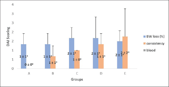
Fig. 1-rvs.jpg" alt="">
Fig. 1. Mean DAI scores of BALB/c mice after 9 days of DSS administration (p > 0.05). Groups consisted of A (control group), B (3% DSS), C (3.5% DSS), D (4% DSS), and E (4.5% DSS). Data are presented as mean ± SD (n=6 per group). The distinct letters above the bars indicate significant differences (p < 0.05). Statistical analysis was performed using one-way analysis of variance followed by LSD post hoc tests.
NEC histological score after DSS induction
The NEC histological score evaluation revealed that groups D and E (with DSS induction at 4% and 4.5%) recorded the highest NEC histological scores of ≥2. This indicates that ileal damage has occurred, marking the beginning of the NEC process (Table 2). Statistical analysis of the NEC histological score in the ileal tissue revealed a significant difference between the treatment groups (p=0.037; Fig. 2).
Table 2. Results of the NEC histological score calculation as a determinant of DSS dose.
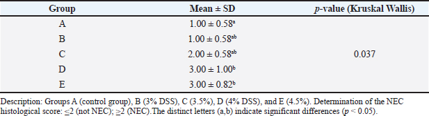
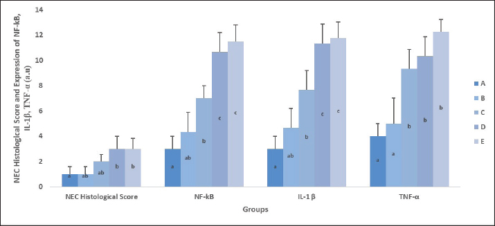
Fig. 2. Mean histological NEC scores (p=0.037), NF-кB (p=0.001), IL-1β (p=0.000), and TNF-α (p=0.000) expression in ileal tissue across treatment groups in BALB/c mice. Groups consisted of A (control group), B (3% DSS), C (3.5% DSS), D (4% DSS), and E (4.5% DSS). Bars represent mean ± SD (n=6 per group). The distinct letters above the bars indicate significant differences (p < 0.05). Statistical analysis was performed using one-way analysis of variance followed by LSD post hoc tests.
Based on histological observations, group A (control group) did not show any ileal structural changes that indicated damage. Groups B, C, D, and E showed varying degrees of ileal structural abnormalities after DSS induction at different doses. Early mucosal damage was observed in group B (treatment 1; DSS 3%), characterized by shortened villi, irregular epithelial arrangement, and activation of the local inflammatory response. The administration of 3.5% DSS in group C resulted in more pronounced mucosal damage compared with that in group B, characterized by shorter villi, a more irregular epithelial appearance, and increased inflammatory cell infiltration. Group D (DSS 4%) showed a more severe degree of mucosal damage than groups B and C. The villi appeared thinned and disconnected, and inflammatory cell infiltration extended to the submucosal layer. Degenerative changes in the epithelium were visible at high magnification, including abnormal crypt formation or crypt enlargement, as well as signs of active inflammation. Group E (DSS, 4.5%) showed the most severe damage among all the treatment groups. The villi were barely visible, indicating extensive mucosal erosion. Massive infiltration of inflammatory cells, accompanied by widening of the interstitial spaces and crypt distortion (Fig. 3).
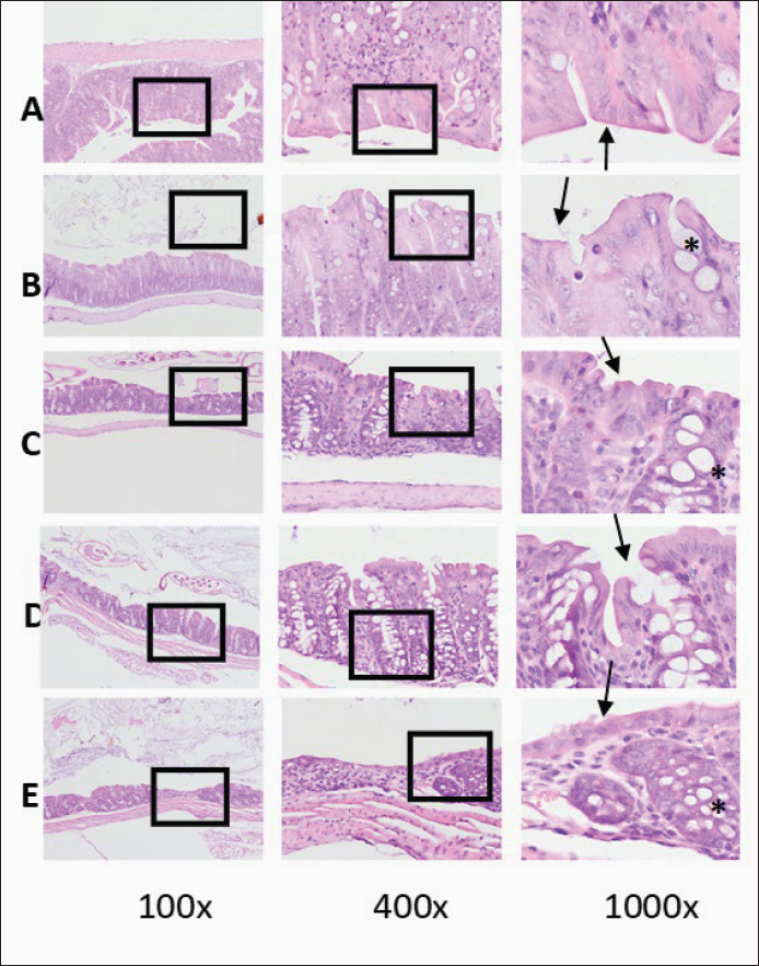
Fig. 3. Ileum tissue stained with H&E. The image shows the tissue area markers that are the focus of observation (black boxes). The structure of the crypt tissue is indicated by a black arrow, and the villi is indicated by a black star symbol. Inflammatory cells are clearly visible throughout the area, especially in groups D and E, at 1,000 magnification. Group A (control group): received only mineral water; Group B: was administered 3% (w/v) DSS; Group C: was given 3.5% (w/v) DSS; Group D: was provided 4% (w/v) DSS, and Group E: received 4.5% (w/v) DSS. DSS was administered in drinking water.
NF-κB expression in ileal tissue
After 9 days of exposure to DSS, ileal tissues were collected on day 10 and analyzed for NF-кB protein expression using immunohistochemistry and quantitative image analysis. NF-кB expression progressively increased with higher DSS concentrations. The control group (A) exhibited the lowest mean expression (3.00 ± 1.00 pmol/l), whereas the DSS-treated groups showed graded upregulation: 4.33 ± 1.53 pmol/l (3% DSS), 7.00 ± 1.00 pmol/l (3.5% DSS), 10.67 ± 1.53 pmol/l (4% DSS), and 11.50 ± 1.29 pmol/l (4.5% DSS) (Fig. 2).
Statistical analysis using one-way analysis of variance showed significant differences among groups (p=0.000). Post-hoc testing confirmed that groups D and E (4% and 4.5% DSS, respectively) had significantly higher NF-кB expression than the control (group A) and 3% DSS group (group B). In contrast, group C (3.5% DSS) displayed an intermediate but significant increase compared with group A. No significant difference was observed between groups D and E, indicating that NF-κB activation reached its maximal response at DSS concentrations of 4%.
Microscopic evaluation supported the quantitative data (Fig. 4). NF-кB staining was faint and restricted to occasional nuclei within the lamina propria in the control group. In contrast, the DSS-treated groups showed progressively stronger cytoplasmic and nuclear staining, particularly in the epithelial cells and submucosal immune populations.
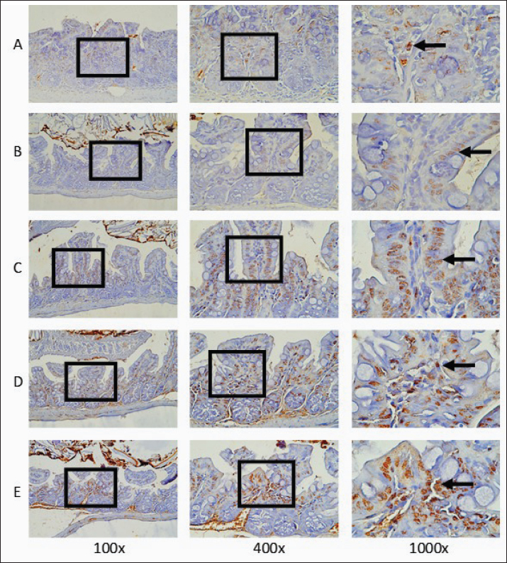
Fig. 4. Representative immunohistochemical images showing the localization of NF-кB in ileal tissue. Images were captured at 100×, 400×, and 1,000× magnification from five random fields per section. Progressive nuclear staining (black arrow) from the control group to the 4.5% DSS group demonstrates enhanced NF-кB activation and epithelial inflammatory signaling. Group A (control group), group B (3% DSS induction), group C (3.5% DSS induction), group D (4% DSS induction), and group E (4.5% DSS induction).
IL-1β expression in ileal tissue
This study measured the expression of IL-1β, a protein associated with the inflammatory process, IL-1β, in mice. The results of IL-1β expression measurements are presented in TTable 3. Group A (control group) had the lowest expression, whereas group E (4.5% DSS) had the highest expression (Figs. 2 and 5). A significant increase was seen at DSS concentrations of 3.5% compared with the control and 3% DSS groups (p=0.000).
Table 3. Mean of IL-1β expression (a.u).
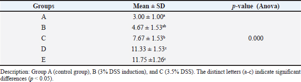
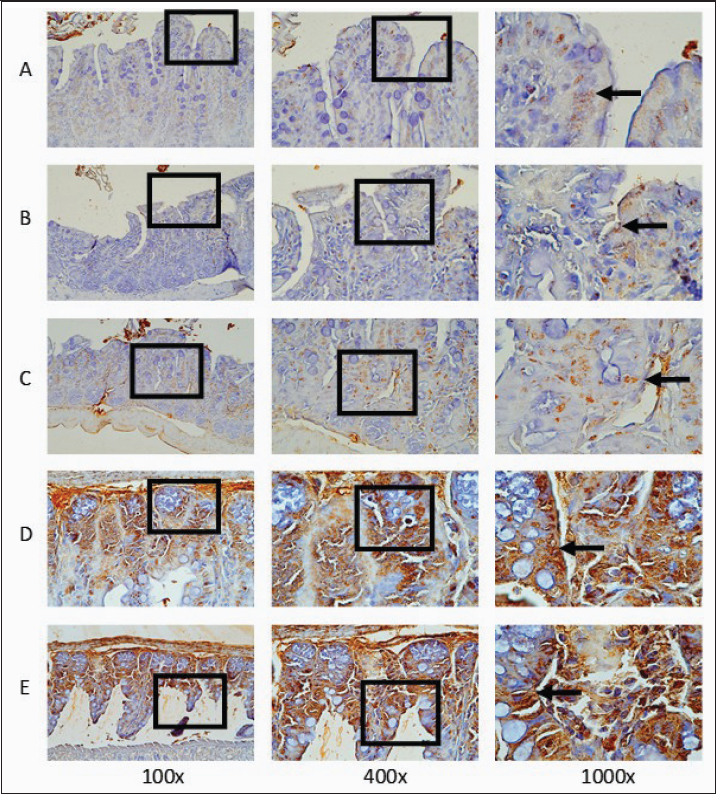
Fig. 5. Histopathological images of ileal tissue in IL-1β expression measurements, with 100, 400, and 1,000× magnification. The box in the image indicates the tissue area marker that is the focus of observation. Areas showing target protein expression in the intestinal epithelial or crypt layer. The arrow indicates the specific location of cells or structures that express the target protein (in brown). Group A (control group), group B (3% DSS induction), group C (3.5% DSS induction), group D (4% DSS induction), and group E (4.5% DSS induction).
TNF-α expression in ileal tissue
The control group (A) exhibited the lowest average level (4.00 ± 1.00 pmol/l), while the DSS-treated groups showed progressively higher mean values: 5.00 ± 1.53 pmol/l (3% DSS), 9.33 ± 1.00 pmol/l (3.5% DSS), 10.33 ± 1.53 pmol/l (4% DSS), and 12.25 ± 1.29 pmol/l (4.5% DSS) (Fig. 2). Statistical analysis using one-way analysis of variance demonstrated significant differences among groups (p=0.000). Post-hoc comparisons indicated that groups C, D, and E (3.5%–4.5% DSS) had significantly higher TNF-α expression compared with the control (A) and 3% DSS (B) groups.
In control tissues, TNF-α immunostaining appeared faint and limited to scattered macrophages and endothelial cells within the lamina propria. In contrast, DSS-treated mice exhibited strong cytoplasmic and perinuclear staining localized to epithelial cells and submucosal inflammatory infiltrates. The 4% and 4.5% DSS groups displayed the most intense staining, characterized by the accumulation of TNF-α-positive cells, epithelial erosion, and crypt destruction, which are indicative of severe mucosal inflammation (Fig. 6).
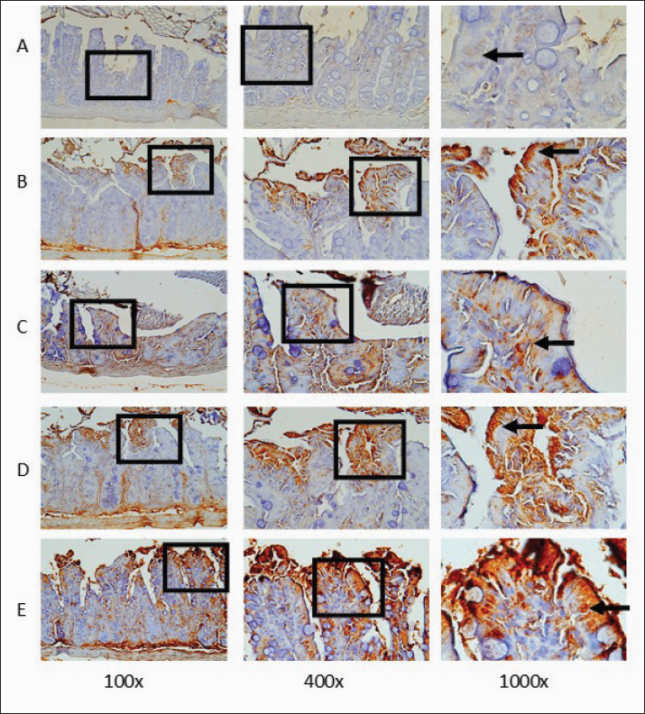
Fig. 6. Representative histological sections showing TNF-α immunostaining in the ileal tissue. Images were captured at 100×, 400×, and 1,000× magnifications from five random fields per section. The most intense TNF-α staining (black arrow) was observed in the epithelial and submucosal areas in the 4% and 4.5% DSS groups, which is consistent with pronounced mucosal inflammation and epithelial damage. Group A is the control group; Group B is treatment 1 with 3% DSS induction; Group C is treatment 2 with 3.5% DSS induction; Group D is treatment 3 with 4% DSS induction; and Group E is treatment 4 with 4.5% DSS induction.
Discussion
This study established a graded DSS-induced enterocolitis model in adult BALB/c mice to replicate intestinal injury of variable severity. Unlike neonatal models that combine hypoxia and formula feeding, this adult model provides greater experimental control and reproducibility, facilitating the study of inflammatory mechanisms in the ileum, a key site of NEC pathology. The use of adult BALB/c mice represents a limitation of this study, as the developmental stage significantly influences the host response to intestinal injury. The mechanisms driving intestinal inflammation and repair are fundamentally different between adults and neonates. Neonates exhibit a state of “physiological intestinal immaturity,” characterized by heightened TLR4 signaling, which can precipitate an exaggerated inflammatory response when combined with microbial colonization (Mihi and Good, 2019). Furthermore, the neonatal immune system is biased toward an anti-inflammatory Th2 phenotype, yet this can paradoxically lead to a deficient response to bacterial translocation, contributing to the systemic spread of inflammation (Cuenca et al., 2013).
Our study does not address these developmental differences directly. Instead, we used adult mice to establish a robust, reproducible, and dose-dependent model of NEC-like epithelial injury. This approach was intentionally chosen to optimize the DSS in a system with lower inter-individual variability, allowing for a clear assessment of histopathological and inflammatory outcomes without the confounding variables of neonatal development, such as maternal separation, litter variability, and the high fragility of neonatal pups. We acknowledge that the mechanisms we have identified, particularly the upregulation of NF-κB, IL-1β, and TNF-α, are likely more representative of a mature, adult inflammatory response. However, the core endpoint of our study is optimizing DSS concentration to achieve a standardized level of epithelial damage and inflammation, which is a prerequisite for future experiments.
The use of DSS as both an osmotic agent and epithelial barrier directly interferes with the intestinal epithelial barrier. This disruption mainly occurs due to its toxic impact on colonic and ileal epithelial cells, resulting in sterile inflammation that histologically resembles the acute inflammatory phase of NEC and effectively replicates the clinical and pathological features of NEC (Eichele and Kharbanda, 2017). Similar to the findings of Blum et al. (2024), this model allows detailed evaluation of inflammation, cytokine activation, and tissue remodeling across different DSS concentrations, representing an important refinement in NEC research methodology. The absence of significant differences in water intake among treatment groups indicates that variations in DAI values, including body weight, were primarily due to intestinal inflammation rather than dehydration or reduced fluid consumption. This finding validates the obtained clinical data and confirms that DSS exposure did not alter the hydration behavior of the animals. Consequently, the observed changes in DAI values reflect genuine disease activity rather than systemic artifacts.
Determining the optimal DSS concentration is essential for ensuring consistent disease induction while maintaining animal survival. Previous studies have reported considerable interstrain variability in DSS susceptibility. Wu et al. (2022) demonstrated that 5% DSS induced severe colitis and mortality in BALB/c mice within 7 days, whereas C57BL/6 mice developed more acute symptoms with earlier onset of weight loss onset. Lin et al. (2025) observed that DSS-induced weight loss typically depends on strain and molecular weight. In the current study, weight loss followed a similar pattern, with progressive reduction observed after day 6, peaking at 4% and 4.5% DSS exposure. The magnitude of weight loss and DAI elevation paralleled the severity of inflammation, supporting dose-dependent disease progression. Bleeding was not detected in our BALB/c cohort. Although no bleeding was detected in any group, the combination of weight loss and stool alterations reflected increased intestinal permeability and inflammatory activity with higher DSS exposure. Almutary et al. (2023) emphasized that DSS concentration directly determines the intensity of inflammation and epithelial damage. Concentrations between 3% and 4% are considered optimal for producing measurable inflammation without excessive mortality.
Consistent with the theoretical framework, the results of this study demonstrated a decline in clinical parameters in mice induced with higher DSS concentrations compared with the control group. These findings confirm that the 4% DSS concentration is the lowest level associated with the development of NEC, indicating that 4% represents the threshold for sustained inflammation with manageable systemic impact, while the 4.5% DSS concentration suggests excessive tissue damage. Higher concentrations (4.5%) resulted in excessive morbidity and mortality. In contrast, lower DSS concentrations (3% and 3.5%) did not produce the expected manifestations of NEC, either in terms of body weight loss or inflammatory protein expression in ileal tissue. Therefore, the ideal DSS dosage for inducing NEC is 4%, confirming that 4% DSS is the most appropriate dose for inducing reproducible NEC-like lesions in BALB/c mice. Earlier DSS models have primarily focused on colonic inflammation, whereas this study emphasizes ileal pathology resembling NEC. These findings indicate that 4% DSS provides an optimal balance between reproducible disease induction and animal welfare, supporting its use as the preferred concentration for the establishment of a murine NEC-like model.
By integrating clinical, cellular, and molecular parameters (DAI, NEC histological score, TNF-α, and NF-кB), the present study provides a comprehensive evaluation of disease severity. Moreover, unlike several studies reviewed by Duess et al. (2023) in the acute colitis model, our DSS model demonstrated sustained inflammation without rapid mortality, which is advantageous for studying subacute or chronic stages of NEC. Sahoo et al. (2023) also reported that DSS not only causes damage to the epithelium but also leads to systemic alterations in oxidative stress indicators, such as elevated levels of nitric oxide and reactive oxygen species, alongside a simultaneous reduction in glutathione. These oxidative and inflammatory events may synergistically intensify tissue injury (Bezerra et al., 2023), further supporting the selection of 4% DSS as the optimal concentration for studying early NEC progression.
These findings demonstrate a clear dose-dependent activation of NF-κB signaling that aligns with the observed clinical and histopathological features of intestinal injury induced by DSS. The observed increase in NF-кB activity is consistent with the activation of the TLR4– myeloid differentiation primary response 88 (MyD88)–NF-кB signaling cascade, in which DSS disrupts epithelial integrity and microbiota homeostasis, leading to the engagement of PRR. This activation stimulates the transcription of pro-inflammatory cytokines such as TNF-α and IL-1β, promoting mucosal inflammation and epithelial damage, which are characteristic of NEC-like pathology. Therefore, in this experimental model, NF-кB serves not only as a biomarker of inflammation but also as a central regulatory mediator in the molecular cascade driving intestinal necrosis and immune activation (Wu et al., 2022).
Analysis of molecular markers provided mechanistic confirmation of DSS-induced intestinal inflammation. The NF-кB-signaling pathway is central to intestinal immune activation and was markedly upregulated in this study, particularly at DSS concentrations of 4% and 4.5%. The 4% and 4.5% DSS groups exhibited intense nuclear localization, indicating NF-κB translocation and transcriptional activation, which are hallmarks of canonical inflammatory signaling. NF-кB activation occurs when DSS disrupts epithelial integrity, allowing luminal bacteria and endotoxins to penetrate the mucosa (Guo et al., 2024). These microbial components, particularly LPS from gram-negative bacteria, activate PRRs such as TLR4. The resulting TLR4–MyD88–NF-кB signaling cascade triggers the transcription of proinflammatory cytokines, including TNF-α and IL-1β (Zamyatina and Heine, 2020). Our study indicates that DSS concentrations ≥ 3.5% significantly increased IL-1β expression. However, a concentration of 3.5% is equivalent to 3%, and 3% matches the control group. Therefore, we opted for a 4% dose in this study.
The mean IL-1β expression in the control group (A) was not significantly different from that in the 3% DSS group (B). However, starting from DSS 3.5% (C) to 4.5% (E), there was a significant increase that differed significantly from the control group. This means that DSS doses ≥ 3.5% began to trigger inflammation, and doses of 4% and 4.5% significantly increased the expression of IL-1β as a marker of inflammation in ileal tissue.
In our model, both NF-кB and TNF-α expression increased, with peak activation observed at 4% DSS. TNF-α is a cytokine that amplifies intestinal inflammation and epithelial injury. To evaluate the downstream effects of NF-кB activation on proinflammatory signaling, the expression of TNF-α was examined in the ileal tissues of mice exposed to different concentrations of DSS. These findings are consistent with those of Liu et al. (2022), who reported that NF-кB overactivation amplifies inflammatory signaling and epithelial apoptosis. Excessive NF-кB stimulation contributes to the sustained release of TNF-α, leading to epithelial barrier breakdown, vascular leakage, and leukocyte infiltration (Ng et al., 2025), which are pathological hallmarks of NEC-like lesions. TNF-α acts as a downstream effector of NF-кB and a pivotal mediator of mucosal injury (Peng et al., 2020). Elevated TNF-α expression in our study coincided with histological evidence of epithelial erosion, crypt destruction, and submucosal immune infiltration (Salem et al., 2024). While TNF-α is essential for normal host defense, sustained overexpression intensifies mucosal damage through caspase activation and intestinal epithelial cell apoptosis (Subramanian et al., 2020). The concurrent upregulation of TNF-α and NF-кB observed in our 4% DSS group supports their interdependent roles in inflammation. NF-кB directly regulates TNF-α transcription, while TNF-α in turn activates NF-кB through a positive feedback loop, perpetuating epithelial damage and inflammation (Guo et al., 2024). NF-кB translocation and transcriptional activation are hallmarks of canonical inflammatory signaling. These findings demonstrate a clear dose-dependent activation of NF-кB signaling that aligns with the clinical and histopathological features of DSS-induced intestinal injury.
In our study, no significant differences were observed among groups C, D, and E, suggesting that TNF-α upregulation reached a plateau at a concentrations of 3.5% DSS. These findings indicate that DSS exposure beyond this level activates the TNF-α-mediated inflammatory response in the ileal mucosa. DSS administration induces a robust proinflammatory response mediated by TNF-α, closely associated with increased DAI scores and histopathological damage. The concurrent elevation of TNF-α and NF-κB expression supports their synergistic role in amplifying the inflammatory cascade, contributing to epithelial barrier disruption and NEC-like pathology.
Our findings of elevated NF-κB, IL-1β, and TNF-α are consistent with the well-established central role of this signaling axis in NEC. However, NEC pathogenesis involves a more intricate network of inflammatory pathways. In many experimental models, the TLR4/MyD88 pathway is the initial trigger for this cascade. The engagement of TLR4 by LPS from translocating gut bacteria, initiates a downstream signaling cascade that culminates in the activation of NF-κB (Nighot et al., 2017; Aucoin et al., 2026). Therefore, the elevated NF-κB activity we observed, induced by DSS, is is likely a direct consequence of upstream TLR4/MyD88 activation.
Furthermore, the maturation and activation of IL-1β and IL-18 are tightly regulated by the NOD-, LRR-, and pyrin domain-containing protein 3 (NLRP3) inflammasome. This multiprotein complex serves as an essential sensor for cellular stress and damage, particularly in response to DSS-induced damage. Upon activation, NLRP3 triggers caspase-1 cleavage, which in turn processes pro-IL-1β and pro-IL-18 into their active, secreted forms (Fusco et al., 2020). The significant upregulation of IL-1β observed in our 4% and 4.5% DSS groups strongly suggests the concurrent activation of the NLRP3 inflammasome, although it was not directly measured. DSS-induced colitis models are highly dependent on NLRP3 inflammasome signaling for the production of IL-1β and the subsequent recruitment of neutrophils to the injury site (Zhen and Zhang, 2019).
Finally, the histological injury, marked by epithelial erosion, villus blunting, and crypt damage, is the result of extensive programmed cell death or apoptosis. The executioner caspase, caspase-3, plays a nonredundant role in this process. The activation of the TLR4/MyD88 pathway can directly induce intestinal epithelial cell apoptosis via caspase activation (Fusco et al., 2020; Duan et al., 2022). Similarly, the NLRP3 inflammasome, in addition to its role in cytokine secretion, can also contribute to a form of inflammatory cell death known as pyroptosis, which is mediated by gasdermin D and is distinct from, but often coincident with, caspase-3-driven apoptosis (Gong et al., 2025). Severe histological damage in the 4% and 4.5% DSS groups implies a high rate of epithelial cell death, likely involving both caspase-3-dependent apoptosis and gasdermin-D-dependent pyroptosis. Future studies using this optimized model will incorporate immunohistochemistry for TLR4, MyD88, NLRP3, and cleaved caspase-3 to fully delineate these interconnected pathways.
In summary, the current study demonstrates that 4% DSS administered for 9 days effectively induces reproducible NEC-like lesions in mice. This concentration optimally triggers NF-кB and TNF-α activation without excessive mortality, providing a robust and ethically viable model for investigating NEC pathogenesis and potential therapeutic interventions. Nevertheless, the BALB/c model remains valuable because its moderate response enables longer observation of disease development and recovery, especially in studies investigating immune modulation or therapeutic interventions. To further elucidate the inflammatory cascade, future studies should integrate additional molecular readouts, including TLR4, MyD88, and NLRP3 inflammasome markers. Longitudinal experiments assessing oxidative stress and epithelial regeneration would also clarify how DSS-induced inflammation transitions from the acute to chronic stages.
Conclusion
This study successfully developed a mouse model of NEC using DSS. The 4% DSS concentration is the most effective dose because it produces consistent clinical symptoms, histological damage, and increased inflammatory mediators in the ileal tissue. This model provides a clearer standard method for NEC studies in adult mice and allows for the regulation of inflammatory severity.
Acknowledgments
The author is grateful to Wibi Riawan and Yohana Hartya Dwi Frastari for their valuable assistance in research.
Conflict of interest
The authors declare no conflicts of interest related to this study.
Funding
This research was funded by the Faculty of Medicine, Universitas Brawijaya (grant number 7169/7/UN10.F0701/B/PT.01.05.1/2025).
Authors’ contributions
E.S. conceptualized the study, designed the experimental framework, conducted the research, and drafted the manuscript. L.E.F., K.S., and H.K. were responsible for data curation, validation, and supervision. L.E.F. also performed critical editing and an extensive review of the manuscript. A.H. contributed to data interpretation, critical discussion, and manuscript revision. All authors approved the final version of the manuscript.
Data availability
All data were provided in the manuscript.
References
Adamkova, P., Hradicka, P., Kupcova Skalnikova, H., Cizkova, V., Vodicka, P., Farkasova Iannaccone, S., Kassayova, M., Gancarcikova, S. and Demeckova, V. 2022. Dextran sulfate sodium acute colitis rat model: a suitable tool for advancing our understanding of immune and microbial mechanisms in the pathogenesis of inflammatory bowel disease. Vet. Sci. 9(5), 1–23.
Alganabi, M., Lee, C., Bindi, E., Li, B. and Pierro, A. 2019. Recent advances in understanding necrotizing enterocolitis version 1; referees: 2 approved. F1000Research 8(0), F1–F8.
Almutary, A.G., Alnuqaydan, A.M., Almatroodi, S.A. and Tambuwala. 2023. Comparative analysis of the effect of different concentrations of dextran sodium sulfate on the severity and extent of inflammation in experimental ulcerative colitis. Appl. Sci. 13(5), 3233.
Alqahtani, A. 2024. Necrotizing enterocolitis among very low birthweight neonates: a systematic review and meta-analysis. Egypt. J. Chem. 67(13), 817–823.
Alsaied, A., Islam, N. and Thalib, L. 2020. Global incidence of necrotizing enterocolitis: a systematic review and meta-analysis. BMC Pediatrics. 20(1), 344.
Aucoin, M., Servantes, B., Saroukhani, S., Alcorn, J.L., Chang, M.L., Findley, T.O. and Koochak, H. 2026. Blood TLR4, MD2 and MYD88 elevation predicts necrotizing enterocolitis: evidence from human patients and a mouse model. AJMS 371, S384–S385.
Bezerra, F.S., Lanzetti, M., Nesi, R.T., Nagato, A.C., Silva, C.P.E., Kennedy-Feitosa, E., Melo, A.C., Cattani-Cavalieri, I., Porto, L.C. and Valenca, S.S. 2023. Oxidative stress and inflammation in acute and chronic lung injuries. Antioxidants 12(3), 548.
Blum, L., Vincent, D., Boettcher, M. and Knopf, J. 2024. Immunological aspects of necrotizing enterocolitis models: a review. Front. Immunol. 15, 1434281.
Chen, S.Y., Zhou, Q.Y.J., Chen, L., Liao, X., Li, R. and Xie, T. 2022. The aurantii fructus immaturus flavonoid extract alleviates inflammation and modulate gut microbiota in dss-induced colitis mice. Front. Nutr. 9, 1–10.
Cho, S.X., Berger, P.J., Nold-Petry, C.A. and Nold, M.F. 2016. The immunological landscape in necrotising enterocolitis. Expert Rev. Mol. Med. 18, 1–17.
Chougule, P.R., Sangaraju, R., Patil, P.B., Qadri, S.S.Y.H., Panpatil, V.V., Ghosh, S., Mungamuri, S.K., Bhanoori, M. and Sinha, S.N. 2023. Effect of ethyl gallate and propyl gallate on dextran sulfate sodium (DSS)-induced ulcerative colitis in c57bl/6 j mice: preventive and protective. Inflammopharmacology 31(4), 2103–2120.
Cuenca, A.G., Wynn, J.L., Moldawer, L.L. and Levy, O. 2013. Role of innate immunity in neonatal infection. Am. J. Perinatol. 30(2), 105.
Duan, T., Du, Y., Xing, C., Wang, H.Y. and Wang, R.F. 2022. Toll-like receptor signaling and its role in cell mediated immunity. Front. Immunol. 13, 812774.
Duess, J.W., Sampah, M.E., Lopez, C.M., Tsuboi, K., Scheese, D.J., Sodhi, C.P. and Hackam, D.J. 2023. Necrotizing enterocolitis, gut microbes, and sepsis. Gut Microbes 15(1), 2221470.
Eichele, D.D. and Kharbanda, K.K. 2017. Dextran sodium sulfate colitis murine model: an indispensable tool for advancing our understanding of inflammatory bowel diseases pathogenesis. World J. Gastroenterol. 23(33), 6016.
Falo, C., Benitez, R., Caro, M., Morell, M., Forte-Lago, I., Hernandez-Cortes, P., Sanchez-Gonzalez, C., O’Valle, F., Delgado, M. and Gonzalez-Rey, E. 2021. The neuropeptide cortistatin alleviates neuropathic pain in experimental models of peripheral nerve injury. Pharmaceutics 13(7), 947.
Fusco, R., Siracusa, R., Genovese, T., Cuzzocrea, S. and Di Paola, R. 2020. Focus on the role of NLRP3 inflammasome in diseases. Int. J. Mol. Sci. 21(12), 4223.
Garcia-Carbonell, R., Yao, S.J., Das, S. and Guma, M. 2019. Dysregulation of intestinal epithelial cell RIPK pathways promotes chronic inflammation in the IBD gut. Front. Immunol. 10, 1094.
Glasser, J.G. 2021. Abdominal compartment syndrome complicating necrotizing enterocolitis: a case report. Ann. Med. Surg. 71, 102961.
Golubkova, A. and Hunter, C.J. 2023. Development of the neonatal intestinal barrier, microbiome, and susceptibility to NEC. Microorganisms 11(5), 1247.
Gomart, A., Vallée, A. and Lecarpentier, Y. 2021. Necrotizing enterocolitis: lPS/TLR4-induced crosstalk between canonical TGF-β/Wnt/β-Catenin pathways and PPARγ. Front. Pediatr. 9, 713344.
Gong, L.L. Zhou, H.J., Zhao, Q.M., Xu, N., Huang, F.C., Su, L.Y. and Li, W.L. 2025. Molecular mechanism of NLRP3 inflammasome in inflammatory diseases and tumors. Immun. Inflamm. Dis. 13(7), e70213.
Guo, Q.Q., Jin, Y., Chen, X., Ye, X., Shen, X., Lin, M., Zeng, C., Zhou, T. and Zhang, J. 2024. NF-κB in biology and targeted therapy: new insights and translational implications. Signal. Transduct. Target. Ther. 9(1), 53.
Isaac, U.E. Oyo-Ita, E., Igwe, N.P. and Ije, E.L. 2023. Preparation of histology slides and photomicrographs: indispensable techniques in anatomic education. Anat. J. Afr. 12(1), 2252–2262.
Kaminsky, L.W., Al-Sadi, R. and Ma, T.Y. 2021. IL-1β and the intestinal epithelial tight junction barrier. Front. Immunol. 12, 767456.
Kapoor, G., Prakash, S., Jaiswal, V. and Singh, A.K. 2025. Chronic inflammation and cancer: key pathways and targeted therapies. Cancer Invest. 43(1), 1–23.
Kefale, D., Asferie, W.N., Aytenew, T.M., Masresha, G.A., Wondim, M., Tassew, M.A., Abie, M., Setargew, K.H., Eshetie, Y. and Kebede, S.D. 2025. Burden and its associated factors of neonatal necrotizing enterocolitis among neonates admitted in neonatal intensive care unit in Ethiopia. A systematic review and meta-analysis. Int. J. Afr. Nurs. Sci. 23, 100899.
Li, D. and Wu, M. 2021. Pattern recognition receptors in health and disease. Signal. Transduct. Target. Ther. 6(1), 1–24.
Lin, Y.C., Pan, S.M., Chang, F.Y. and Yang, F.Y. 2025. Transcranial ultrasound stimulation ameliorates dextran sulfate sodium-induced colitis and behavioral disorders by suppressing the inflammatory response in the brain. Brain. Commun. 7, 1–13.
Liu, D., Zhong, Z. and Karin, M. 2022. NF-κB: a double-edged sword controlling inflammation. Biomedicines 10(6), 1250.
Mendez, Y.S., Khan, F.A., Perrier, G.V. and Radulescu, A. 2020. Animal models of necrotizing enterocolitis. World. J. Pediatr. Surg. 3(1), 1–8.
Mihi, B. and Good, M. 2019. Impact of toll-like receptor 4 signaling in necrotizing enterocolitis: the state of the science. Clin. Perinatol. 46(1), 145–157.
Moak, R., Boone, N., Eidson, N., Rohrer, A., Engevik, M., Williams, K. and Chetta, K. 2023. Exploring the links between necrotizing enterocolitis and cow’s milk protein allergy in preterm infants: a narrative review. Front. Pediatr. 1, 1274146.
Ng, C.T., Fong, L.Y., Tan, J.J. and Abdullah, M.N.H. 2025. Endothelial barrier disruptive effect of IFN-ɣ and TNF-α: synergism of pro-inflammatory cytokines. Cytokine 190, 156922.
Nighot, M., Al-Sadi, R., Guo, S., Rawat, M., Nighot, P., Watterson, M.D. and Ma, T.Y. 2017. Lipopolysaccharide-induced increase in intestinal epithelial tight permeability is mediated by toll-like receptor 4/myeloid differentiation primary response 88 (Myd88) activation of myosin light chain kinase expression. Am. J. Pathol. 187, 2698.
Panpetch, W., Hiengrach, P., Nilgate, S., Tumwasorn, S., Somboonna, N., Wilantho, A., Chatthanathon, P., Prueksapanich, P. and Leelahavanichkul, A. 2020. Additional Candida albicans administration enhances the severity of dextran sulfate solution induced colitis mouse model through leaky gut-enhanced systemic inflammation and gut-dysbiosis but attenuated by Lactobacillus rhamnosus L34. Gut Microbes 11(3), 465–480.
Peng, C., Ouyang, Y., Lu, N. and Li, N. 2020. The NF-κB. 2020. Signaling pathway, the microbiota, and gastrointestinal tumorigenesis: recent advances. Front. Immunol. 11, 1387.
Sahoo, D.K., Heilmann, R.M., Paital, B., Patel, A., Yadav, V.K., Wong, D. and Jergens, A.E. 2023. Oxidative stress, hormones, and effects of natural antioxidants on intestinal inflammation in inflammatory bowel disease. Front. Endocrinol. 14, 1217165.
Salem, M.B., El-Lakkany, N.M., Seif El-din, S.H., Hammam, O.A. and Samir, S. 2024. Diosmin alleviates ulcerative colitis in mice by increasing Akkermansia muciniphila abundance, improving intestinal barrier function, and modulating the NF-κB and Nrf2 pathways. Heliyon 10(6), e27527.
Shu, L.Z., Ding, Y.D., Xue, Q.M., Cai, W. and Deng, H. 2023. Direct and indirect effects of pathogenic bacteria on the integrity of intestinal barrier. Therap. Adv. Gastroenterol. 16, 1–17.
Subramanian, S., Geng, H. and Tan, X.D. 2020. Cell death of intestinal epithelial cells in intestinal diseases. Acta. Physiologica. Sinica. 72(3), 308–324.
Tatiya-Aphiradee, N., Chatuphonprasert, W. and Jarukamjorn, K. 2020. Oxidative stress exacerbates dextran sulfate sodium-induced ulcerative colitis in ICR mice. Biologia 75(11), 2063–2071.
Virgilio, T., Latino, I., Pizzichetti, C., Chahine, K., Capucetti, A., Detotto, C., Bergadano, A. and F Gonzalez, S. 2025. Impact of prolonged isoflurane or ketamine-xylazine anesthesia with or without buprenorphine and oxygen on mouse vitals and immune responses. Lab. Anim. (NY). 54(10), 270–277.
Wu, X. Shu, F., Jiang, M., Wang, J., Tang, H., Fang, C., Li, W. and Fu, C. 2022. Sanhuang xiexin decoction ameliorates DSS-induced colitis in mice by regulating intestinal inflammation, intestinal barrier, and intestinal flora. J. Ethnopharmacol. 297, 115537.
Xiong, T., Maheshwari, A., Neu, J., Ei-Saie, A. and Pammi, M. 2020. An overview of systematic reviews of randomized-controlled trials for preventing necrotizing enterocolitis in preterm infants. Neonatology 117(1), 46–56.
Xiong, X., Wu, L., Liu, X., Wang, J., Xiao, J., Chen, K., Zhuansun, D., Meng, X., Feng, J. and Chen, X. 2025. Comprehensive characterization of the oxidative stress profiles in neonatal necrotizing enterocolitis. Int. J. Med. Sci. 22(9), 2139–2154.
Yang, C. and Merlin, D. 2024. Unveiling colitis: a journey through the dextran sodium sulfate-induced model. Inflamm. Bowel Dis. 30, 844–853.
Zamyatina, A. and Heine, H. 2020. Lipopolysaccharide recognition at the crossroads of TLR4 and caspase-4/11 mediated inflammatory pathways. Front. Immunol. 12, 649442.
Zhang, X. and Hartmann, P. 2023. How to calculate sample size in animal and human studies. Front. Med. 10, 1215927.
Zhen, Y. and Zhang, H. 2019. NLRP3 Inflammasome and inflammatory bowel disease. Front. immunol. 10, 276
Description: Groups A (control group), B (3% DSS), C (3.5%), D (4% DSS), and E (4.5%). Determination of the NEC histological score: ≤2 (not NEC); ≥2 (NEC).The distinct letters (a,b) indicate significant differences (
p < 0.05).
 Fig. 2-rvs.jpg" alt="">
Fig. 3-rvs.jpg" alt="">
Fig. 4-rvs.png" alt="">
Description: Group A (control group), B (3% DSS induction), and C (3.5% DSS). The distinct letters (a-c) indicate significant differences (
p < 0.05).

Fig. 6-rvs.png" alt="">








