





| Research Article | ||
Open Vet. J.. 2026; 16(7): 4746-4759
Open Veterinary Journal, (2026), Vol. 16(7): 4746-4759 Research Article Erythrocyte and platelet indices in response to alternative topical approaches to surgically induced wounds: An experimental study in ratsNadža Kapo-Dolan1*, Nedim Čović2 and Muhamed Katica11Department of Clinical Veterinary Sciences, Veterinary Faculty, University of Sarajevo, Sarajevo, Bosnia and Herzegovina 2Department of Transformational Processes in Sport, Faculty of Sport and Physical Education, University of Sarajevo, Sarajevo, Bosnia and Herzegovina *Corresponding Author: Nadža Kapo-Dolan. Department of Clinical Veterinary Sciences, Veterinary Faculty, University of Sarajevo, Sarajevo, Bosnia and Herzegovina. Email: nadzakapo [at] gmail.com Submitted: 08/01/2026 Revised: 27/05/2026 Accepted: 08/06/2026 Published: 20/07/2026 © 2025 Open Veterinary Journal
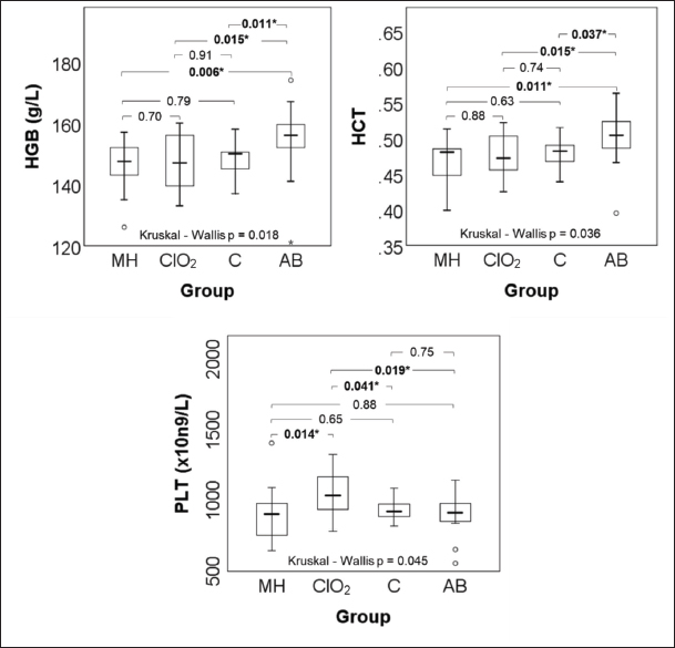
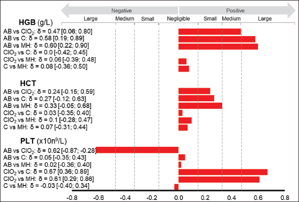
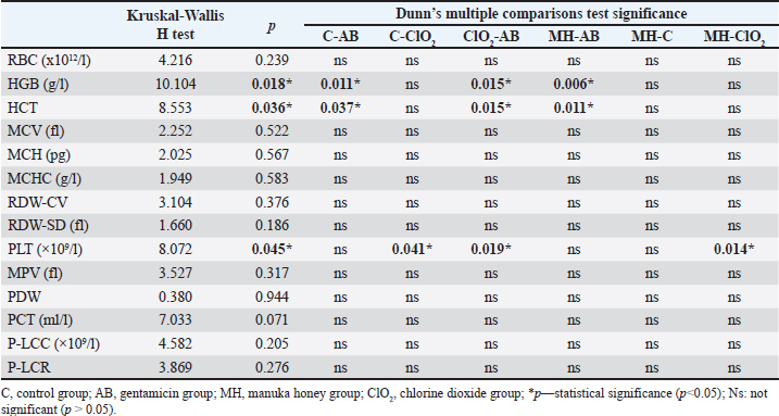
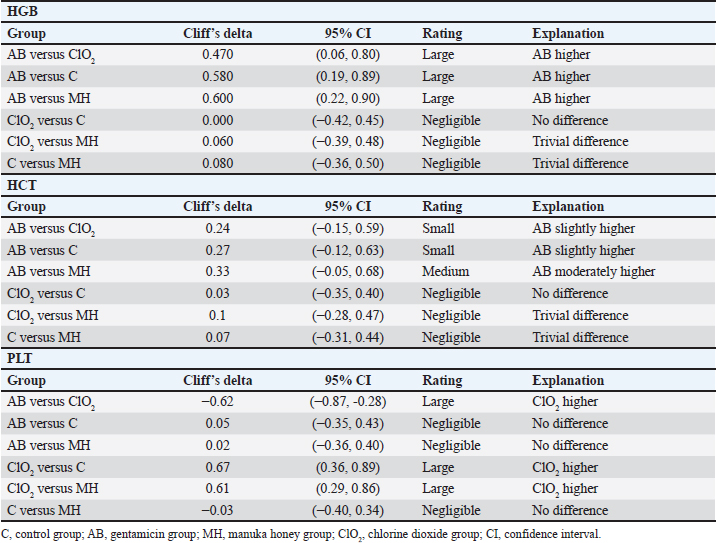
ABSTRACTBackground: Wound healing is a complex physiological process that involves hemostasis, inflammation, proliferation, and remodeling. Erythrocyte and platelet parameters, along with erythrocyte morphology, including poikilocytosis, are valuable indicators of systemic responses to injury and therapeutic interventions. Manuka honey and chlorine dioxide (ClO2) are known for their antimicrobial and reparative properties; however, their systemic hematological effects following topical application remain poorly characterized. Aim: This study aimed to evaluate systemic hematological responses in Wistar rats with surgically induced wounds following the topical application of manuka honey and ClO2, compared with gentamicin treatment and untreated controls, focusing on erythrocyte and platelet parameters and poikilocytosis. Methods: Sixty Wistar rats were allocated into four groups: control (C, n=15), gentamicin (AB, n=15), manuka honey (MH, n=14), and chlorine dioxide (ClO2, n=15). Under full anesthesia, a 4-cm abdominal incision was surgically induced in all rats, followed by topical treatment twice daily for 14 days. On day 14, peripheral blood samples were collected for hematological analysis, including red blood cell count, hemoglobin concentration (HGB), hematocrit (HCT), platelet count (PLT), plateletcrit, and poikilocytosis. Statistical comparisons were performed using the Kruskal–Wallis test; significance was set at p < 0.05. Results: Most hematological parameters remained within the expected physiological ranges. Significant intergroup differences were observed for HGB, HCT, and PLT. Post hoc Dunn comparisons showed that HGB was significantly higher in the AB group than in the C (p=0.011), ClO2 (p=0.015), and MH (p=0.006) groups. HCT was significantly higher in the AB group than in the C (p=0.037), ClO2 (p=0.015), and MH (p=0.011) groups. PLT was significantly higher in the ClO2 group than in the C (p=0.041), AB (p=0.019), and MH (p=0.014) groups. Poikilocytosis was minimal in all groups, primarily absent to mild, with the most frequent forms being anulocytes and stomatocytes. Conclusion: At the evaluated endpoint, the topical application of manuka honey and ClO2 did not result in marked systemic hematological disturbances in Wistar rats with surgical wounds. ClO2 had the most pronounced effect on platelet parameters among the tested treatments. Minimal poikilocytosis further supports the relative hematological stability observed at the evaluated endpoint. These findings provide valuable insights into systemic responses to topical treatments applied to surgically induced wounds and support further studies incorporating leukocyte profiling and complementary hematological, immunological, and histopathological assessments. Keywords: Chlorine dioxide, Hematology, Manuka honey, Wound. IntroductionWounds are injuries caused by various mechanical factors, such as trauma, laceration, incision, or contusion (Ranjani et al., 2010). Their healing is a complex and precisely coordinated physiological process that restores tissue integrity and enables the repair of damaged structures (Hanks and Spodnick, 2005; Ranjani et al., 2010). This biologically complex cascade involves hundreds of interconnected events, from wound formation to complete closure (Goldman, 2004; Öztürk and Ermertcan, 2011), and is classically described through four phases: hemostasis, inflammation, proliferation, and remodeling (Hanks and Spodnick, 2005; Araújo et al., 2010). Hematological parameters provide essential information about the body’s response to injury, deprivation, and/or stress (Ihedioha et al., 2004), making them important indicators of both physiological and pathological status in humans and animals (Adeneye et al., 2006; Kapo-Dolan et al., 2025). Special importance is also attributed to poikilocytosis, since abnormal erythrocyte morphology is often associated with specific diseases, thereby facilitating the monitoring of pathogenesis, diagnosis, and treatment (Christopher et al., 2014). Poikilocytes may arise due to biochemical alterations, toxins, or mechanical damage to erythrocytes, and regardless of the cause, they may reduce erythrocyte survival and contribute to anemia (Harvey, 2012; Marks, 2013). Antimicrobial resistance (AMR) represents a global health problem and a significant economic burden (Catalano et al., 2022; Mba et al., 2022). Although AMR has historically received greater emphasis in human medicine, increasing attention has recently been directed toward its importance in veterinary medicine and animal health (Vercelli et al., 2022; Zhao et al., 2025). This fact further emphasizes the urgent need for the development of alternative therapeutic approaches in wound management (Mba et al., 2022). Manuka honey is a monofloral honey produced exclusively from the nectar of the Leptospermum scoparium tree, native to New Zealand and southeastern Australia (El-Senduny et al., 2021; Wang et al., 2024). Its effectiveness is primarily attributed to methylglyoxal (MGO), which exhibits strong antimicrobial activity, including activity against antibiotic-resistant strains (Saikaly and Khachemoune, 2017; Eroglu et al., 2018; Repellin et al., 2021). In addition to its antimicrobial effects, manuka honey also exhibits antioxidant, anticancer, immunomodulatory, anti-inflammatory, and reparative properties, making it a suitable agent in wound therapy (Wang et al., 2024). Recent studies suggest that honey-based dressings may improve selected outcomes in chronic wound management, particularly wound-healing time and percentage wound closure, although results remain heterogeneous across studies (Tang et al., 2024). Alongside manuka honey, the disinfectant chlorine dioxide (ClO2) has also garnered significant attention as a strong and broad-spectrum antimicrobial agent effective against bacteria, fungi, and viruses (Kapo-Dolan et al., 2025). It is characterized by high oxidative capacity, pronounced biocidal activity, good biocompatibility, and is less expensive than manuka honey, making it a viable candidate for wound therapy (Dudek-Wicher et al., 2025). While ClO2 solution has demonstrated antimicrobial efficacy, its potential therapeutic use should be interpreted with caution because safety considerations may vary depending on the formulation and concentration applied (Ma et al., 2017; Dudek-Wicher et al., 2025). In the present study, manuka honey and ClO2 were selected because both represent non-conventional topical agents with documented antimicrobial activity and potential applicability in wound management (Wang et al., 2024; Dudek-Wicher et al., 2025). Although their mechanisms of action differ, they have attracted interest as possible alternatives or adjuncts to conventional topical antimicrobial therapy. Although the therapeutic potential of manuka honey and ClO2 has been well established, most previous studies have examined their effects only through oral administration (Aliyu et al., 2012; Ahmed et al., 2017; Nassar et al., 2020; Suartha et al., 2022; Touzani et al., 2022; Chinko et al., 2023; Umogbai et al., 2023; Hussein and Kadhem, 2025), and the reported systemic hematological alterations reflect this route of exposure. However, information regarding hematological alterations after topical application on wounds—particularly in experimental models—is limited. Preliminary data from a small experimental study suggest that short-term topical treatment with manuka honey and ClO2 may induce mild changes in red blood cell (RBC) and platelet parameters (Kapo-Dolan et al., 2025). These observations highlight the need for comprehensive investigations to better understand the potential systemic effects of topical wound therapies, which are increasingly used in both clinical and experimental settings. This study aimed to comprehensively evaluate systemic hematological responses in Wistar rats with surgically induced wounds, wherein the incision was performed under full anesthesia, following the topical application of ClO2 and manuka honey, and to comparatively analyze the effects of these treatments against a non-treated control group. In this way, the study sought to provide new insights into the potential systemic implications of topical application of antimicrobial and biologically active agents, thereby addressing the existing gap in the literature on hematological responses to topical wound therapy. Materials and MethodsAnimal modelThe study included 60 clinically healthy adult Wistar rats of both sexes, aged 2–3 months, with a body weight range of 180–400 g, which had not been previously used in experimental procedures. The rats had free access to food and water during the experiment, and 12-hour rotations of light and dark. The ambient temperature was maintained between 20°C and 23°C and humidity 60% ± 10% (Katica and Gradaščević, 2017). General experimental procedure and study groupsThe 60 rats were allocated into four groups based on the treatment administered. Group allocation was performed manually before the start of the experimental procedures, with the aim of achieving comparable group sizes and distributing male and female rats as evenly as possible across groups. No computer-generated randomization sequence, randomization software, or randomization seed was used. No formal allocation concealment was performed. All animals were housed and maintained under identical environmental conditions to minimize potential confounding. Each experimental group consisted of 15 animals (total n=60). No predefined criteria for animal exclusion or data points were established before the study. During the experimental period, one animal from the MH group died before blood sampling due to causes unrelated to the experimental treatment and was, therefore, excluded from the final hematological analysis. The final number of animals included in the statistical analysis was n=14 for the MH group, whereas the remaining groups each included 15 animals. The first group C served as the surgical control and received no topical treatment. The second group AB was treated with gentamicin ointment (Bosnalijek 1 mg/g, Bosnia and Herzegovina). The third group, MH, received manuka honey MGO 550+ (Manuka Health, Te Awamutu, New Zealand; Batch No: OMH030728), and the fourth group, ClO2, was treated with ClO2 (120 ppm, ITR d.o.o., Bosnia and Herzegovina). Study designSurgical procedure and treatment administrationBefore the surgical intervention, all the test animals were acclimatized to the experimental conditions. General anesthesia was induced by intramuscular injection of 5 mg/kg xylazine hydrochloride 2% (2% Xylazin, CpPharma, Bergdorf, Germany) and 60 mg/kg ketamine hydrochloride (International B.V., Netherlands). Once surgical anesthesia was confirmed, the rats’ abdominal hair was carefully removed to ensure clear visibility and access to the surgical site. Under full anesthesia, a 4-cm longitudinal incision was made through skin and subcutaneous tissue along the median abdominal line, and the wounds were left open. After 24 hours from incision, each animal received its designated topically administered treatment, which was administered twice daily for 14 consecutive days. Manuka honey was applied in a small amount sufficient to cover the entire wound surface with a thin layer and was gently spread using sterile gloves. The exact amount was not weighed during the experiment; however, the applied amount was estimated to be approximately 1–2 ml per wound per application. According to the product labeling, the MGO 550+ rating corresponds to approximately 550 mg/kg of MGO. Chlorine dioxide solution was applied using a spray applicator, with two sprays administered per application to ensure wound surface coverage. The exact delivered volume per spray was not measured during the experiment. Gentamicin ointment was applied in a thin layer to cover the wound surface. The exact amount was not weighed during the experiment; approximately 0.1–0.2 g per wound per application was used. According to the manufacturer, the gentamicin formulation contains paraffin oil, white petrolatum, methylparahydroxybenzoate (E218), and propylparahydroxybenzoate (E216) as excipients. No occlusive dressing or bandaging was used following treatment application due to the behavioral tendency of rats to remove such materials immediately. Blinding during treatment administration and daily animal monitoring were not feasible because the appearance, consistency, and mode of application of the topical interventions differed visibly. Therefore, the personnel involved in treatment administration and animal monitoring were aware of group allocation. Blood smear evaluation for poikilocytosis was not performed under blinded conditions. Animals were monitored continuously during anesthesia and throughout the recovery period until full awakening. Animals were observed at least once daily during the experimental period to assess general health status, behavior, and wound condition. Welfare monitoring included feeding and drinking behavior, posture, locomotor activity, social interaction, vocalization, wound appearance, and potential signs of pain, discomfort, or distress. No formal pain scoring system was applied; however, the animals were clinically monitored throughout the study for signs indicative of pain or compromised welfare. Given the minor nature of the surgical incision and the analgesic effect of the ketamine-xylazine anesthetic protocol, no additional postoperative analgesia was administered. Humane endpoints included severe or persistent distress, marked reduction in food or water intake, impaired locomotion, and wound complications requiring intervention. If any of these endpoints had been reached, the affected animal would have been removed from the experiment and provided with appropriate veterinary care. Hematological proceduresOn day 14 of the experiment, peripheral blood samples were taken by tail vein puncture in ethylenediaminetetraacetic acid vacutainers with a volume of 3 ml. The puncture site had been previously disinfected using standard disinfectants. Hematological parameters were analyzed using a Mindray BC-20S automated hematology analyzer. The following 14 parameters were determined: (RBC, ×10¹²/l), hemoglobin concentration (HGB, g/l), hematocrit (HCT, L/l), mean corpuscular volume (MCV, fl), mean corpuscular hemoglobin (MCH, pg), mean corpuscular hemoglobin concentration (MCHC, g/l), red cell distribution width–coefficient of variation (RDW-CV) and standard deviation (RDW-SD, fl), platelet count (PLT, ×109/l), mean platelet volume (MPV, fl), platelet distribution width (PDW), plateletcrit (PCT, ml/l), platelet-large cell count (P-LCC, ×109/l), and platelet-large cell ratio (P-LCR). Following the hematological evaluation, all rats were humanely euthanized via intraperitoneal injection of ketamine (200 mg/kg BW) and xylazine (20 mg/kg BW), in accordance with the AVMA Guidelines for the Euthanasia of Animals (Underwood and Anthony, 2020). Quantification of poikilocytotic RBCBlood smears were prepared from the same blood samples used for hematological analysis. The smears were air-dried and stained according to standard laboratory practice using the Diff-Quick staining method. Poikilocytes were evaluated semiquantitatively (Christopher et al., 2014). For each stained smear, 2,000 erythrocytes were manually counted and morphologically characterized under a microscope at 1,000 × magnification. Poikilocytes were identified based on standard morphological criteria and counted in representative single-layer fields where approximately half of the erythrocytes were in contact but not overlapping. The number and type of poikilocytes were recorded and expressed as a percentage of the total erythrocyte count. Poikilocytosis was classified as follows: absent (0%), rare (0.05%–0.5%), mild (>0.5%–3%), moderate (>3%–10%), and pronounced (>10%). Statistical analysisAll statistical analyses were conducted to evaluate differences in erythrocyte and platelet indices between the four experimental groups: MH (n=14), ClO2 (n=15), AB (n=15), and C (n=15). Data are presented as mean ± standard deviation or median ± interquartile range. As the data did not meet the criteria for normality of data distribution using the Kolmogorov–Smirnov test, nonparametric methods were applied. Overall group comparisons were performed using the Kruskal–Wallis test. For variables showing significant overall differences, post hoc pairwise comparisons were performed using Dunn’s test. The effect size estimates for significant variables were expressed as Cliff’s delta with corresponding 95% confidence intervals. The magnitude of Cliff’s delta was interpreted as negligible (<0.147), small (<0.33), medium (<0.474), and large (≥0.474). The significance level was set at p < 0.05. Statistical analysis was performed using SPSS software (version 21.0 IBM), STATISTICA (StatSoft, Ver. 10.0), and Excel (Microsoft Corp.). The statistician was blinded to the treatment groups. Ethical approvalThis study was approved by the Ethics Committee of the Veterinary Faculty of the University of Sarajevo (Approval No. 07-03-101-3/26, dated March 10, 2026). The study was reported in accordance with the ARRIVE 2.0 guidelines. ResultsTable 1 presents the overall intergroup comparisons of erythrocyte and platelet indices using the Kruskal–Wallis H test and Dunn’s post hoc test. The Kruskal–Wallis H test presents the overall intergroup statistic, while the corresponding p-values indicate overall group significance. Pairwise post hoc comparisons are presented in the rightmost columns. Parameters showing significant overall group differences (p < 0.05) are further described by specific intergroup comparisons in the following text. Overall, significant differences were identified for HGB, HCT, and PLT. Post hoc Dunn comparisons showed that HGB values were significantly higher in the AB group than in the C (p=0.011), ClO2 (p=0.015), and MH (p=0.006) groups. HCT was significantly higher in the AB group than in the MH (p=0.011), ClO2 (p=0.015), and C (p=0.037) groups. PLT was significantly higher in the ClO2 group than in the MH (p=0.014), C (p=0.041), and AB (p=0.019) groups. None of the other hematological parameters showed statistically significant differences among the groups. Effect size estimates for pairwise comparisons of the significant variables are summarized in Table 2. Graphical distributions of HGB, HCT, and PLT across experimental groups are shown in Figure 1, while the magnitude and direction of pairwise differences are illustrated in Figure 2. Box plot representations of the remaining erythrocyte and platelet indices are provided in Supplementary Figure S1.
Fig. S1. Box plot representation of erythrocyte and platelet indices in Wistar rats across the manuka honey (MH, n=14), chlorine dioxide (ClO2, n=15), control (C, n=15), and gentamicin (AB, n=15) groups after 14 days of topical treatment. Parameters shown include red blood cell count (RBC), mean corpuscular volume (MCV), mean corpuscular hemoglobin (MCH), mean corpuscular hemoglobin concentration (MCHC), red cell distribution width coefficient of variation (RDW-CV), red cell distribution width standard deviation (RDW-SD), mean platelet volume (MPV), plateletcrit (PCT), platelet-large cell count (P-LCC), platelet distribution width (PDW), and platelet-large cell ratio (P-LCR). Overall intergroup comparisons were assessed using the Kruskal–Wallis test.
Fig. 2. Effect size estimates (Cliff’s delta with 95% confidence intervals) for significant hematological variable pairwise comparisons.
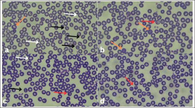
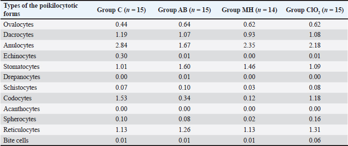
Fig. 1. Box plot representation of significant hematological variables (HGB, HCT, and PLT) across experimental groups. Asterisks indicate statistically significant pairwise differences (*p < 0.05). Analysis of poikilocytosis revealed a generally low degree of erythrocyte shape alterations across all experimental groups, predominantly classified as rare to mild, without the presence of moderate or pronounced changes. The most frequently observed forms were anulocytes (C=2.84%, AB=1.67%, MH=2.35%, and ClO2=2.18%) and stomatocytes (C=1.01%, AB=1.60%, MH=1.46%, and ClO2=1.09%) (Table 3). Original microscopic images of individual poikilocytotic forms of erythrocytes from four experimental groups of rats are shown in Figure 3. Descriptive statistics and individual raw hematological data are presented in Supplementary Tables S1–S5.
Fig. 3. Original microscopic images of individual poikilocytotic forms of erythrocytes in the four experimental groups of rats: a) AB group, b) C group, c) MH group, and d) ClO2 group. Arrows indicate specific cell types: anulocytes (white), stomatocytes (black), target cells (red), and reticulocytes (orange). DiscussionIn this study, the systemic hematological responses of Wistar rats to topical application of manuka honey and ClO2 were investigated to better understand the systemic effects of topical treatments applied to surgically induced wounds. Although most erythrocyte-related parameters remained within the expected physiological ranges, measurable intergroup differences were observed in selected parameters. The most pronounced changes were observed in erythrocyte and platelet parameters. HGB values differed significantly among groups, with post hoc analysis showing higher values in the AB group than in the C (p=0.011), ClO2 (p=0.015), and MH (p=0.006) groups. Despite these differences, the values remained within the physiological range, suggesting that the magnitude of the observed effect was limited. Accordingly, the findings may reflect a modest intergroup variation in the systemic hematological response on day 14 rather than a pronounced treatment-related hematological disturbance. On the contrary, toxicological and experimental studies involving the oral administration of honey, including manuka and tualang honey, have described a mild “potentiating” effect of honey on HGB, RBC, PCV, and PLT (Ahmed et al., 2017; Aliyu et al., 2012; Hussein and Kadhem, 2025), as well as stabilizing or anti-anemic effects, particularly at higher concentrations (Aliyu et al., 2012). In addition, several studies have confirmed that oral honey administration may increase PCV, HGB, and RBC values, which are attributed to the mineral and antioxidant composition of honey that stimulates hematopoiesis (James et al., 2009; Hussein and Kadhem, 2025). Studies have also reported that dark, phenolic-rich honeys, such as manuka honey, may enhance erythropoiesis by stimulating bone marrow activity (Mousavi Kiasari et al., 2020). HCT was significantly higher in the AB group than in the MH (p=0.011), ClO2 (p=0.015), and C groups (p=0.037). As most values remained within expected physiological ranges, this finding is best interpreted as a modest intergroup variation in systemic hematological response at day 14. Akinbami et al. (2013) reported that inflammatory and systemic pathological conditions may affect erythrocyte parameters and the overall hematological profile, with abnormalities, such as reduced HGB, PCV, and RBC values, being associated with active inflammation or systemic disease. Therefore, the elevated HCT in the AB group may indicate a difference in systemic hematological response related to treatment. The most notable changes were observed in the platelet parameters. Healthy rodents generally exhibit higher PLT values than larger animal species, which is considered a physiological adaptation that enables a rapid hemostatic response (Božić, 2012; Kampfmann et al., 2012). The total PLT was significantly higher in the ClO2 group than in the MH (p=0.014), C (p=0.041), and AB (p=0.019) groups. These findings may reflect a modest platelet-associated hematological response observed at the evaluated endpoint. However, because most values remained within the expected physiological ranges and no additional inflammatory or coagulation markers were assessed, their biological significance should be interpreted cautiously. Increased platelet values may reflect a modest platelet-associated hematological response following surgically induced injury, consistent with the known physiological role of platelets in hemostatic responses (Božić, 2012). The observed increase in platelet parameters may also be viewed in the context of the broader physiological role of platelets in hemostatic and tissue-related responses following injury (Kim et al., 2025). Given the strong antimicrobial and oxidative activity of ClO2, the observed increase in platelet parameters may additionally reflect systemic hematological responses associated with surgically induced tissue injury, because platelets play a key role in hemostasis, tissue repair, and immune activation. Although the available literature does not describe the systemic hematological effects following topical application of ClO2, the recorded elevation in platelet values is consistent with its known oxidative and antimicrobial properties. These findings should nevertheless be interpreted cautiously because the safety profile of ClO2 remains dependent on the formulation and concentration used, and broader toxicity concerns remain relevant in the literature. Analysis of poikilocytosis showed that all groups exhibited a low degree of erythrocyte shape alterations, mostly ranging from rare to mild, without moderate or pronounced deviations. The most frequently observed forms were anulocytes (C=2.84%, AB=1.67%, MH=2.35%, and ClO2=2.18%) and stomatocytes (C=1.01%, AB=1.60%, MH=1.46%, and ClO2=1.09%), corresponding to a mild degree of poikilocytosis. Other forms, such as ovalocytes, dacrocytes, codocytes, and reticulocytes, were observed at low and uniform frequencies across the groups and were classified as rare to mild changes. Echinocytes, drepanocytes, acanthocytes, and bite cells were present only in trace amounts or were nearly undetectable. The absence of moderate or pronounced morphological alterations indicates that topical treatment with manuka honey and chlorine dioxide was not associated with evident erythrocyte morphological changes indicative of marked hemolytic stress or membrane damage at the evaluated endpoint. Overall, these findings support the relative stability of the erythrocyte population and the absence of pronounced systemic morphological disturbances under the conditions of the present study. Several limitations of this study should be acknowledged. Hematological parameters were evaluated at a single time point on day 14, which does not allow assessment of the temporal dynamics of systemic hematological responses. Transient early or intermediate hematological changes following wound creation and topical treatment cannot be excluded. A further limitation of the present study is the absence of a non-incised baseline group, which limits the differentiation between hematological changes associated with the surgical procedure and those related to topical treatment. In addition, the study focused on erythrocyte and platelet indices and did not include tissue-level wound healing parameters, such as planimetry, histological scoring, or tensile strength. Leukocyte profiling, including differential leukocyte counts, was not included in the present analysis, limiting the interpretation of systemic inflammatory responses. Several formulation-related parameters relevant to reproducibility were not independently assessed. The pH, sterility status, and stability of the applied products were not measured over the 14-day treatment period. With regard to dosing, the exact amount of manuka honey and gentamicin ointment applied per wound was not weighed, and the delivered volume of ClO2 per spray wasn‘t determined. Therefore, topical dosing could only be reported approximately for manuka honey and gentamicin ointment and descriptively for ClO2. Blood smear evaluation for poikilocytosis was not blinded, which may represent a potential source of observer bias. Although statistically significant differences were identified for selected variables, most values remained within expected physiological ranges, and the magnitude of these differences appears to be limited, suggesting a modest biological impact. Considering the topical route of administration, systemic absorption of applied treatments is expected to be minimal, especially in the case of ClO2, which may explain the limited magnitude of the observed systemic hematological changes. Finally, sex-stratified or sex-adjusted analyses were not performed, although both male and female rats were included and distributed as evenly as possible across the experimental groups during allocation. The study was not specifically designed or statistically powered to evaluate sex-related differences, and the limited number of animals within each treatment-by-sex subgroup did not allow reliable interpretation of sex-specific effects. Therefore, future studies should address potential sex-related differences with an appropriate sample size and predefined sex-based analysis. ConclusionBased on the obtained results, no marked systemic hematological disturbances were observed at the evaluated endpoint following topical application of manuka honey and ClO2 in Wistar rats with surgically induced wounds. Most parameters remained stable, whereas HGB, HCT, and PLT showed significant intergroup differences. Chlorine dioxide exerted the most pronounced effect on the platelet-related parameters. Minimal poikilocytosis further supports the observed relative hematological stability across the experimental groups. The obtained results provide valuable insights into the systemic hematological responses associated with the application of different topical wound therapies. Although the analysis in this study primarily focused on erythrocyte and platelet parameters, future research could include a more detailed evaluation of the leukocyte profile and integration of hematological, immunological, and histopathological indicators. Such a multidisciplinary approach would enable a comprehensive understanding of the mechanisms of action and safety profile of alternative topical treatments. AcknowledgmentsNone. Conflict of interestThe authors declare no conflicts of interest. FundingThis research was conducted as part of a project supported by the Federal Ministry of Education and Science of the Federation of Bosnia and Herzegovina during the 2025/2026 period (Decision No. 05-35-3131-2/25). Authors’ contributionsConception—M.K., N.K.D.; Design—M.K., N.K.D.; Supervision—M.K., N.K.D.; Materials—M.K., N.K.D.; Data Collection and Processing—N.Č., M.K., N.K.D.; Interpretation—N.K.D., M.K., N.Č.; Literature Review—N.K.D., M.K.; Writing—N.K.D.; Critical Review—M.K. Data availabilitySupporting data and supplementary material are available in the manuscript. ReferencesAdeneye, A.A., Ajagbonna, O.P., Adeleke, T.I. and Bello, S.O. 2006. Preliminary toxicity and phytochemical studies of the stem bark aqueous extract of Musanga cecropioides in rats. J. Ethnopharmacol. 105(3), 374–379; doi:10.1016/j.jep.2005.11.027 Ahmed, S., Sulaiman, S.A. and Othman, N.H. 2017. Oral administration of Tualang and Manuka honeys modulates breast cancer progression in Sprague-Dawley rats model. Evid. Based. Complement. Alternat. Med. 2017, 5904361; doi:10.1155/2017/5904361 Akinbami, A., Popoola, A., Adediran, A., Dosunmu, A., Oshinaike, O., Adebola, P. and Ajibola, S. 2013. Full blood count pattern of pre-chemotherapy breast cancer patients in Lagos, Nigeria. Caspian. J. Int. Med. 4(1), 574–579. Aliyu, M., Odunola, O.A., Owumi, S.E., Gbadegesin, M.A., Choudhary, M.I., Farooq, A.D., Rasheed, H., Erukainure, O.L. and Ahmed, S. 2012. Daily consumption of honey: effects on male Wistar albino rats. IJFSNPH 1(2), 66–74. Araújo, L.U., Grabe-Guimarães, A., Mosqueira, V.C.F., Carneiro, C.M. and Silva-Barcellos, N.M. 2010. Profile of wound healing process induced by allantoin. Acta. Cir. Bras. 25(5), 460–466; doi:10.1590/s0102-86502010000500014 Božić, T. 2012. Poremećaji bele krvne slike i hemostaze. In Patološka fiziologija domaćih životinja. Božić, T. 2nd, Belgrade, Serbia: Naučna KMD, pp: 71–116. Catalano, A., Iacopetta, D., Ceramella, J., Scumaci, D., Giuzio, F., Saturnino, C., Aquaro, S., Rosano, C. and Sinicropi, M.S. 2022. Multidrug resistance (MDR): a widespread phenomenon in pharmacological therapies. Molecules 27(3), 616; doi:10.3390/molecules27030616 Chinko, B.C., Pughikumo, D.T., Obia, O., Udeh, W.C. and Hart, V.O. 2023. Honey attenuates phenylhydrazine-induced hematotoxicity and oxidative stress in male Wistar rats. IBRR 14(3), 10–18; doi:10.9734/ibrr/2023/v14i3308 Christopher, M.M., Hawkins, M.G. and Burton, A.G. 2014. Poikilocytosis in rabbits: prevalence, type, and association with disease. PLoS One. 9(11), e112455; doi:10.1371/journal.pone.0112455 Dudek-Wicher, R., Brożyna, M., Paleczny, J., Mączyńska, B., Dudek, B., Migdał, P., Dołowacka-Jóźwiak, A., Fischer, J. and Junka, A. 2025. Antimicrobial properties and toxicity challenges of chlorine dioxide used in alternative medicine. Sci. Rep. 15(1), 18267; doi:10.1038/s41598-025-01852-z El-Senduny, F.F., Hegazi, N.M., Abd Elghani, G.E. and Farag, M.A. 2021. Manuka honey, a unique mono-floral honey. A comprehensive review of its bioactives, metabolism, action mechanisms, and therapeutic merits. Food Biosci. 42, 101038; doi:10.1016/j.fbio.2021.101038 Eroglu, O., Deniz, T., Kisa, U., Comu, F.M., Kaygusuz, S. and Kocak, O.M. 2018. The effect of different types of honey on healing infected wounds. J. Wound Care 27(10), S18–S25; doi:10.12968/jowc.2018.27.Sup10.S18 Goldman, R. 2004. Growth factors and chronic wound healing: past, present, and future. Adv. Skin. Wound. Care. 17(1), 24–35; doi:10.1097/00129334-200401000-00012 Hanks, J. and Spodnick, G. 2005. Wound healing in the veterinary rehabilitation patient. Vet. Clin. North Am. Small Anim. Pract. 35(6), 1453-1471; doi:10.1016/j.cvsm.2005.08.005 Harvey, J.W. 2012. Veterinary hematology: a diagnostic guide and color atlas. St. Louis, MO: Elsevier Saunders. Hussein, D.Y. and Kadhem, W.M. 2025. Effect of honey on blood and biochemical parameters of rats with induced anemia. Agric. Biotechnol. J. 17(1), 231–246; doi:10.22103/jab.2025.24690.1646 Ihedioha, J.I., Okafor, C. and Ihedioha, T.E. 2004. The haematological profile of the Sprague-Dawley outbred albino rat in Nsukka, Nigeria. Anim. Res. Int. 1(2), 125–132; doi:10.4314/ari.v1i2.40755 James, O.O., Mesubi, M.A., Usman, L.A., Yeye, S.O., Ajanaku, K.O., Ogunniran, O., Ajani, O. and Siyanbola, T.O. 2009. Physical characterisation of some honey samples from North-Central Nigeria. Int. J. Phys. Sci. 4(9), 464–470. Kampfmann, I., Bauer, N., Johannes, S. and Moritz, A. 2012. Differences in hematologic variables in rats of the same strain but different origin. Vet. Clin. Pathol. 41(2), 228–234; doi:10.1111/j.1939-165X.2012.00427.x Kapo-Dolan, N., Kapo, N., Čović, N., Ališah, A. and Katica, M. 2025. Evaluation of the hematological response to various therapeutic approaches in the management of open wounds: an experimental study in rodents. Veterinaria 74(1), 28–38; doi:10.51607/22331360.2025.74.S2.28 Katica, M. and Gradaščević, N. 2017. Hematologic profile of laboratory rats fed with bakery products. Int J Res GRANTHAALAYAH 5(5), 221–231; doi:10.29121/granthaalayah.v5.i5.2017.1853 Kim, S., Yang, S. and Kim, S. 2025. Role of platelet derivatives and their therapeutic potential in wound healing. Biocell 49(12), 2335–2364; doi:10.32604/biocell.2025.072418 Ma, J.W., Huang, B.S., Hsu, C.W., Peng, C.W., Cheng, M.L., Kao, J.Y., Way, T.D., Yin, H.C. and Wang, S.S. 2017. Efficacy and safety evaluation of a chlorine dioxide solution. Int. J. Environ. Res. Public. Health. 14(3), 329; doi:10.3390/ijerph14030329 Marks, P.W. 2013. Hematologic manifestations of liver disease. Semin. Hematol. 50(3), 216–221; doi:10.1053/j.seminhematol.2013.06.003 Mba, I.E., Okeke, O.P., Sharndama, H.C., Osondu-Chuka, G.O., Ukuomadu, J. and Ugwu, C. 2022. Antimicrobial resistance: revisiting the mechanisms of resistance. Access Microbiol. 4(5), 577; doi:10.1099/acmi.ac2021.po0053 Mousavi Kiasari, S.A., Nobahar, M., Ghorbani, R. and Tamaddon, M.R. 2020. The effect of thyme honey on anemia in hemodialysis patients. Nurs. Pract. Today. 7(2), 151–160; doi:10.18502/npt.v7i2.2738 Nassar, A.M.K., Salim, Y.M.M., Eid, K.S.A., Shaheen, H.M., Saati, A.A., Hetta, H.F., Elmistekawy, A. and Batiha, G.E.S. 2020. Ameliorative effects of honey, propolis, pollen, and royal jelly mixture against chronic toxicity of sumithion insecticide in white albino rats. Molecules 25(11), 2633; doi:10.3390/molecules25112633 Öztürk, F. and Ermertcan, A.T. 2011. Wound healing: a new approach to the topical wound care. Cutan. Ocul. Toxicol. 30(2), 92–99; doi:10.3109/15569527.2010.539586 Ranjani, M., Rajan, S. and Brindha, P. 2010. Antioxidant and antibacterial potentials of aloe vera juice extract against wound isolates. J. Pure. Appl. Microbiol. 4(2), 733–739. Available via https://www.cabdirect.org/cabdirect/abstract/20103368586 Repellin, R.L., Pitt, K.A., Lu, M., Welker, J., Noland, E.L. and Stanley, B.J. 2021. The effects of a proprietary Manuka honey and essential oil hydrogel on the healing of acute full-thickness wounds in dogs. Vet. Surg. 50(8), 1634–1643; doi:10.1111/vsu.13711 Saikaly, S.K. and Khachemoune, A. 2017. Honey and wound healing: an update. Am. J. Clin. Dermatol. 18(2), 237–251; doi:10.1007/s40257-016-0247-8 Suartha, I.N., Sudimartini, L.M., Jayanti, P.D. and Wijayanti, N.P.A.D. 2022. Effect of oral administration of honey on hemato-biochemical parameters of dogs with atopic dermatitis. World. Vet. J. 12(2), 197–202; doi:10.54203/scil.2022.wvj25 Tang, Y., Chen, L. and Ran, X. 2024. Efficacy and safety of honey dressings in the management of chronic wounds: an updated systematic review and meta-analysis. Nutrients 16(15), 2455; doi:10.3390/nu16152455 Touzani, S., Al-Waili, N., Imtara, H., Aboulghazi, A., Hammas, N., Falcão, S., Vilas-Boas, M., Arabi, I.E., Al-Waili, W. and Lyoussi, B. 2022. Arbutus unedo honey and propolis ameliorate acute kidney injury, acute liver injury, and proteinuria via hypoglycemic and antioxidant activity in streptozotocin-treated rats. Cell. Physiol. Biochem. 56(1), 66–81; doi:10.33594/000000496 Umogbai, O.O., Ogli, S.A., Agaba, E.I. and Yongo, M.A. 2023. Natural honey pre-treatment protect against immune suppression in cyclophosphamide exposed Wistar rats. Highl. Med. Res. J. 22(1), 20–26. Underwood, W. and Anthony, R. 2020. AVMA guidelines for the euthanasia of animals: 2020 Edition. American Veterinary Medical Association. Schaumburg, IL: American Veterinary Medical Association. Vercelli, C., Gambino, G., Amadori, M. and Re, G. 2022. Implications of Veterinary Medicine in the comprehension and stewardship of antimicrobial resistance phenomenon. From the origin till nowadays. Vet. Anim. Sci. 16, 100249; doi:10.1016/j.vas.2022.100249 Wang, S., Qiu, Y. and Zhu, F. 2024. An updated review of functional ingredients of Manuka honey and their value-added innovations. Food. Chem. 440, 138060; doi:10.1016/j.foodchem.2023.138060 Zhao, D., Wojnarowski, K., Cholewińska, P. and Palić, D. 2025. Current trends in approaches to prevent and control antimicrobial resistance in aquatic veterinary medicine. Pathogens 14(7), 681; doi:10.3390/pathogens14070681 Table 1. Overall intergroup comparisons of erythrocyte and platelet indices in Wistar rats across the control (C, n=15), gentamicin (AB, n=15), manuka honey (MH, n=14), and chlorine dioxide (ClO2, n=15) groups after 14 days of topical treatment, using the Kruskal–Wallis H test and Dunn’s post hoc test.
Table 2. Cliff’s delta effect sizes for pairwise comparisons of significant hematological variables (HGB, HCT, and PLT) in the control (C, n=15), gentamicin (AB, n=15), manuka honey (MH, n=14), and chlorine dioxide (ClO2, n=15) groups after 14 days of topical treatment.
Table 3. Percentage of poikilocytotic red blood cell forms identified in each group from a total of 2,000 examined erythrocytes.
Table S2. Raw hematological data for AB group (gentamicin group).
Supplementary TableTable S1. Descriptive statistics of erythrocyte and platelet indices in Wistar rats after 14 days of topical treatment with manuka honey (MH), chlorine dioxide (ClO2), gentamicin (AB), or no treatment (control, C).
Table S3. Raw hematological data for C group (control group).
Table S4. Raw hematological data for MH group (manuka honey group).
Table S5. Raw hematological data for ClO2 group (chlorine dioxide group).
| ||


| How to Cite this Article |
| Pubmed Style Kapo-dolan N, Čović N, Katica M. Erythrocyte and platelet indices in response to alternative topical approaches to surgically induced wounds: An experimental study in rats. doi:10.5455/OVJ.2026.v16.i7.54 Web Style Kapo-dolan N, Čović N, Katica M. Erythrocyte and platelet indices in response to alternative topical approaches to surgically induced wounds: An experimental study in rats. https://www.openveterinaryjournal.com/?mno=306130 [Access: July 15, 2026]. doi:10.5455/OVJ.2026.v16.i7.54 AMA (American Medical Association) Style Kapo-dolan N, Čović N, Katica M. Erythrocyte and platelet indices in response to alternative topical approaches to surgically induced wounds: An experimental study in rats. doi:10.5455/OVJ.2026.v16.i7.54 Vancouver/ICMJE Style Kapo-dolan N, Čović N, Katica M. Erythrocyte and platelet indices in response to alternative topical approaches to surgically induced wounds: An experimental study in rats. doi:10.5455/OVJ.2026.v16.i7.54 Harvard Style Kapo-dolan, N., Čović, . N. & Katica, . M. (2026) Erythrocyte and platelet indices in response to alternative topical approaches to surgically induced wounds: An experimental study in rats. doi:10.5455/OVJ.2026.v16.i7.54 Turabian Style Kapo-dolan, Nadža, Nedim Čović, and Muhamed Katica. 2026. Erythrocyte and platelet indices in response to alternative topical approaches to surgically induced wounds: An experimental study in rats. doi:10.5455/OVJ.2026.v16.i7.54 Chicago Style Kapo-dolan, Nadža, Nedim Čović, and Muhamed Katica. "Erythrocyte and platelet indices in response to alternative topical approaches to surgically induced wounds: An experimental study in rats." doi:10.5455/OVJ.2026.v16.i7.54 MLA (The Modern Language Association) Style Kapo-dolan, Nadža, Nedim Čović, and Muhamed Katica. "Erythrocyte and platelet indices in response to alternative topical approaches to surgically induced wounds: An experimental study in rats." doi:10.5455/OVJ.2026.v16.i7.54 APA (American Psychological Association) Style Kapo-dolan, N., Čović, . N. & Katica, . M. (2026) Erythrocyte and platelet indices in response to alternative topical approaches to surgically induced wounds: An experimental study in rats. doi:10.5455/OVJ.2026.v16.i7.54 |


