





| Research Article | ||
Open Vet. J.. 2026; 16(7): 4217-4227 Open Veterinary Journal, (2026), Vol. 16(7): 4217-4227 Research Article Histological evaluation of neutral buffered formalin and Bouin’s fixative: Liver and gills of Nile tilapia (Oreochromis niloticus)Dini Agusti Paramanandi1*, Fajar Shodiq Permata1, Intan Nur Afifah2 and Cynthia Alifianny21Laboratory of Veterinary Anatomy, Histology and Embryology, Faculty of Veterinary Medicine, Universitas Brawijaya, Malang, Indonesia 2Program Study of Veterinary Medicine, Faculty of Veterinary Medicine, Universitas Brawijaya, Malang, Indonesia *Corresponding Author: Dini Agusti Paramanandi. Laboratory of Veterinary Anatomy, Histology and Embryology, Faculty of Veterinary Medicine, Universitas Brawijaya, Malang, Indonesia. Email: paramanandi [at] ub.ac.id Submitted: 13/01/2026 Revised: 24/04/2026 Accepted: 04/06/2026 Published: 02/07/2026 © 2026 Open Veterinary Journal
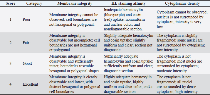
AbstractBackground: The initial fixation step is paramount in histology; the eventual quality of a tissue section hinges largely on the specific fixative chosen and how long the sample remains submerged. Aim: This study aimed to compare the histological quality of the liver and gills of Nile tilapia (Oreochromis niloticus) fixed in 10% neutral-buffered formalin (NBF) and Bouin’s solution over various immersion times (1, 7, 14, and 21 days). Methods: Histological quality, including membrane integrity, staining affinity, and cytoplasmic density, was assessed using hematoxylin–eosin staining and observed under a light microscope at 400× magnification. A qualitative scoring grade (1=poor, 2=fair, 3=good, and 4=excellent) was used, and data were presented as median (interquartile range). Statistical analysis was performed using the nonparametric Friedman test, Wilcoxon signed-rank test, and Mann–Whitney U test (p < 0.05). Results: A 1-day immersion in both NBF and Bouin’s solution yielded the highest histological quality for both organs. In liver tissue, 10% NBF provided superior preservation during the first week of immersion (Days 1–7; p=0.008). However, by Day 21, Bouin’s solution exhibited significantly higher structural stability for the liver than NBF (p=0.032). No significant differences were observed between NBF and Bouin’s solution in gill tissues until Day 14 (p > 0.05). However, a significant divergence occurred on Day 21, when Bouin’s solution outperformed 10% NBF (p=0.008). Conclusion: Under the tested conditions, a 24-hour immersion duration provides the most consistent results for Nile tilapia liver and gills. While 10% NBF is effective for short-term liver preservation, Bouin’s solution offers better resilience for both organs during prolonged immersion. Keywords: Hematoxylin–Eosin, Histology, Immersion duration, Teleost, Tissue preservation. IntroductionNile tilapia (Oreochromis niloticus) is a reliable bioindicator for monitoring aquatic health because of its physiological sensitivity to environmental stressors and pollutants (Camargo and Martinez, 2007). Among its biological conditions, the liver and gills are the most critical organs for evaluation. The liver is a multifunctional organ central to detoxification, vitellogenin production, and carbohydrate and fat metabolism (Sales et al., 2017). Similarly, the gills are vital for maintaining homeostasis, including osmoregulation, ion balance, and gas exchange through the lamellar structure (Aliza, 2014). Histopathological changes in these specific organs serve as essential biomarkers for assessing the impact of external agents on fish health (Hadi and Alwan, 2012). Therefore, gaining a clear histological quality is crucial for accurate analysis and pathological diagnosis (Ostrander, 2005 ). Histopathological assessment can help characterize fish health status and is increasingly used as a reliable biomarker for aquaculture quality monitoring (Guelmamene et al., 2025). However, the accuracy assessment influences by the tissue preservation quality. Poor fixation can induce significant artifacts that obscure cellular details or mimic pathological lesions (Wolf and Wheeler, 2018). Fish organs, such as the liver and gills, have a complex structure and a high metabolic role that require optimized fixation protocols to keep their cellular shape as close to the “life” state as possible (Ulucan et al., 2019). Despite the importance of Nile tilapia (O. niloticus) in the ecosystem, comprehensive research on the impact of fixative selection and immersion duration remains limited (Wolf and Wheeler, 2018; Guelmamene et al., 2025). Optimal tissue preservation through fixation is mandatory to ensure high-quality evaluation. Fixation is the critical initial step that is designed to prevent autolysis-induced tissue degradation that leads to irreversible cell death (Pratiwi et al., 2019; Fitri and Cahayani, 2020). Fixative solutions preserve cellular morphology and prevent the loss of intracellular molecules by hardening and stabilizing the tissue (Suvarna et al., 2013). In histology, the 2 most frequently used fixatives are 10% neutral buffered formalin (NBF) and Bouin’s solution. 10 percent NBF is widely known for its ability to stabilize tissues through cross-linking with proteins. On the contrary, Bouin’s solution, which contains picric acid, is often preferred for its ability to minimize tissue shrinkage, preserve delicate structures, and facilitate easier trimming (Copper et al., 2018). However, selecting an inappropriate fixative or miscalculating the immersion time often results in histological artifacts, such as tissue brittleness or loss of cellular detail, which compromise diagnostic accuracy (Musyarifah and Agus, 2018). While previous studies have evaluated various fixatives and acknowledged the critical role of immersion duration in other teleost models, such as zebrafish (Copper et al., 2018; Ulucan et al., 2019), specific data for Nile tilapia organs—particularly concerning the degradation of histological quality over an extended immersion of up to 21 days—remains sparse. Currently, comparative data regarding the long-term effects of immersion durations on specific teleost organs are limited. The 1-, 7-, 14-, and 21-day intervals selected in this study were specifically designed to simulate various field conditions, ranging from immediate laboratory processing to the delayed fixation often encountered in remote aquaculture sites. Consequently, this study was designed to analyze and compare the histological quality of the liver and gills of Nile tilapia preserved in 10% NBF and Bouin’s solutions across various immersion intervals to determine the most effective protocol for these specific organs. Materials and MethodsAnimal preparation and ethical proceduresIn this study, 10 Nile tilapia (O. niloticus) with a minimum total length of 20 cm were used. The fish were collected from a local pond and acclimated for 1–2 hours before the procedure. Euthanasia was performed by submerging the fish in clove oil solution (eugenol) at a concentration of 0.17 ml/l, following the guidelines of Leary et al. (2020). Experimental design and tissue sampling protocolLiver and gill tissues were harvested immediately after euthanasia during necropsy. The fish were divided into two fixative groups: 10% NBF (n=5) and Bouin’s solution (n=5). The liver and gills from each fish were trimmed into equivalent sections of approximately 1.0 × 1.0 × 0.5 cm to ensure optimal fixative penetration and evaluate the effect of fixation kinetics. Subsamples were then assigned to one of 4 immersion durations: 1, 7, 14, or 21 days. This experimental design was specifically chosen to minimize inter-individual biological variation, ensuring that any observed changes in histological quality were primarily due to the fixative’s performance over time rather than physiological differences between individual fish. The experimental unit was defined as the individual fish (n=5 per fixative group). To evaluate the effect of immersion duration, tissue subsamples collected from the same fish on Days 1, 7, 14, and 21 were treated as repeated measurements (dependent observations). Fixation procedureThe harvested organs were immersed in 2 fixative solutions: 10% NBF and Bouin’s solution. Both fixatives were prepared in-house in the laboratory according to the standard histological formulations. The 10% NBF was prepared by buffering 10% formalin with sodium phosphate to reach a neutral pH (7.2–7.4), while Bouin’s solution was prepared using a mixture of saturated picric acid, formaldehyde, and glacial acetic acid. The tissue-to-fixative volume ratio was maintained at 1:10, and the fixation process was conducted at room temperature. For each fixative, the organs were divided into 4 groups based on immersion intervals: 1, 7, 14, and 21 days. Histological processingFollowing the fixation procedure, the samples were subjected to standard histological processing, including graded dehydration in ethanol, clearing in xylene, and infiltration in paraffin wax. Tissues were embedded in paraffin blocks and sectioned at a thickness of 5 µm using a rotary microtome (Leica Biosystems, Wetzlar, Germany). The resulting sections were mounted on glass slides and stained with H&E to facilitate morphological evaluation. Morphological assessment and scoringTo evaluate the morphological quality, histological sections were observed using a light microscope (Olympus Corp., Tokyo, Japan) at 400× magnification. The quality of the tissue was evaluated based on three parameters: membrane integrity, H&E staining affinity, and cytoplasmic density. These specific parameters were selected to ensure data credibility and prevent the misinterpretation of fixation-induced artifacts, which is a critical requirement in fish histopathology. A single trained observer performed histological scoring. To minimize bias, an assistant coded the glass slides so that the observer was blinded to the treatment groups and immersion durations during the microscopic evaluation. The qualitative scoring system for liver and gill tissues was adapted and modified from previous studies conducted by Patil et al. (2015); Copper et al. (2018), and Ulucan et al. (2019). Each preparation was categorized on a four-point scale: 1 (poor), 2 (fair), 3 (good), and 4 (excellent). The detailed rubric for each organ is presented in Tables 1 (liver) and 2 (gills). Membrane integrity was assessed based on the preservation of cellular boundaries and the maintenance of hepatocytes’ characteristic hexagonal or polygonal shape. Furthermore, to ensure diagnostic reliability, staining affinity (H&E) was examined by measuring nuclear hematoxylin clarity, cytoplasmic eosin intensity, and the overall color uniformity and clarity of the slide. The cytoplasmic density profile was also assessed, focusing on the presence of adequate and homogeneous staining, absence of cell shrinkage, and lack of cytoplasmic fragmentation (Speilberg et al., 1993; Pertiwi et al., 2017; Ulucan et al., 2019). This procedure can distinguish technical artifacts from genuine pathological changes by integrating these parameters, maintaining high data credibility as proposed by Wolf and Wheeler (2018) and consistency with the health monitoring standards for Nile tilapia (Guelmamene et al., 2025). For each tissue sample, the individual scores of the three parameters (membrane integrity, staining affinity, and cytoplasmic density) were averaged to calculate a single composite histological score ranging from 1 to 4 before statistical analysis. Table 1. Histological scoring criteria for liver tissue of Nile Tilapia (O. niloticus).
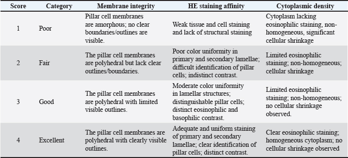
Table 2. Histological scoring criteria for Nile tilapia (O. niloticus) gill tissue.
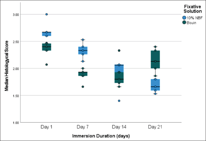
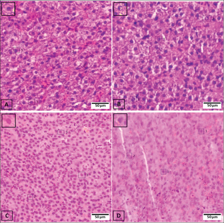
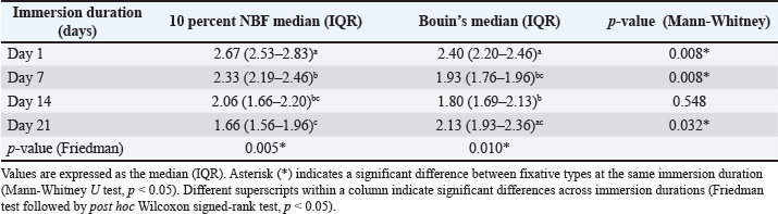
Statistical analysisQuantitative analysis of the histological scores was performed to compare the histological quality across all treatment groups. Data were analyzed using IBM SPSS Statistics software version 26.0 (IBM Corp., Armonk, NY). The data distribution was assessed using the Shapiro–Wilk normality test. The histological scores did not follow a normal distribution (p < 0.05). Consequently, nonparametric statistical methods were used. The Friedman test (appropriate for repeated measures) was used to analyze the temporal differences within each fixative group, followed by the Wilcoxon signed-rank test for post hoc comparisons. In addition, the Mann–Whitney U test was used to compare the histological scores between the 10% NBF and Bouin’s groups at each time point. A p-value of <0.05 (p < 0.05) was considered statistically significant. Ethical approvalThis study was conducted in strict accordance with the ethical guidelines for animal experimentation. The research ethics committee of Universitas Brawijaya, Malang, Indonesia, approved the protocol under the ethical clearance certificate number 101-KEP-UB-2021. ResultsHistological evaluation of the liverNotably, the re-analysis of the temporal data using the Friedman test for repeated measures yielded consistent significance patterns with our initial analysis, confirming that the core biological conclusions of the study were not altered by the choice of statistical model. The histological integrity of the Nile tilapia liver fluctuated notably depending on the fixative and immersion length (refer to the criteria in Table 1). Table 3 presents a comparative summary of the performance of 10% NBF versus Bouin’s solution. The distribution of these scores is further illustrated in the boxplot in Figure 1. Using the Friedman test, immersion duration significantly affected liver histological scores in both the 10% NBF (p=0.005) and Bouin’s solution groups (p=0.010). The 10% NBF-fixed tissues reached their peak quality on Day 1 (2.67 [2.53–2.83]). However, post hoc analysis using the Wilcoxon signed-rank test indicated a significant decline over time, with the score 1.66 (1.56–1.96) by Day 21.
Fig. 1. Comparison of histological scores in liver tissue of Nile tilapia (O. niloticus) between 10% NBF and Bouin’s solution. The boxplots illustrate the distribution of scores across four immersion durations (Days 1, 7, 14, and 21), where each box shows the median and interquartile range. The whiskers extend to show the full range of the scores, providing a visual summary of TSS over time. A similar downward trend was observed with Bouin’s solution, which achieved its highest performance on Day 1 (2.40 [2.20–2.46]). Interestingly, although 10% NBF consistently outperformed Bouin’s solution during the first week (Days 1 and 7, p=0.008, Mann–Whitney U test), a significant shift occurred toward the end of the study. By Day 21, Bouin’s solution demonstrated significantly higher structural stability than the NBF group (2.13 [1.93–2.36] vs. 1.66 [1.56–1.96]; p=0.032). Microscopic observation on Day 1 showed that liver samples fixed in 10% NBF exhibited intact membrane integrity, with distinct hexagonal or polygonal shapes maintained by hepatocytes (Fig. 3A). Conversely, samples subjected to prolonged immersion displayed brittle tissue sections, loss of membrane definition, and reduced H&E absorption (Fig. 3D). While Bouin’s solution showed adequate staining on Day 1 (Fig. 4A), it initially resulted in lower membrane integrity scores compared to 10% NBF. Although the cytoplasmic color intensity in the Bouin-fixed samples remained relatively stable until Day 14, a significant decline in overall structural integrity was observed over time (Table 3; Fig. 4D).
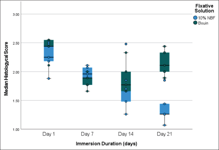
Fig. 2. Comparison of histological scores in Nile tilapia (O. niloticus) gill tissues treated with 10% NBF and Bouin’s solution. These boxplots track changes in tissue quality from Day 1 to Day 21. The central line within each box denotes the median score, whereas the boxes and whiskers display the data’s spread and variability, allowing for a clear comparison between the two fixatives.
Fig. 3. Photomicrographs of liver tissue of Nile tilapia (O. niloticus) fixed in 10% NBF at different immersion intervals (H&E, 400×). (A) Day 1, (B) Day 7, (C) Day 14, and (D) Day 21. The black transparent boxes 1, 2, and 3 represent the areas evaluated for histological scoring, focusing on hepatocyte morphology and sinusoidal spaces. Scale bar=50 µm.
Fig. 4. Photomicrographs of liver tissue of Nile tilapia (O. niloticus) fixed in Bouin’s solution at different immersion intervals (H&E, 400×). (A) Day 1, (B) Day 7, (C) Day 14, and (D) Day 21. The black boxes indicate the representative areas used for scoring. The sections show the hepatocyte arrangement and cytoplasmic staining patterns. Scale bar=50 µm. Table 3. Comparative histological scores of Nile tilapia liver in 10% NBF and Bouin’s solution across different immersion durations.
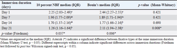
Histological evaluation of the gillsThe gills exhibited a different response to the fixatives compared with the liver tissue, according to the scoring rubric in Table 2. Table 4 presents the detailed comparative scores between 10% NBF and Bouin’s solution for gill tissues, with the visual distribution tracked in Figure 2. Statistical analysis using the Friedman test confirmed that immersion duration significantly affected the histological scores of the gills in both the 10% NBF group (p=0.037) and the Bouin’s group (p=0.006). The highest median score for 10% NBF-fixed gills was recorded on Day 1 (2.25 [2.03–2.49]), decreasing significantly to 1.26 (1.16–1.64) by Day 21. Post hoc analysis using the Wilcoxon signed-rank test showed that this decline was statistically significant. For Bouin’s solution, the highest score was achieved on Day 1 (2.44 [2.27–2.55]), which represented the highest overall quality among all gill treatment groups, before decreasing to 2.11 (1.94–2.38) by Day 21. Table 4. Comparative histological scores of Nile tilapia gills in 10% NBF and Bouin’s solution across different immersion durations.
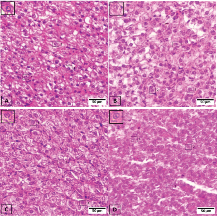
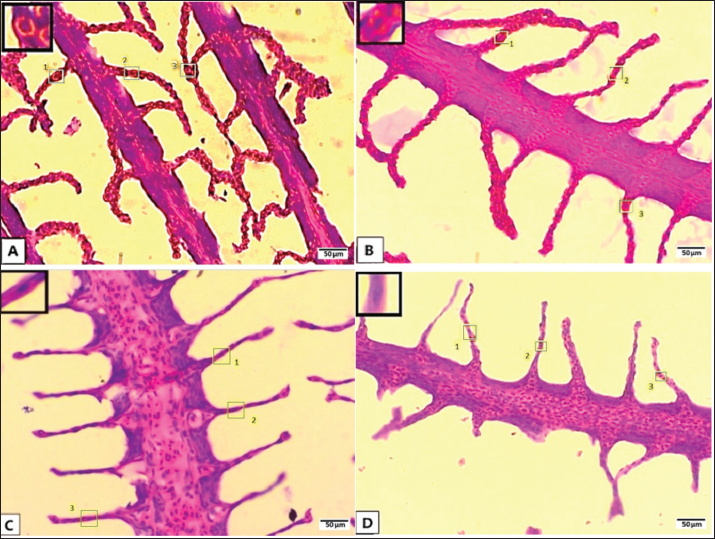
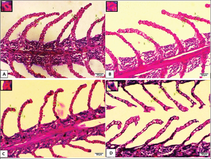
Furthermore, Mann–Whitney U test results showed no significant difference in quality between 10% NBF and Bouin’s solution from Day 1 to Day 14 (p > 0.05). However, a significant difference was noted at Day 21, when Bouin’s solution provided superior preservation compared with 10% NBF (p=0.008) (Table 4). On Day 1, both fixatives displayed well-preserved pillar cells and lamellar structures (Figs. 5A and 6A). The polyhedral shape of the pillar cells remained distinct in the Bouin-fixed samples, complemented by uniform H&E staining (Fig. 6A). However, by the later stages (Days 14 and 21), there was clear evidence of cytoplasmic shrinkage and a loss of homogeneity in both fixative groups. These degraded samples also exhibited a noticeable reduction in nuclear basophilic affinity, indicating a decline in the overall preparation quality over the 21-day period (Figs. 5D and 6D).
Fig. 5. Photomicrographs of gill tissues of Nile tilapia (O. niloticus) fixed in 10% NBF at different immersion intervals (H&E staining, 400×). (A) Day 1, (B) Day 7, (C) Day 14, and (D) Day 21. Boxes 1, 2, and 3 highlight the primary and secondary lamellae evaluated for scoring. Scale bar=50 µm.
Fig. 6. Photomicrographs of gill tissues of Nile tilapia (O. niloticus) fixed in Bouin’s solution at different immersion intervals (H&E staining, 400×). (A) Day 1, (B) Day 7, (C) Day 14, and (D) Day 21. The sections display the structural organization of the gill lamellae and the tissue affinity for H&E staining. Scale bar=50 µm. DiscussionThe findings of this study highlight a crucial principle in histology: tissue quality is not merely a product of the chemical composition of the fixative. Instead, it is also a function of its interaction with the specific architectural properties of the target organ and immersion duration. The results of this study indicate that while 10% NBF and Bouin’s solution both serve as effective primary fixatives, their long-term performance significantly diverges between dense glandular tissues, such as the liver, and delicate, cartilage-supported structures, such as the gills. This study specifically accounts for the nonindependence of samples taken from the same individual fish by using the Friedman test for repeated measures, providing a more robust statistical framework for evaluating temporal changes. Comparative fixative performance in liver tissuesIn the case of liver tissue, 10% NBF showed a clear advantage during the first week of immersion (Days 1–7), outperforming Bouin’s solution with significantly higher morphological scores (Table 3; p=0.008). This early-stage success is hypothesized to stem from the superior buffering capacity of 10% NBF, which maintains the cellular environment stable and close to its in vivo state. By maintaining a neutral pH (7.2–7.4), 10% NBF stabilizes tissue components and minimizes potential fixation artifacts that often occur in NBFs. These findings of optimal preservation at Day 1 (Table 3) align with those of Copper et al. (2018) who identified a 24-hour 10% NBF immersion as the “gold standard” protocol for nuclear clarity. It is widely suggested in the literature that 10% NBF stabilizes the hepatocyte cytoskeleton through the formation of methylene bridges between proteins; however, while this theoretical mechanism preserves structural integrity in short intervals, our data suggest that it may not prevent long-term distortion. A significant turning point was encountered on Day 21. At this stage, the histological quality of 10% NBF-fixed livers dropped to its lowest point (p=0.005), becoming significantly inferior to that of Bouin’s-fixed samples (Table 3; p=0.032). This decline may contribute to progressive, irreversible tissue hardening. In contrast to the results seen with 10% NBF, the initial performance of Bouin’s solution on liver samples was suggested to be hindered by its acidic components. The low pH (1.5–2.0) is hypothesized to induce protein denaturation, which may affect subcellular structures and potentially fail to fix the cytoplasm properly, leading to the “muddy” appearance observed in early intervals (Khristian and Inderiati, 2017). However, the liver tissues preserved in Bouin’s maintained a higher degree of structural stability compared to the 10% NBF group by Day 21. This suggests a compelling tradeoff: the picric acid within the solution appears to create a more stable long-term matrix for dense tissues, while the initial acid hydrolysis might cause some loss of fine detail (Suvarna et al., 2013). Temporal divergence in gill histologyThe specific chemistry of the fixative played a secondary role when examining the gills of Nile Tilapia compared to the sheer length of immersion. Statistical analysis using the Friedman test revealed no significant divergence between 10% NBF and Bouin’s performance from Day 1 to Day 14 (Table 4; p > 0.05), with both groups reaching their quality peak at Day 1. The slight numerical advantage of Bouin’s at the 24-hour mark may be attributed to the rapid nuclear penetration of the solution and the hypothesized ability of acetic acid to soften cartilaginous elements, facilitating better sectioning. These high-quality results at the 24-hour mark are consistent with the observations of Ulucan et al. (2019) regarding optimal primary fixation. However, a significant divergence emerged on Day 21 (p=0.008), where Bouin’s solution significantly outperformed 10% NBF. The general architecture of the gills appeared to be more resilient over time than that of the liver, possibly due to the internal cartilaginous support that helps hold the tissue framework together. Nevertheless, the severe decline in 10% NBF-fixed gills by Day 21 (median: 1.26) indicates that prolonged formalin exposure is particularly detrimental to the delicate lamellar epithelium. As Wolf and Wheeler (2018) noted, maintaining these minute details is necessary for accurate aquatic health monitoring, as researchers must be able to distinguish technical artifacts from true biological reactions. Practical implications for aquaculture laboratoriesThe results of this study offer several practical recommendations for aquaculture diagnostic laboratories: (1) Prioritizing processing: when dealing with high sample volumes, laboratories should prioritize processing liver samples over more resilient structures like gills because the liver shows a sharper decline in quality after Day 7 in 10% NBF. (2) Fixative selection for fieldwork: for field collections where samples cannot reach a lab within 2 weeks, Bouin’s solution appears to be a more reliable “long-term” preservative for both liver and gills, despite its initial acidic artifacts. (3) Artifact recognition: pathologists should be cautioned that a “shattered” tissue appearance or loss of membrane definition in 21-day samples may be a result of over-fixation rather than a pathological condition of the fish. This study primarily serves to confirm and extend the existing literature on teleost tissue preservation. Previous studies, such as those by Madi et al. (2016) and Ulucan et al. (2019) have documented the general effects of various fixatives on fish tissues, while Copper et al. (2018) highlighted the 24-hour 10% NBF immersion as a reliable standard for zebrafish. Our descriptive findings corroborate these earlier reports regarding NBF’s short-term efficacy. However, this study expands upon these foundations by explicitly detailing the progressive histological degradation in Nile Tilapia over an extended 21-day period. By doing so, we demonstrate that while 24-hour NBF fixation is favorable, Bouin’s solution offers a distinct advantage for long-term resilience when immediate processing is not feasible—a practical scenario often encountered in aquaculture field studies. However, the relatively small sample size (n=5 per group) limits the overall statistical power of this study. Therefore, this work should be considered as an exploratory pilot study. Future investigations with larger sample sizes and formal a priori power analyses are recommended to validate and expand these preliminary findings. ConclusionIn conclusion, our results show that fixative type and immersion duration significantly influence the histological quality of Nile Tilapia tissues. Under these experimental conditions, a 24-hour fixation period provided the most consistent morphology for both the aforementioned organs. While 10% NBF was effective for liver samples during the first week, Bouin’s solution offered better structural resilience over 21 days, particularly by limiting the over-fixation artifacts observed with 10% NBF. The gills appeared more robust than the liver but still showed a quality decline in 10% NBF over time. These findings suggest that processing samples within shorter timeframes is beneficial to avoid misinterpreting technical artifacts as pathological lesions in aquaculture health assessments. AcknowledgmentsThe authors sincerely thank the Faculty of Veterinary Medicine, Universitas Brawijaya, for providing the laboratory facilities and support necessary to conduct this research. FundingThe Faculty of Veterinary Medicine, Universitas Brawijaya, funded this research through the DPP SPP Research Grant (Grant No: 7/UN10F13.06/PN/2021). Authors' contributionsDini conceived and designed the study, secured funding, performed the laboratory experiments, wrote the original manuscript draft, and provided critical revisions. Fajar supervised the research progress and contributed to data interpretation and final manuscript editing. Intan and Cynthia performed the laboratory experiments, collected and scored the data, and assisted in the drafting of the manuscript. All authors have reviewed, discussed, and agreed to the final version of the manuscript for submission. Conflict of interestThe authors have no conflicts of interest related to this study. Data availabilityAll data supporting the findings of this study are available in the manuscript. ReferencesAliza, D. 2014. Behavioral and gill profiles of Nile Tilapia (Oreochromis niloticus) experiencing density stress. J. Med. Vet. 8, 80–83. Camargo, J.A. and Martinez, C.B.R. 2007. Histopathology of gills, kidney and liver of a Neotropical fish Prochilodus lineatus exposed to benzene. Neotrop. Ichthyol. 5, 323–330. Copper, J.E., Budgeon, L.R., Foutz, C.A., van Rossum, D.B., Vanselow, D.J., Hubley, M.J., Clark, D.P., Mandrell, D.T. and Cheng, K.C. 2018. Comparative analysis of fixation and embedding techniques for optimized histological preparation of zebrafish. Comparative Biochem. Physiol. Part. C. 208, 38–46. Fitri, L.E. and Cahayani, W.A. 2020. Malaria pathology: histological, immunological, and ultrastructural review. Malang, Indonesia: UB Press. Guelmamene, R., Elgroud, R., Bennoune, O. and Aloui, N. 2025. Quality monitoring of fish through histological assessment of their health status: proposal for a New Scoring System. Vet. Ital. 61(4); doi: 10.12834/VetIt.3767.35661.4 Hadi, A.A. and Alwan, S.F. 2012. Histopathological changes in gills, liver and kidney of fresh water fish, Tilapia zillii, exposed to aluminum. Int. J. Pharm. Life Sci. 3, 1571–1581. Khristian, E. and Inderiati, D. 2017. Cyto-histotechnology. Jakarta, Indonesia: Ministry of Health of the Republic of Indonesia. Leary, S., Underwood, W., Anthony, R., Cartner, S., Johnson, C.L. and Patterson-Kane, E. 2020. AVMA guidelines for the euthanasia of animals: 2020 edition. Washington, D.C.: American Veterinary Medical Association, pp, 1–121. Madi, S.C., Silva, M.S. and Castro, A.L. 2016. Effect of fixatives and time of fixation on the morphology of the liver of Nile Tilapia (Oreochromis niloticus). Int. J. Morphol. 34, 1345–1350. Musyarifah, Z. and Agus, S. 2018. The fixation process in histopathological examination. J. Kesehat. Andalas. 7, 443–453. Ostrander, G.K. 2005. Techniques in aquatic toxicology. Boca Raton, FL: CRC Press / Lewis Publishers. Patil, S., Rao, R., Ganavi, B. and Majumdar, B. 2015. Natural sweeteners as fixatives in histopathology: a longitudinal study. J. Nat. Sci. Biol. Med. 6, 67–70. Pertiwi, S.L., Zainuddin. and Rahmi, E. 2017. Histological description of the respiratory system of snakehead fish (Channa striata). J. Ilm. Mhs. Vet. 1, 291–298. Pratiwi, Y.N., Durachim, A., Mahmud, D. and Gusnandjar, A. 2019. Comparison of fixation using cane sugar and neutral buffered formalin on cell integrity. J. Ris. Kesehat. 11, 190–197. Sales, C.F., Silva, R.F., Amaral, M.G.C., Domingos, F.F.T., Ribeiro, R.I.M.A., Thomé, R.G. and Santos, H.B. 2017. Comparative histology in the liver and spleen of three species of freshwater teleost. Neotrop. Ichthyol. 15, e160135. Speilberg, L., Evensen, Ø., Bratberg, B. and Skjerve, E. 1993. Evaluation of five different immersion fixatives for light microscopic studies of liver tissue in Atlantic salmon (Salmo salar). Dis. Aquat. Organ. 17, 47–55. Suvarna, S.K., Layton, C. and Bancroft, J.D. 2013. Bancroft’s theory and practice of histological techniques. Oxford, UK: Churchill Livingstone Elsevier. Ulucan, A., Yüksel, H., Dörtbudak, M.B. and Yüksel, S. 2019. Comparative examination of commonly used some fixatives with routine histochemical staining’s for the optimal histological appearance in the gill tissue of zebrafish. Kocatepe Vet. J. 12, 158–167. Wolf, J.C. and Wheeler, J.R. 2018. A critical review of histopathological findings associated with endocrine and non-endocrine hepatic toxicity in fish models. Aquatic Toxicol. 197, 60–78. | ||
| How to Cite this Article |
| Pubmed Style Paramanandi DA, Permata FS, Afifah IN, Alifianny C. Histological evaluation of neutral buffered formalin and Bouin’s fixative: Liver and gills of Nile tilapia (Oreochromis niloticus). Open Vet. J.. 2026; 16(7): 4217-4227. doi:10.5455/OVJ.2026.v16.i7.9 Web Style Paramanandi DA, Permata FS, Afifah IN, Alifianny C. Histological evaluation of neutral buffered formalin and Bouin’s fixative: Liver and gills of Nile tilapia (Oreochromis niloticus). https://www.openveterinaryjournal.com/?mno=306584 [Access: June 30, 2026]. doi:10.5455/OVJ.2026.v16.i7.9 AMA (American Medical Association) Style Paramanandi DA, Permata FS, Afifah IN, Alifianny C. Histological evaluation of neutral buffered formalin and Bouin’s fixative: Liver and gills of Nile tilapia (Oreochromis niloticus). Open Vet. J.. 2026; 16(7): 4217-4227. doi:10.5455/OVJ.2026.v16.i7.9 Vancouver/ICMJE Style Paramanandi DA, Permata FS, Afifah IN, Alifianny C. Histological evaluation of neutral buffered formalin and Bouin’s fixative: Liver and gills of Nile tilapia (Oreochromis niloticus). Open Vet. J.. (2026), [cited June 30, 2026]; 16(7): 4217-4227. doi:10.5455/OVJ.2026.v16.i7.9 Harvard Style Paramanandi, D. A., Permata, . F. S., Afifah, . I. N. & Alifianny, . C. (2026) Histological evaluation of neutral buffered formalin and Bouin’s fixative: Liver and gills of Nile tilapia (Oreochromis niloticus). Open Vet. J., 16 (7), 4217-4227. doi:10.5455/OVJ.2026.v16.i7.9 Turabian Style Paramanandi, Dini Agusti, Fajar Shodiq Permata, Intan Nur Afifah, and Cynthia Alifianny. 2026. Histological evaluation of neutral buffered formalin and Bouin’s fixative: Liver and gills of Nile tilapia (Oreochromis niloticus). Open Veterinary Journal, 16 (7), 4217-4227. doi:10.5455/OVJ.2026.v16.i7.9 Chicago Style Paramanandi, Dini Agusti, Fajar Shodiq Permata, Intan Nur Afifah, and Cynthia Alifianny. "Histological evaluation of neutral buffered formalin and Bouin’s fixative: Liver and gills of Nile tilapia (Oreochromis niloticus)." Open Veterinary Journal 16 (2026), 4217-4227. doi:10.5455/OVJ.2026.v16.i7.9 MLA (The Modern Language Association) Style Paramanandi, Dini Agusti, Fajar Shodiq Permata, Intan Nur Afifah, and Cynthia Alifianny. "Histological evaluation of neutral buffered formalin and Bouin’s fixative: Liver and gills of Nile tilapia (Oreochromis niloticus)." Open Veterinary Journal 16.7 (2026), 4217-4227. Print. doi:10.5455/OVJ.2026.v16.i7.9 APA (American Psychological Association) Style Paramanandi, D. A., Permata, . F. S., Afifah, . I. N. & Alifianny, . C. (2026) Histological evaluation of neutral buffered formalin and Bouin’s fixative: Liver and gills of Nile tilapia (Oreochromis niloticus). Open Veterinary Journal, 16 (7), 4217-4227. doi:10.5455/OVJ.2026.v16.i7.9 |


