





| Research Article | ||
Open Vet. J.. 2026; 16(7): 4441-4456 Open Veterinary Journal, (2026), Vol. 16(7): 4441-4456 Research Article Vasculoprotective and antioxidant effects of Moringa oleifera Lam. leaf extract in cisplatin-treated diabetic rats: Role of malondialdehyde and E-selectinAgus Jati Sunggoro1,2,3, Bambang Purwanto1,3*, Paramasari Dirgahayu1,3,4, Brian Wasita1,3,5, Risya Cilmiaty1,3 and Eti Poncorini Pamungkasari1,3,61Doctoral Program of Medical Science, Faculty of Medicine, Universitas Sebelas Maret, Surakarta, Indonesia 2Division of Hematology and Medical Oncology, Department of Internal Medicine, Faculty of Medicine, Universitas Sebelas Maret, Surakarta, Indonesia 3Faculty of Medicine, Universitas Sebelas Maret, Surakarta, Indonesia 4Department of Parasitology and Mycology, Faculty of Medicine, Universitas Sebelas Maret, Surakarta, Indonesia 5Department of Anatomical Pathology, Faculty of Medicine, Universitas Sebelas Maret, Surakarta, Indonesia 6Department of Public Health, Faculty of Medicine, Universitas Sebelas Maret, Surakarta, Indonesia *Corresponding Author: Bambang Purwanto. Doctoral Program of Medical Science, Faculty of Medicine, Universitas Sebelas Maret, Surakarta, Indonesia. Email: bambang_p48 [at] staff.uns.ac.id Submitted: 17/01/2026 Revised: 31/05/2026 Accepted: 13/06/2026 Published: 11/07/2026 © 2025 Open Veterinary Journal
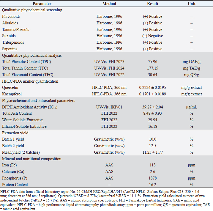
AbstractBackground: Cisplatin is a widely used chemotherapeutic agent in both human and veterinary medicine, particularly in canine osteosarcoma protocols. However, its use still carries the risk of dose-dependent toxicity, one of which is cisplatin-induced vascular toxicity, caused by oxidative stress and endothelial dysfunction. This toxicity can be exacerbated by pre-existing metabolic diseases that have vascular complications, such as diabetes mellitus (DM). Moringa oleifera Lam., a widely studied herbal remedy as an adjuvant therapy, is known to possess high antioxidant and anti-inflammatory properties. However, its vasculoprotective potential, especially in a clinically relevant DM-cisplatin model, remains unexplored. Aim: This study examined the vasculoprotective effects of M. oleifera Lam. leaf extract (MOLE) in reducing cisplatin-induced vascular toxicity in DM rat models, specifically focusing on the mechanisms of oxidative stress and endothelial activation biomarkers. Methods: Thirty-five male Sprague-Dawley rats were randomly divided into seven groups (n=5 each). Type 2 DM (T2DM) was induced using nicotinamide–streptozotocin, followed by cisplatin (8 mg/kg BW) administration. Treatment groups received MOLE at 350, 700, and 1050 mg/kg BW for 14 days, while positive controls received erythropoietin. Serum malondialdehyde (MDA), E-selectin, NF-κB, and C-reactive protein levels were quantified using ELISA, and systolic blood pressure (SBP) was measured. Results: Cisplatin-treated diabetic rats showed significant elevations in MDA, E-selectin, NF-κB, CRP, and SBP compared to controls (p < 0.001). MOLE treatment produced dose-dependent improvements, with the highest dose (1050 mg/kg BW) reducing MDA by 83.8%, E-selectin by 83.0%, NF-κB by 90.4%, and CRP by 69.0%. SBP decreased from 213.0 ± 5.05 to 97.8 ± 3.34 mmHg (p < 0.001). Strong correlations were observed between SBP and all biomarkers (r > 0.96, p < 0.001). Conclusion: MOLE reduced cisplatin-induced vascular toxicity in a diabetic rat model through antioxidant and anti-inflammatory mechanisms. These preclinical findings, obtained in a rodent model of T2DM, are hypothesis-generating and warrant further investigation in species-specific models, including canine studies, before any translational application in veterinary oncology is considered. Keywords: Cisplatin-induced toxicity, Diabetes mellitus, Endothelial dysfunction, Moringa oleifera Lam., Oxidative stress. IntroductionCancer remains a major health challenge in both human and veterinary medicine, with incidence and mortality rates continuing to rise worldwide. In companion animals, cancer is a leading cause of death, particularly in geriatric dogs, with approximately 50% of dogs over 10 years of age experiencing some form of neoplasia (Withrow et al., 2013; Sung et al., 2021). Among the several current cancer therapy options, cisplatin (cis-diamminedichloroplatinum (II)) remains one of the most widely used chemotherapeutic agents and is considered effective across a wide range of species. In veterinary oncology, cisplatin is the primary treatment for canine osteosarcoma, bladder transitional cell carcinoma, squamous cell carcinoma, ovarian carcinoma, and mesothelioma and is also used intralesionally to treat equine skin tumors such as sarcoid and melanoma (Chun et al., 1996; Knapp et al., 2000; Brown et al., 2019). Cisplatin has a cytotoxic mechanism of action by forming DNA-platinum adducts that inhibit DNA replication and transcription, ultimately causing apoptosis of rapidly dividing cells (Dasari and Tchounwou, 2014). Although considered to have high antineoplastic efficacy, the clinical use of cisplatin in human patients and animals also still considers the risk of dose-dependent toxicity, especially nephrotoxicity, cardiotoxicity, neurotoxicity, and vascular injury (Dugbartey et al., 2016). In animals, especially dogs, cisplatin-induced kidney toxicity has been reported in several cases and requires extensive fluid diuresis protocols before and after administration, while in cats, the use of cisplatin is contraindicated because it causes fatal pulmonary edema (Gustafson et al., 2013). Vascular toxicity has increasingly come to the fore as a major complication of cisplatin treatment in both humans and animals, primarily caused by oxidative stress, endothelial inflammation, and impaired regulation of vascular tone (Dugbartey et al., 2016). This toxicity is exacerbated in individuals with pre-existing metabolic disorders (Saito et al., 2022). Diabetes mellitus, a common metabolic disease in humans, also occurs in companion animals with a reported prevalence of 0.3%–1.3% in dogs, although canine diabetes is predominantly insulin-dependent (type 1), which differs pathophysiologically from the more common type 2 diabetes mellitus (T2DM) in humans. The nicotinamide–streptozotocin model used in the present study most closely reflects human T2DM pathophysiology. Nevertheless, the shared downstream pathological features of hyperglycemia-induced oxidative stress and endothelial dysfunction are common across diabetes types regardless of upstream etiology (Catchpole et al., 2005; Kim and Yun, 2025), creating a clinically relevant concern for veterinary patients receiving cisplatin-based chemotherapy. Cisplatin causes vascular injury in several vital organs, such as the heart, kidneys, and liver, where oxidative stress and inflammation play a significant role. Cisplatin-induced vascular toxicity is closely associated with increased reactive oxygen species (ROS) and impaired antioxidant defenses, which subsequently damage the vascular endothelium and impair NO-dependent vasodilation (McSweeney et al., 2021). Cisplatin also significantly increases malondialdehyde (MDA) levels, a marker of increased lipid peroxidation and known as a major end product and biomarker of oxidative injury. MDA also decreases glutathione and other antioxidant enzymes, such as SOD, CAT, and GPx (Ma et al., 2017). In diabetic models, oxidative stress and MDA are positively correlated with endothelial cell-selective adhesion molecules, linking lipid peroxidation to endothelial activation (An et al., 2023). These findings suggest that cisplatin-induced ROS (as indicated by increased MDA) can trigger endothelial activation and increased expression of adhesion molecules such as E-selectin, which mediate leukocyte adhesion and trigger vascular inflammation, and play an important role in the pathogenesis of vascular injury (Zhang et al., 2024). Transcription factors are crucial components of intracellular signaling complexes, given their role in integrating various environmental inputs (including ROS and NO) and translating them into appropriate cellular responses. Among the many transcription factors, nuclear factor kappa B (NF-κB) has been studied in various experimental models and is implicated in pro-inflammatory cytokine production and endothelial dysfunction (Scioli et al., 2020). Both diabetes and cisplatin induce ROS formation, which subsequently activates the mitogen-activated protein kinases (MAPK) and IκB kinase pathways, resulting in the activation of NF-κB as a transcription factor, increasing the expression of pro-inflammatory cytokines and adhesion molecules such as E-selectin, and ultimately leading to endothelial activation, leukocyte recruitment, barrier opening, and downstream tissue injury (Volpe et al., 2018; Liu et al., 2023; Wang et al., 2023). On this basis, therapeutic agents that have a mechanism of action to reduce oxidative stress and inhibit the NF-κB signaling pathway will be a promising option to prevent cisplatin-induced vascular toxicity, especially in patients with underlying metabolic diseases such as diabetes. In the realm of both human and veterinary medicine, herbal remedies with natural ingredients that have high antioxidant and anti-inflammatory content are increasingly in demand as adjunctive therapies to reduce the side effects of treatments such as chemotherapy, one of which is M. oleifera Lam. (Chiș et al., 2023; Pareek et al., 2023). Moringa oleifera Lam. leaf extract (MOLE) is considered to contain antioxidants such as polyphenols, flavonoids, and vitamins A, C, and E, which can fight various free radicals, further inhibit lipid peroxidation, and prevent oxidative damage in various organs (Fatema et al., 2020). In the context of diabetes, the flavonoid content, such as quercetin and phenolics in MOLE, has antioxidant properties that produce a scavenging effect on ROS released from mitochondria, thereby protecting pancreatic beta cells and maintaining stable blood sugar levels (Al-Malki and El Rabey, 2015). Previous study about antioxidant and anti-inflammatory effects of herbal medicine in the context of chemotherapy-induced toxicity has been conducted, which showed the occurrence of acute vascular injuries of vital organs 24 hours after a single administration of cisplatin (6 mg/kg body weight), but this study was conducted in a normal rat model without metabolic diseases, such as diabetes, and further examined the protective effect of A. millefolium extract as an anti-inflammatory agent (Eslamifar and Sabbagh, 2020). To date, no studies have examined the vasculoprotective effects of MOLE in a dual-injury model combining diabetes and cisplatin-induced toxicity. This gap is clinically relevant, as cisplatin-treated patients with concurrent metabolic comorbidities represent a particularly vulnerable population in both human and veterinary oncology. Therefore, this study aimed to address this gap by investigating the vasculoprotective and antioxidant effects of MOLE in a diabetic rat model treated with cisplatin. We hypothesized that MOLE would ameliorate vascular damage by reducing oxidative stress, inhibiting NF-κB-mediated endothelial activation, and improving vascular function in a dose-dependent manner. In particular, we evaluated its effects on key biomarkers including MDA, E-selectin, NF-κB p65, C-reactive protein (CRP), and systolic blood pressure (SBP). The findings of this preclinical study may provide preliminary evidence to inform future species-specific investigations into supportive care strategies in animals undergoing cisplatin-based chemotherapy. Materials and methodsPlant material and authenticationFresh leaves of M. oleifera Lam. (Moringaceae) were collected from Batu, Malang, East Java, Indonesia, in June 2025. The plant was taxonomically authenticated by a botanist at the UPT Laboratorium Herbal Materia Medica Batu, East Java Provincial Health Office. A voucher specimen (No. 250605.P.G.P.745) was deposited at the herbarium of the laboratory. The leaves were washed, shade-dried at 40°C–50°C, and ground into a fine powder (50 mesh). Extract preparationEthanolic extraction was performed using cold maceration with 70% ethanol (1:10 w/v) for 72 hours under intermittent agitation. The filtrate was concentrated using a rotary evaporator and subsequently freeze–dried to obtain MOLE, which was stored at 4°C in airtight amber containers. Two independent batches were prepared using the same protocol to assess reproducibility. The extraction yield was 10.0% (Batch 1) and 12.5% (Batch 2) w/w, giving a mean of 11.25% ± 1.77% (w/w; %RSD=15.71%), which is within acceptable limits for crude botanical extracts prepared by maceration. Quantitative high-performance liquid chromatography–photodiode array (HPLC–PDA) marker analysis (official laboratory report No. 26-03/MH-KSO/Rep/LHA/017; ArcTM HPLC, Zorbax Eclipse Plus C18, 250 × 4.6 mm, 5 μm; detection at 366 nm) confirmed two pharmacologically relevant flavonols: quercetin 0.2224 ± 0.0195 mg/g (%RSD=8.77%; r²=0.9994; retention time 5.937 minutes) and kaempferol 0.1701 ± 0.0189 mg/g (%RSD=11.11%; r²=0.9995; retention time 6.720 minutes). Both compounds are documented NF-κB inhibitors, providing molecular-level plausibility for the observed anti-inflammatory effects. Batch 1 extract was used for all animal treatments. Dose selection was based on previous studies demonstrating the safety and efficacy of M. oleifera Lam. at doses ranging from 200 to 1,000 mg/kg BW in rodent models (Verma et al., 2009; Soliman et al., 2020), with the highest dose (1050 mg/kg) selected to establish dose-response relationships. Before daily administration, MOLE was freshly suspended in distilled water at the required concentration for each dose group and administered via oral gavage using a stainless steel feeding needle. The normal control group (K1) received an equivalent volume of distilled water via the same route and procedure to ensure comparable handling stress across all groups Phytochemical characterizationPhytochemical analysis was performed at the UPT Laboratorium Herbal Materia Medica Batu (Certificate No. 400.7.21.4/2437/102.20/2025). Qualitative screening confirmed the presence of flavonoids, alkaloids, tannins/phenols, triterpenoids, and saponins. Quantitative analysis revealed total phenolic content of 75.96 mg gallic acid equivalent (GAE)/g extract, total flavonoid content of 30.64 mg quercetin equivalent (QE)/g extract, and total tannin content of 177.15 mg tannic acid equivalent (TAE)/g extract. The antioxidant activity determined by the DPPH assay showed an IC₅₀ value of 39.27 ± 2.04 μg/mL (certificate No. 400.7.21.4/2439/102.20/2025). Mineral analysis identified a high intrinsic iron concentration of 113 ppm, along with substantial amounts of calcium (2.6%) and phosphorus (1878 ppm). The protein content was 16.2%. The detailed phytochemical profile is presented in Table 1. Table 1. Phytochemical, physicochemical, and mineral characterization of MOLE.
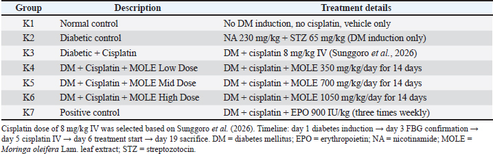
Experimental animalsThirty-five male Sprague-Dawley rats (8–10 weeks old, 180–200 g) were obtained and housed at the PAU Laboratory, Universitas Gadjah Mada, under standard laboratory conditions (22°C ± 2°C, 55% ± 10% humidity, and a 12-hours light/dark cycle) with ad libitum access to food and water. All rats survived until the end of the experimental period, and no premature deaths or humane endpoints were recorded during the study. Sample size determinationSample size determination was conducted using two complementary approaches. First, an a priori power analysis was performed using G*Power software (version 3.1.9.7), informed by effect sizes obtained from our preliminary dose-finding study. Based on our preliminary dose-finding study, which demonstrated large treatment effects on oxidative stress and inflammatory biomarkers, we adopted an effect size of Cohen’s d=2.0 for the a priori power calculation. While this represents a large effect size by conventional criteria (Cohen, 1988), it was considered appropriate and justified by the magnitude of differences observed in our preliminary data between cisplatin-treated diabetic rats and MOLE-treated groups, as well as consistent with effect sizes reported in comparable in vivo studies investigating natural product interventions against cisplatin-induced organ toxicity (Soliman et al., 2020; Kilany et al., 2025). To further mitigate the risk of underpowering, the final sample size was increased to five animals per group, exceeding the minimum of four derived from the power analysis. With a significance level of α=0.05 and a statistical power of 0.80, the minimum required sample size was calculated to be four animals per group for a two-tailed comparison. Second, sample size adequacy was independently verified using the resource equation method, which is recommended for multi-group animal experiments when reliable estimates of variance are unavailable (Charan and Kantharia, 2013; Arifin and Zahiruddin, 2017). In this approach, the error degrees of freedom (E) are calculated as the total number of animals minus the total number of experimental groups. In the present study, E=35−7=28, exceeding the recommended minimum threshold of 10 and indicating sufficient power to detect biologically meaningful differences across multiple treatment groups. Based on these considerations, a sample size of five rats per group (total N=35) was selected. This group size is consistent with established practices in preclinical pharmacological and toxicological studies investigating cisplatin-induced organ toxicity and natural product protective effects in rodent models (da Silva Faria et al., 2015; Soliman et al., 2020; Kilany et al., 2025). Importantly, the chosen sample size adheres to the principles of the 3Rs (replacement, reduction, and refinement) and complies with the ARRIVE 2.0 guidelines for ethical animal research (Percie du Sert et al., 2020). Group allocationRats were randomly assigned to seven groups (n=5 each) using a computer-generated randomization sequence (Microsoft Excel RAND function): K1 (Normal Control: no DM induction, no cisplatin, vehicle only); K2 (Diabetic Control: NA 230 mg/kg + STZ 65 mg/kg); K3 (Diabetic + Cisplatin: DM + cisplatin 8 mg/kg IV); K4 (DM + Cisplatin + MOLE Low Dose: DM + cisplatin + MOLE 350 mg/kg/day for 14 days); K5 (DM + Cisplatin + MOLE Mid Dose: DM + cisplatin + MOLE 700 mg/kg/day for 14 days); K6 (DM + Cisplatin + MOLE High Dose: DM + cisplatin + MOLE 1050 mg/kg/day for 14 days); and K7 (Positive Control: DM + cisplatin + erythropoietin 900 IU/kg three times weekly). The experimental design is summarized in Table 2. Table 2. Experimental group design and treatment protocols.
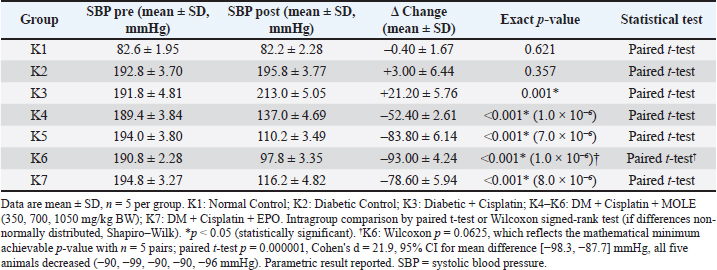
Induction of T2DM and cisplatin injuryAfter a 7-day acclimatization period with ad libitum access to standard pellet diet and filtered water, T2DM was induced on day 1 by intraperitoneal nicotinamide (NA; 230 mg/kg), followed by intravenous streptozotocin (STZ; 65 mg/kg) 15 minutes later. On day 3, fasting blood glucose (FBG) was measured after an overnight fast; rats with FBG > mg/dL (8.3 mmol/L) were confirmed diabetic and included in the study (Furman, 2021). Following a 2-day stabilization period, cisplatin (8 mg/kg BW) was administered as a single intravenous (IV) injection via the lateral tail vein on day 5, performed by a trained laboratory technician. No injection-site complications, acute adverse reactions, or mortality related to the IV administration procedure were observed in any animal. Vascular oxidative injury was induced by a single intravenous injection of cisplatin at 8 mg/kg BW. Dose selection was informed by our preliminary dose-finding study (Sunggoro et al., 2026), which demonstrated that both 6 and 8 mg/kg BW produced moderate, non-lethal hematological toxicity in the same streptozotocin–nicotinamide-induced T2DM Sprague-Dawley rat model with no mortality at either dose. The higher dose (8 mg/kg BW) was selected for the present study to ensure maximal induction of oxidative stress and vascular inflammatory responses—the primary endpoints of interest—while remaining within the established safety range confirmed by Sunggoro et al. (2026). Treatment protocolFollowing cisplatin administration, MOLE or erythropoietin (EPO) treatment was initiated 24 hours later (on day 6) and continued for 14 consecutive days. MOLE was freshly suspended in distilled water at the required concentration before each administration once daily via oral gavage using a stainless steel feeding needle. EPO was administered subcutaneously at a dose of 900 IU/kg BW/day three times weekly. This model represents a post-cisplatin therapeutic (restorative) intervention, rather than a preventive approach. EPO was selected as a positive control based on its documented pleiotropic effects beyond erythropoiesis, including anti-inflammatory, anti-apoptotic, and tissue-protective properties mediated through non-erythropoietic receptors (Brines and Cerami, 2012). All animals were fasted overnight and sacrificed on day 19 for blood collection and biochemical analysis. Diabetic status was reconfirmed at sacrifice; all rats in the diabetic groups maintained FBG > 150 mg/dL at the time of sacrifice, confirming sustained hyperglycemia throughout the experimental period. Measurement of SBPSBP was measured before and after the treatment period using a non-invasive tail–cuff plethysmography system (CODA, Kent Scientific, Torrington, CT). Prior to data collection, all rats underwent 3–5 days of acclimatization sessions with the restraint device and tail–cuff to minimize stress-induced measurement artifacts. Measurements were performed in a quiet, temperature-controlled room (22°C ± 2°C) at a fixed daily time window (09:00–12:00) to reduce circadian variability. Each recording session consisted of five consecutive inflation cycles; the first two were discarded as familiarization cycles, and the final three were averaged as the recorded value for that session. All measurements were performed by a single trained technician who was blinded to group allocation throughout. The intra-group coefficient of variation (CV) for SBP measurements, calculated from individual animal data (n=5 per group), was ≤ 4.2% across all groups and time points, confirming acceptable measurement reproducibility. Sample collection and serum preparationAt the end of the treatment period (day 19), final body weights were recorded for all groups. Rats were then fasted overnight and anesthetized via intraperitoneal administration of ketamine (80 mg/kg) and xylazine (10 mg/kg). Approximately 5 ml of blood was collected by cardiac puncture into plain Vacutainer tubes. After allowing the samples to clot for 30 minutes, they were centrifuged at 3,000 rpm at 4°C for 15 minutes. The resulting serum was aliquoted and stored at −80°C until biochemical analysis. Biochemical and immunological measurementsSerum levels of E-selectin, NF-κB p65, and CRP were quantified using commercially available rat-specific enzyme-linked immunosorbent assay (ELISA) kits (Elabscience Biotechnology Inc., Wuhan, China): Rat E-Selectin/CD62E ELISA Kit (Cat. No. E-EL-R0265; sensitivity: 0.19 ng/mL), Rat NF-κB p65 ELISA Kit (Cat. No. E-EL-R0674; sensitivity: 0.10 ng/mL), and Rat CRP ELISA Kit (Cat. No. E-EL-R0506; sensitivity: 0.47 ng/mL). Serum MDA levels, a biomarker of lipid peroxidation, were determined using the thiobarbituric acid reactive substances (TBARS) method with a commercially available assay kit (TBARS Assay Kit, Cat. No. 10009055, Cayman Chemical Company, Ann Arbor, MI, USA). MDA concentrations were calculated from a standard curve and expressed as nmol/mL. All assays were performed in duplicate according to the manufacturers’ protocols, and optical density was measured using a microplate reader (BioTek Epoch™, BioTek Instruments Inc., Winooski, VT, USA). Intra-assay and inter-assay coefficients of variation were maintained below 10% and 15%, respectively. Standard curves for ELISA assays achieved correlation coefficients (r²) ≥ 0.99. Outcome assessors performing all biochemical and immunological measurements were blinded to group allocation throughout the analytical process. Statistical analysisData are presented as mean ± standard deviation (SD). Data normality and homogeneity of variance were assessed using the Shapiro–Wilk and Levene’s tests, respectively. Differences among groups were analyzed using one-way analysis of variance (ANOVA) followed by Tukey’s honestly significant difference (HSD) post hoc test for normally distributed data, which inherently controls the family-wise error rate across all pairwise comparisons, thereby reducing the risk of type I errors from multiple group comparisons. For non-normally distributed data, the Kruskal–Wallis test was applied as an omnibus test of overall group differences. It should be noted that formal pairwise post hoc comparisons with correction for multiplicity were not performed for non-parametric data; therefore, specific between-group differences identified for these variables should be interpreted with caution. Intragroup comparisons of pre- and post-treatment SBP were evaluated using a paired t-test or the Wilcoxon signed-rank test, as appropriate. Correlations between SBP and biochemical markers (MDA, E-selectin, NF-κB, and CRP) were assessed using Spearman’s rank correlation analysis. Spearman correlations were performed at two levels: using group mean values (N=7 groups) and using individual animal data (N=35). Ninety-five percent confidence intervals for correlation coefficients were calculated using the Fisher z-transformation. For key between-group comparisons, 95% confidence intervals for mean differences and Cohen’s d effect sizes were calculated. Exact p-values are reported throughout; where software output indicated p < 0.0001, values are reported as such per GraphPad Prism 9.0 reporting convention. Statistical significance was defined as p < 0.05. All statistical analyses were performed using GraphPad Prism software (version 9.0). Ethical approvalAll procedures adhered to the ARRIVE guidelines and were approved by the Health Research Ethics Committee, Faculty of Medicine, Universitas Sebelas Maret (Approval No. 129/UN27.06.11/KEP/EC/2025) and the Research Ethics Commission, Faculty of Veterinary Medicine, Universitas Gadjah Mada (Approval No. 68/EC-FKH/Int./2025). ResultsAll 35 rats survived until the end of the study, with no premature deaths recorded. Final body weights at the time of sacrifice showed expected variations among groups: normal control rats (K1) maintained stable body weight, while diabetic groups (K2–K7) exhibited lower body weight compared to K1, consistent with the catabolic effects of NA-STZ-induced T2DM and cisplatin administration. However, body weight differences among the treatment groups (K4–K7) were not statistically significant (p > 0.05), indicating that the observed treatment effects on biomarkers were not confounded by differential body weight changes. Fasting blood glucose levels confirmed sustained diabetic status (FBG > 150 mg/dL) in all rats of the diabetic groups (K2–K7) at the time of sacrifice. Effect of MOLE on SBPBaseline SBP values were comparable across all groups before intervention (p>0.05). After cisplatin administration, the diabetic–cisplatin group (K3) showed a significant increase in SBP compared to the normal control (K1) and the untreated diabetic group (K2) (213.0 ± 5.05 vs. 82.2 ± 2.28 and 195.8 ± 3.77 mmHg, respectively; p<0.001), confirming the hypertensive effect of cisplatin in diabetic patients (Table 3). Table 3. Effect of MOLE on cisplatin-induced changes in SBP in diabetic rats.
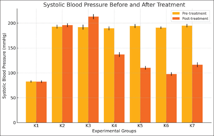
Therapy with MOLE produced a significant and dose-dependent reduction in SBP across all K4–K6 groups (−52.4, −83.8, and −93.0 mmHg, respectively), with the highest dose (1050 mg/kg BW, K6) showing the greatest antihypertensive effect (97.8 ± 3.34 mmHg), approaching normal values. The positive control group receiving EPO (K7) also showed a significant reduction in SBP (−78.6 ± 5.94 mmHg), although the effect was less pronounced compared to the high-dose MOLE group (Fig. 1). Further intragroup analysis confirmed these findings, with a significant increase in SBP in the diabetic–cisplatin group (K3) (p=0.001), while the MOLE (K4–K6) and EPO (K7) groups showed a significant reduction (p<0.001). These data indicate that MOLE exerts potent antihypertensive and vascular protective effects in cisplatin-treated diabetic rat models, especially at higher doses.
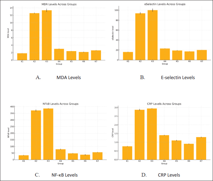
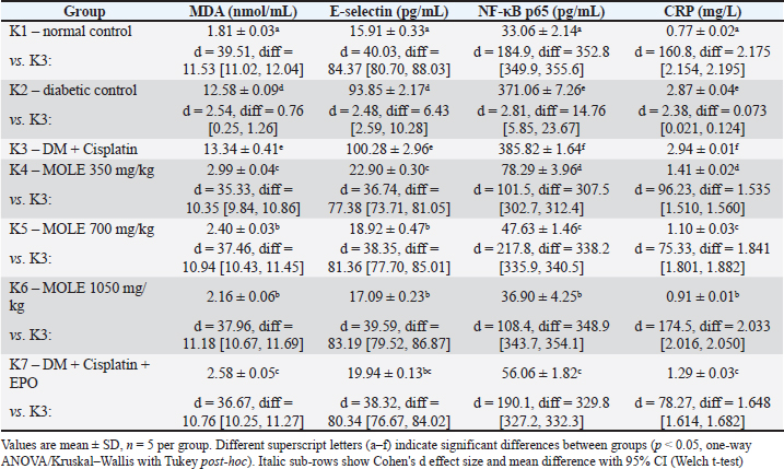
Fig. 1. Pre- and post-treatment systolic blood pressure in cisplatin-induced diabetic rats following M. oleifera Lam. extract administration. K1: Normal Control; K2: Diabetic Control; K3: Diabetic + Cisplatin; K4–K6: DM + Cisplatin + MOLE (350, 700, 1050 mg/kg BW); and K7: DM + Cisplatin + EPO. Effects of MOLE on oxidative stress, endothelial activation, and vascular inflammationDiabetic control rats (K2) and cisplatin-treated diabetic rats (K3) showed substantial increases in MDA (12.58 ± 0.09 and 13.34 ± 0.41 nmol/mL), E-selectin (93.85 ± 2.17 and 100.28 ± 2.96 nmol/mL), NF-κB (371.06 ± 7.26 and 385.82 ± 1.64 nmol/mL), and CRP (2.87 ± 0.04 and 2.94 ± 0.01 mg/l) compared to normal controls (K1) (p < 0.001). K3 showed consistently higher values than K2 across all biomarkers (MDA: p=3.88 × 10⁻³; E-selectin: p=4.41 × 10⁻³; NF-κB: p=2.18 × 10⁻³; CRP: p=5.51 × 10⁻³), confirming the additive effect of cisplatin on diabetes-induced vascular injury (Table 4; Fig. 2).
Fig. 2. Effects of M. oleifera Lam. leaf extract on oxidative stress, endothelial activation, and vascular inflammation in cisplatin-treated diabetic rats. (A) Serum malondialdehyde (MDA) levels, (B) E-selectin levels, (C) NF-κB levels, and (D) C-reactive protein (CRP) levels. K1: Normal Control; K2: Diabetic Control; K3: Diabetic + Cisplatin; K4–K6: DM + Cisplatin + MOLE (350, 700, 1050 mg/kg BW); and K7: DM + Cisplatin + EPO. Table 4. Post-treatment serum levels of oxidative stress and endothelial dysfunction biomarkers (mean ± SD) with effect sizes and 95% confidence intervals versus K3.
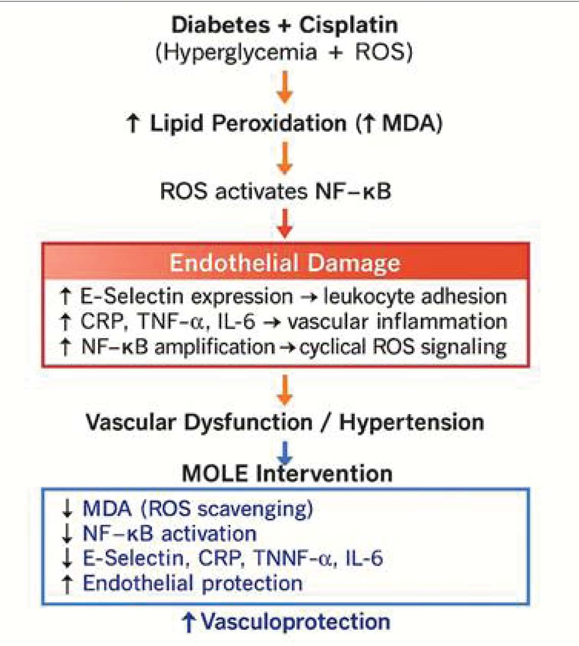
MOLE treatment resulted in significant and dose-dependent improvements in all studied biomarkers. High-dose MOLE (1050 mg/kg BW, K6) demonstrated the greatest therapeutic effect, reducing MDA levels to 2.16 ± 0.06 nmol/mL (an 83.8% decrease from K3) and E-selectin levels to 17.09 ± 0.23 nmol/mL (an 83.0% decrease), approaching normal values. NF-κB and CRP levels also decreased significantly to 36.90 ± 4.25 mg/L and 0.91 ± 0.01 mg/L, respectively, indicating a >90% reduction in vascular inflammatory signals compared with the untreated diabetic–cisplatin group (K3). The EPO (K7) treatment group, used as a positive control, showed a moderate decrease in inflammatory markers but was less effective compared to the high-dose MOLE group, particularly in suppressing oxidative stress (MDA) and endothelial adhesion molecule (E-selectin) expression. These findings suggest that MOLE exerts notable vascular protective effects by targeting multiple pathological pathways, including including inhibition of lipid peroxidation, reduction of endothelial activation, and reduction of the NF-κB-mediated inflammatory cascade (Fig. 4).
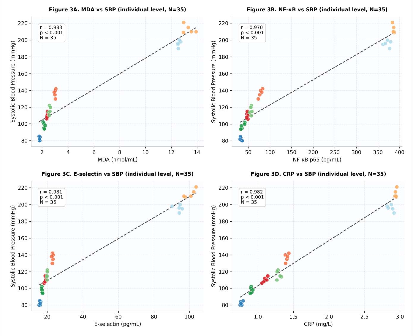
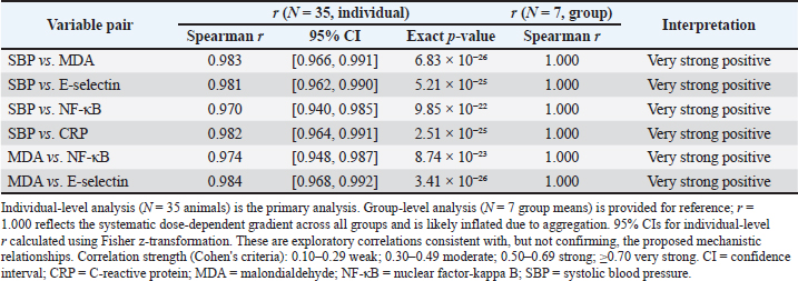
Fig. 4. Proposed mechanistic model illustrating how diabetes and cisplatin synergistically induce oxidative stress, lipid peroxidation, NF-κB activation, endothelial damage, vascular inflammation, and hypertension, as well as the inhibitory and vasculoprotective effects of MOLE. Moringa oleifera Lam. attenuates cisplatin-induced vascular injury in diabetic rats by reducing lipid peroxidation (↓MDA), inhibiting NF-κB activation, lowering endothelial adhesion molecule expression (↓E-selectin), and suppressing systemic inflammation (↓CRP, TNF-α, IL-6). These actions preserve endothelial integrity and protect against vascular dysfunction and hypertension. ROS, reactive oxygen species; MDA, malondialdehyde; NF-κB, nuclear factor-kappa B; CRP, C-reactive protein; TNF, tumor necrosis factor; IL, interleukin; MOLE, Moringa oleifera Lam. leaf extract. Correlation analysis between oxidative stress, endothelial activation, and vascular dysfunctionExploratory Spearman correlation analysis was performed at two levels: (1) using group mean values (N=7 groups) and (2) using individual animal data (N=35). At the individual level, very strong positive associations were observed between all biomarker pairs: SBP versus MDA r=0.983 (95% CI 0.966–0.991, p=6.83 × 10⁻²⁶); SBP versus E-selectin r=0.981 (95% CI 0.962–0.990, p=5.21 × 10⁻²⁵); SBP versus NF-κB r=0.970 (95% CI 0.940–0.985, p=9.85 × 10⁻²²); SBP versus CRP r=0.982 (95% CI 0.964–0.991, p=2.51 × 10⁻²⁵); MDA versus NF-κB r=0.974 (95% CI 0.948–0.987, p=8.74 × 10⁻²³); and MDA versus E-selectin r=0.984 (95% CI 0.968–0.992, p=3.41 × 10⁻²⁶) (Table 5). Group-level correlations (N=7) yielded r=1.000 for all pairs (p < 0.001), reflecting the systematic dose-dependent gradient across all groups. The near-identical individual- and group-level coefficients confirm that the strong associations are not attributable to aggregation bias but reflect genuine biological associations across individual animals. Nevertheless, these remain exploratory analyses consistent with, but not confirming, the proposed mechanistic relationships between oxidative stress, endothelial activation, and vascular dysfunction. Individual-level scatter plots are presented in Figure 3.
Fig. 3. Exploratory Spearman correlations between oxidative stress and inflammatory biomarkers with systolic blood pressure at the individual animal level (N=35). (A) MDA versus SBP: r=0.983, 95% CI [0.966, 0.991], p=6.83×10⁻²⁶. (B) NF-κB versus SBP: r=0.970, 95% CI [0.940, 0.985], p=9.85×10⁻²². (C) E-selectin versus SBP: r=0.981, 95% CI [0.962, 0.990], p=5.21×10⁻²⁵. (D) CRP versus SBP: r=0.982, 95% CI [0.964, 0.991], p=2.51×10⁻²⁵. Each dot represents one animal (n=5 per group, 7 groups). Dashed line shows linear regression. Correlations are exploratory and should not be interpreted as confirming direct causal relationships. MDA, malondialdehyde; NF-κB, nuclear factor-kappa B; CRP, C-reactive protein; and SBP, systolic blood pressure. Table 5. Exploratory Spearman correlation analysis between SBP and vascular biomarkers—individual-level (N=35) and group-level (N=7).
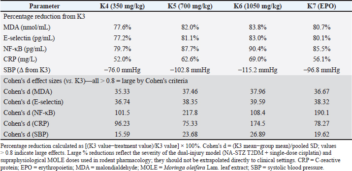
Summary of MOLE treatment efficacyTable 6 summarizes the percentage reductions relative to the untreated diabetic–cisplatin group (K3) to comprehensively illustrate the therapeutic effects of MOLE on all measured parameters. MOLE therapy demonstrated a clear dose-dependent protective effect, with the highest dose (1050 mg/kg BW, K6) consistently producing the greatest reductions in all biomarkers. Results in K6 demonstrated an 83.8% reduction in MDA, 83.0% in E-selectin, 90.4% in NF-κB, and 69.0% in CRP, accompanied by a 115.2 mmHg reduction in SBP compared to K3. It is noteworthy that the therapeutic output of high-dose MOLE (K6) showed greater reductions in the measured parameters compared with EPO (K7) in all parameters, indicating notable vascular protective efficacy. The medium-dose MOLE (700 mg/kg, K5) also demonstrated substantial protective effects, while the low dose (350 mg/kg, K4) provided moderate but still significant improvements. These findings collectively indicate that MOLE exerts a comprehensive and dose-dependent vascular protective effect through simultaneous reduction of oxidative stress, endothelial activation, and systemic inflammation. Table 6. Summary of MOLE treatment effects: percentage reduction from diabetic–cisplatin group (K3) and Cohen's d effect sizes.
DiscussionThis study examined whether MOLE could protect against vascular injury in rats carrying two simultaneous insults: T2DM induced by the nicotinamide–streptozotocin protocol and acute cisplatin administration. Across all dose groups, MOLE reduced circulating MDA, E-selectin, NF-κB p65, and CRP in a dose-dependent manner, and the highest dose brought SBP close to normal values. These patterns are consistent with antioxidant and anti-inflammatory activity, though the study relied on serum biomarkers and blood pressure rather than tissue histology or direct pathway assays. Any mechanistic interpretation must therefore be regarded as preliminary. The K3 group—diabetic rats given cisplatin—showed the highest values across all biomarkers, consistently exceeding the diabetic control (K2). Prior work has shown that cisplatin acutely raises HbA1c and provokes endothelial dysfunction on top of existing diabetic vasculopathy (Cameron et al., 2016), which helps explain why K3 was worse than K2 across every parameter. Both diabetes and cisplatin promote ROS generation through overlapping but distinct pathways; when combined, they appear to drive a more severe inflammatory response than either condition alone, as reflected in the K3 data (Clasen et al., 2021; An et al., 2023; Katanić Stanković et al., 2023). This additive pattern supports the rationale for a dual-injury model, though the relative contribution of each insult cannot be separated from these data. MOLE reduced MDA levels across all treatment doses, most markedly at 1050 mg/kg BW. This is consistent with reduced lipid peroxidation, an expected consequence of the extract’s polyphenolic load. HPLC–PDA analysis confirmed quercetin (0.2224 ± 0.0195 mg/g) and kaempferol (0.1701 ± 0.0189 mg/g) as the dominant flavonol constituents. Both compounds have well-characterized NF-κB inhibitory activity: quercetin blocks IκB-α phosphorylation and thereby prevents NF-κB p65 from entering the nucleus, while kaempferol also targets the p38 MAPK and JNK pathways, which generate TNF-α through NF-κB-independent routes (Kashyap et al., 2022). The high tannin content (177.15 mg TAE/g) adds a further layer of anti-inflammatory potential. The falls in circulating NF-κB p65 and E-selectin after MOLE treatment are therefore consistent with attenuation of NF-κB-mediated endothelial activation—but this interpretation is inferential. The study did not measure NF-κB nuclear translocation, phospho-IκB-α, or E-selectin expression in vascular tissue. Immunohistochemistry and Western blotting in future work will be needed to move from biomarker association to mechanistic confirmation. The observed fall in E-selectin is relevant because elevated E-selectin promotes leukocyte adhesion to the endothelium and is a recognized marker of endothelial activation and vascular inflammatory risk (Zhang et al., 2024). The parallel fall in CRP after MOLE treatment is also consistent with reduced NF-κB transcriptional activity, since CRP is an acute-phase reactant whose hepatic synthesis is driven partly by NF-κB-dependent cytokine production in the diabetic state (Ming-Kuen Tang et al., 2021). The fall in SBP—from 213.0 ± 5.05 mmHg in K3 to 97.8 ± 3.34 mmHg at the highest MOLE dose (K6)—was the most clinically tangible outcome in this study. Exploratory Spearman correlations using individual animal data (N=35) showed strong positive associations between SBP and all biomarkers (r=0.970–0.984, p < 0.001 throughout). This pattern is consistent with the idea that the antihypertensive effect reflects, at least in part, reduced endothelial inflammation and improved nitric oxide bioavailability—a mechanism reported previously for MOLE in resistance artery preparations and hypertensive rat models (Chichioco-Hernandez, 2019; Aekthammarat et al., 2020). However, because all biomarkers moved in the same dose-dependent direction, these correlations cannot be treated as independent mechanistic evidence. They are exploratory, and the most that can be said is that they are consistent with the proposed pathway. EPO was chosen as the positive control rather than a conventional antioxidant such as N-acetylcysteine because this study’s primary interest was vasculoprotection and endothelial anti-inflammatory signalling, not radical scavenging per se. EPO acts through non-erythropoietic receptors on endothelial and vascular smooth muscle cells to produce anti-inflammatory, anti-apoptotic, and tissue-protective effects (Brines and Cerami, 2012)—a multi-target profile that more closely resembles the proposed mechanism of MOLE than simple antioxidant supplementation would. K6 outperformed K7 across every parameter, which is consistent with a broader polyphenolic anti-inflammatory effect at the doses used. That said, EPO was used at a fixed dose without optimization, and the comparison must be read cautiously. Adding N-acetylcysteine or another antioxidant as a second comparator in future studies would sharpen the mechanistic picture. The percentage reductions in K6 relative to K3 (77%–90% across biomarkers) look large, and they are—but the context matters. These values arise from a worst-case dual-injury baseline in K3 combined with supraphysiological oral doses in rodent pharmacology; they do not reflect what one would expect in a clinical setting. The Cohen’s d values for the K3 versus K6 comparison (MDA d=37.96; E-selectin d=39.59; NF-κB d=108.43; CRP d=174.49; and SBP d=26.89) are enormous by any conventional benchmark, again reflecting the severity of the experimental conditions rather than a realistic clinical effect size. With only five animals per group, sampling variability can further inflate apparent magnitudes. Similar large reductions after MOLE treatment in cisplatin-exposed animals have been reported by others (Ahmed et al., 2020; Kilany et al., 2025; Anggraini et al., 2025; Sidharta et al., 2025), which provides contextual plausibility, but confirmatory studies with larger groups are needed before these numbers can be relied upon. Translating rodent doses to a clinical context requires caution. Using body surface area normalization (Reagan-Shaw et al., 2008), the rat doses of 350, 700, and 1050 mg/kg correspond to approximate human equivalent doses of roughly 3.9, 7.9, and 11.9 g/day for a 70-kg adult, compared with 1.9, 3.8, and 5.7 g/day for a 20-kg dog. The lowest dose is within the range that might be feasible as a food supplement; the higher doses would be difficult to administer routinely in clinical practice. These figures are rough estimates, and pharmacokinetic studies in the target species will be needed before any dosing proposal can be made. On the model side, the nicotinamide–streptozotocin protocol produces a T2DM phenotype—partial beta-cell destruction with preserved residual insulin secretion—that maps reasonably onto human T2DM (Furman, 2021), but not onto canine diabetes, which is predominantly insulin-dependent (Kim and Yun, 2025). The downstream vascular consequences of hyperglycemia, oxidative stress, and endothelial dysfunction are nevertheless shared across diabetes subtypes, so the oxidative injury pathway studied here may have some relevance beyond the rodent T2DM model. These results should be treated as preliminary, hypothesis-generating data—a starting point for species-specific work, not a basis for clinical recommendations. Several limitations of this study should be acknowledged. First, the sample size of five rats per group, while supported by a priori power analysis (Cohen’s d=2.0, α=0.05, power=0.80) and consistent with established preclinical practice, limits generalizability and may amplify apparent treatment effects through sampling variability. Second, the absence of vascular histopathological and immunohistochemical assessment precludes direct morphological confirmation of endothelial protection. The biomarkers measured (MDA, E-selectin, NF-κB p65, CRP) are circulating serum markers and do not provide direct evidence of tissue-level pathway modulation; vascular histopathology, immunohistochemical staining for NF-κB nuclear translocation, or Western blot confirmation of phosphorylated NF-κB would be needed to establish direct mechanistic evidence. Third, the 14-day treatment period represents a short-term intervention; long-term efficacy and safety remain to be determined. Fourth, the nicotinamide–streptozotocin rat model reflects human T2DM and does not replicate the predominantly insulin-dependent diabetes of dogs, the primary veterinary target species of interest. Species-specific differences in drug metabolism, pharmacokinetics, and pathophysiology preclude direct extrapolation to canine patients. Fifth, this study did not assess potential interactions between MOLE and cisplatin’s antitumor efficacy, which is critical for any proposed adjunctive therapy in oncology settings and must be addressed before clinical translation. Sixth, correlations were performed on group-level means as a primary analysis; individual-level analyses (N=35) were performed as supplementary and yielded near-identical results, confirming robustness. Seventh, only one batch was used for all animal experiments; inter-batch variability data are available, but longitudinal batch stability was not assessed. In summary, this study provides preliminary preclinical evidence consistent with vasculoprotective activity of MOLE in a diabetic rat model treated with cisplatin, mediated through mechanisms involving antioxidant and anti-inflammatory pathways. Validation through vascular histopathology, mechanistic pathway studies, pharmacokinetic profiling in target species, and assessment of MOLE–cisplatin antitumor interaction will be necessary before these findings can inform clinical or veterinary practice. ConclusionMoringa oleifera Lam. leaf extract (MOLE) reduced cisplatin-induced vascular toxicity in a diabetic rat model, as indicated by dose-dependent reductions in circulating MDA, E-selectin, NF-κB p65, and CRP levels, accompanied by normalization of SBP. These findings are consistent with antioxidant and anti-inflammatory mechanisms of vascular protection, supported by the presence of the NF-κB inhibitors quercetin and kaempferol identified by HPLC–PDA analysis. As preclinical observations obtained in a rodent model of T2DM, these results are hypothesis-generating and warrant further species-specific investigation—particularly in dogs receiving cisplatin-based chemotherapy for osteosarcoma, transitional cell carcinoma, and other malignancies—to establish safe and effective dosing protocols and to assess the potential of MOLE as adjunctive therapy in clinical veterinary and human oncology settings. Future studies should incorporate vascular histopathology, pharmacokinetic profiling, and assessment of MOLE–cisplatin antitumor interaction to fully characterize the safety and efficacy of this intervention. AcknowledgmentsThe authors thank all the staff of the Animal Research Facility Center for Food and Nutrition Studies, Gadjah Mada University, for their support in this study. FundingThis research received no specific grant from any funding agency in the public, commercial, or not-for-profit sectors. Authors’ contributionsAJS, PD, BP, BW, EPP, and RC designed the study protocol and interpreted the results. AJS performed the experiments, analyzed the data, drafted the manuscript, and prepared the figures. All authors reviewed and approved the final manuscript. Conflicts of interestThe authors declare no conflict of interest. Data availabilityAll data supporting the findings of this study are available within the manuscript or from the corresponding author upon reasonable request. ReferencesAcuram, L.K. and Chichioco Hernandez, C.L. 2019. Anti-hypertensive effect of Moringa oleifera Lam. Cogent Biol. 5(1), 1596526; doi:10.1080/23312025.2019.1596526 Aekthammarat, D., Tangsucharit, P., Pannangpetch, P., Sriwantana, T. and Sibmooh, N. 2020. Moringa oleifera leaf extract enhances endothelial nitric oxide production leading to relaxation of resistance artery and lowering of arterial blood pressure. Biomed. Pharmacother. 130, 110605. Ahmed, N.F., Sadek, K.M., Soliman, M.K., Khalil, R.H., Khafaga, A.F., Ajarem, J.S., Maodaa, S.N. and Allam, A.A. 2020. Moringa oleifera leaf extract repairs the oxidative misbalance following sub-chronic exposure to sodium fluoride in Nile tilapia Oreochromis niloticus. Animals 10(4), 626. Al-Malki, A.L. and El Rabey, H.A. 2015. The antidiabetic effect of low doses of Moringa oleifera Lam. seeds on streptozotocin induced diabetes and diabetic nephropathy in male rats. Biomed. Res. Int. 1(1), 381040. Anggraini, N.W.P., Sulistyowati, S., Soetrisno, S., Purwanto, B. and Dirgahayu, P. 2025. Effects of Moringa oleifera leaves on CRP, MDA, and sFlt-1 levels in a mouse model of preeclampsia. Open Vet. J. 15(4), 1784. An, Y., Xu, B.T., Wan, S.R., Ma, X.M., Long, Y., Xu, Y. and Jiang, Z.Z. 2023. The role of oxidative stress in diabetes mellitus-induced vascular endothelial dysfunction. Cardiovasc. Diabetol. 22(1), 237. Arifin, W.N. and Zahiruddin, W.M. 2017. Sample size calculation in animal studies using resource equation approach. Malays. J. Med. Sci. 24(5), 101–105. Brines, M. and Cerami, A. 2012. The receptor that tames the innate immune response. Mol. Med. 18(1), 486–496; doi:10.2119/molmed.2011.00414 Brown, A., Kumar, S. and Tchounwou, P.B. 2019. Cisplatin-based chemotherapy of human cancers. J. Cancer Sci. Ther. 11, 97. Cameron, A.C., Touyz, R.M. and Lang, N.N. 2016. Vascular complications of cancer chemotherapy. Can. J. Cardiol. 32, 852–862. Catchpole, B., Ristic, J.M., Fleeman, L.M. and Davison, L.J. 2005. Canine diabetes mellitus: can old dogs teach us new tricks?. Diabetologia 48, 1948–1956. Charan, J. and Kantharia, N.D. 2013. How to calculate sample size in animal studies?. J. Pharmacol. Pharmacother. 4(4), 303–306. Chiș, A., Noubissi, P.A., Pop, O.L., Mureșan, C.I., Fokam Tagne, M.A., Kamgang, R., Fodor, A., Sitar-Tăut, A.V., Cozma, A., Orășan, O.H., Hegheș, S.C., Vulturar, R. and Suharoschi, R. 2023. Bioactive compounds in Moringa oleifera: mechanisms of action, focus on their anti-inflammatory properties. Plants 13(1), 20. Chun, R., Knapp, D.W., Widmer, W.R., Glickman, N.W., DeNicola, D.B. and Bonney, P.L. 1996. Cisplatin treatment of transitional cell carcinoma of the urinary bladder in dogs: 18 cases (1983-1993). J. Am. Vet. Med. Assoc. 209, 1588–1591. Clasen, S.C., Dinh, P.C., Hou, L., Fung, C., Sesso, H.D. and Travis, L.B. 2021. Cisplatin, environmental metals, and cardiovascular disease: an urgent need to understand underlying mechanisms. Cardio-Oncology 7, 34. Da Silva Faria, M.C., Dos Santos, N.A., Rodrigues, M.A.C., Rodrigues, J.L., Junior, F.B. and Dos Santos, A.C. 2015. Effect of diabetes on biodistribution, nephrotoxicity and antitumor activity of cisplatin in mice. Chem. Biol. Interact. 229, 119–131. Dasari, S. and Tchounwou, P.B. 2014. Cisplatin in cancer therapy: molecular mechanisms of action. Eur. J. Pharmacol. 740, 364–378. Dugbartey, G.J., Peppone, L.J. and De Graaf, I.A.M. 2016. An integrative view of cisplatin-induced renal and cardiac toxicities: molecular mechanisms, current treatment challenges and potential protective measures. Toxicology 371, 58–66. Eslamifar, Z. and Sabbagh, S. 2020. A histopathological study of cisplatin-induced acute vascular injuries in vital organs and protective effect of Achillea millefolium. J. Pharm. Res. Int. 32(10), 56–69. Fatema, F., Nahar, S. and Akhter, Q.S. 2020. Cyclophosphamide induced anemia in Wistar rats: can it be prevented by Sajna (Moringa oleifera)?. Bangladesh. J. Physiol. 15(1), 11–16. Furman, B.L. 2021. Streptozotocin‐induced diabetic models in mice and rats. Curr. Protoc. 1(4), 78; doi:10.1002/cpz1.78 Gustafson, D.L., Page, R.L., Withrow, S.J., Vail, D.M. and Page, R.L. 2013. Cancer chemotherapy.In Withrow and MacEwen's Small Animal Clinical Oncology. Withrow, S.J., Vail, D.M. and Page, R.L International Diabetes Federation. 2021. IDF Diabetes Atlas. 10th ed. International Diabetes Federation, Brussels. Kashyap, P., Kumar, S., Riar, C.S., Jindal, N., Baniwal, P., Guiné, R.P.F., Correia, P.M.R., Mehra, R. and Kumar, H. 2022. Recent advances in drumstick (Moringa oleifera) leaves bioactive compounds: composition, health benefits, bioaccessibility, and dietary applications. Antioxidants 11(2), 402. Katanić Stanković, J.S., Selaković, D. and Rosić, G. 2023. Oxidative damage as a fundament of systemic toxicities induced by cisplatin—the crucial limitation or potential therapeutic target?. Int. J. Mol. Sci. 24, 14574. Kilany, O., Aldesouky, M. and Abdelnaeim, N.S. 2025. Cardioprotective effects of Moringa oleifera in cisplatin-intoxicated rats. Egypt. J. Vet. Sci. 56(11), 2821–2831; doi:10.21608/ejvs.2024.296784.2169 Kim, I. and Yun, J.H. 2025. Comparative pharmacological and pharmaceutical perspectives on antidiabetic therapies in humans, dogs, and cats. Pharmaceutics 17(9), 1098. Knapp, D.W., Glickman, N.W., Widmer, W.R., DeNicola, D.B., Adams, L.G., Kuczek, T., Bonney, P.L., DeGortari, A.E., Han, C. and Glickman, L.T. 2000. Cisplatin versus cisplatin combined with piroxicam in a canine model of human invasive urinary bladder cancer. Cancer. Chemother. Pharmacol. 46, 221–226. Liu, J., Han, X., Zhang, T., Tian, K., Li, Z. and Luo, F. 2023. Reactive oxygen species (ROS) scavenging biomaterials for anti-inflammatory diseases: from mechanism to therapy. J. Hematol. Oncol. 16(1), 116. Ma, X., Yan, L., Zhu, Q. and Shao, F. 2017. Puerarin attenuates cisplatin-induced rat nephrotoxicity: the involvement of TLR4/NF-κB signaling pathway. PLos One 12(2), 171612. Mcsweeney, K.R., Gadanec, L.K., Qaradakhi, T., Ali, B.A., Zulli, A. and Apostolopoulos, V. 2021. Mechanisms of cisplatin-induced acute kidney injury: pathological mechanisms, pharmacological interventions, and genetic mitigations. Cancers 13(7), 1572. Ming-kuen Tang, P., Zhang, Y.Y., Shuk-chun Hung, J., Yat-fai Chung, J., Huang, X.R., To, K.F. and Lan, H.Y. 2021. DPP4/CD32b/NF-κB circuit: a novel druggable target for inhibiting CRP-driven diabetic nephropathy. Mol. Ther. 29(1), 365–375. Pareek, A., Pant, M., Gupta, M.M., Kashania, P., Ratan, Y., Jain, V., Pareek, A. and Chuturgoon, A.A. 2023. Moringa oleifera: an updated comprehensive review of its pharmacological activities, ethnomedicinal, phytopharmaceutical formulation, clinical, phytochemical, and toxicological aspects. Int. J. Mol. Sci. 24, 2098. Percie Du Sert, N., Hurst, V., Ahluwalia, A., Alam, S., Avey, M.T., Baker, M., Browne, W.J., Clark, A., Cuthill, I.C., Dirnagl, U., Emerson, M., Garner, P., Holgate, S.T., Howells, D.W., Karp, N.A., Lazic, S.E., Lidster, K., MacCallum, C.J., Macleod, M., Pearl, E.J., Petersen, O.H., Rawle, F., Reynolds, P., Rooney, K., Sena, E.S., Silberberg, S.D., Steckler, T. and Würbel, H. 2020. The ARRIVE guidelines 2.0: updated guidelines for reporting animal research. PLos Biol. 18(7), e3000410; doi:10.1371/journal.pbio.3000410 Reagan-Shaw, S., Nihal, M. and Ahmad, N. 2008. Dose translation from animal to human studies revisited. FASEB. J. 22(3), 659–661; doi:10.1096/fj.07-9574LSF Saito, Y., Sakamoto, T., Takekuma, Y., Kobayashi, M., Okamoto, K., Shinagawa, N., Shimizu, Y., Kinoshita, I. and Sugawara, M. 2022. Diabetes mellitus degenerates cisplatin-induced nephrotoxicity in short hydration method: a propensity score-matching analysis. Sci. Rep. 12, 21819. Scioli, M.G., Storti, G., D’Amico, F., Rodríguez Guzmán, R., Centofanti, F., Doldo, E., Céspedes Miranda, E.M. and Orlandi, A. 2020. Oxidative stress and new pathogenetic mechanisms in endothelial dysfunction: potential diagnostic biomarkers and therapeutic targets. J. Clin. Med. 9(6), 1995. Sidharta, B.R., Purwanto, B., Wasita, B., Widyaningsih, V., Soetrisno, S., Cilmiaty, R. and Ardyanto, T. 2025. The effect of Moringa oleifera ethanol extract on improving cisplatin induced liver cells damages. Asian Pac. J. Cancer Prev. 26(6), 2175–2183. Soliman, M.M., Aldhahrani, A., Alkhedaide, A., Nassan, M.A., Althobaiti, F. and Mohamed, W.A. 2020. The ameliorative impacts of Moringa oleifera leaf extract against oxidative stress and methotrexate-induced hepato-renal dysfunction. Biomed. Pharmacother. 128, 110259. Sung, H., Ferlay, J., Siegel, R.L., Laversanne, M., Soerjomataram, I., Jemal, A. and Bray, F. 2021. Global cancer statistics 2020: GLOBOCAN estimates of incidence and mortality worldwide for 36 cancers in 185 countries. CA. Cancer J. Clin. 71(3), 209–249. Sunggoro, A.J., Dirgahayu, P., Purwanto, B., Pamungkasari, E.P., Warsita, B., Cilmiaty, R., Wardhani, D.T.M. and Dewi, Y.H.P.K. 2026. Determination of the optimal cisplatin dose to induce anemia in streptozotocin-nicotinamide-induced type 2 diabetic rats: a preliminary study. Open Vet. J. 16(2), 1090–1111. Verma, A.R., Vijayakumar, M., Mathela, C.S. and Rao, C.V. 2009. In vitro and in vivo antioxidant properties of different fractions of Moringa oleifera leaves. Food Chem. Toxicol. 47, 2196–2201. Volpe, C.M.O., Villar-Delfino, P.H., Dos Anjos, P.M.F. and Nogueira-Machado, J.A. 2018. Cellular death, reactive oxygen species (ROS) and diabetic complications. Cell. Death. &. Dis. 9(2), 119. Wang, S., Liu, Y., Sun, Q., Zeng, B., Liu, C., Gong, L., Wu, H., Chen, L., Jin, M., Guo, J., Gao, Z. and Huang, W. 2023. Triple cross‐linked dynamic responsive hydrogel loaded with selenium nanoparticles for modulating the inflammatory microenvironment via PI3K/Akt/NF‐κB and MAPK signaling pathways. Adv. Sci. 10(31), 2303167. Withrow, S.J., Vail, D.M. and Page, R.L. 2013. Withrow and MacEwen’s Small Animal Clinical Oncology. Zhang, J., Huang, S., Zhu, Z., Gatt, A. and Liu, J. 2024. E-selectin in vascular pathophysiology. Front. Immunol. 15, 1401399. | ||
| How to Cite this Article |
| Pubmed Style Sunggoro AJ, Purwanto B, Dirgahayu P, Wasita B, Cilmiaty R, Pamungkasari EP. Vasculoprotective and antioxidant effects of Moringa oleifera Lam. leaf extract in cisplatin-treated diabetic rats: Role of malondialdehyde and E-selectin. doi:10.5455/OVJ.2026.v16.i7.29 Web Style Sunggoro AJ, Purwanto B, Dirgahayu P, Wasita B, Cilmiaty R, Pamungkasari EP. Vasculoprotective and antioxidant effects of Moringa oleifera Lam. leaf extract in cisplatin-treated diabetic rats: Role of malondialdehyde and E-selectin. https://www.openveterinaryjournal.com/?mno=307111 [Access: July 11, 2026]. doi:10.5455/OVJ.2026.v16.i7.29 AMA (American Medical Association) Style Sunggoro AJ, Purwanto B, Dirgahayu P, Wasita B, Cilmiaty R, Pamungkasari EP. Vasculoprotective and antioxidant effects of Moringa oleifera Lam. leaf extract in cisplatin-treated diabetic rats: Role of malondialdehyde and E-selectin. doi:10.5455/OVJ.2026.v16.i7.29 Vancouver/ICMJE Style Sunggoro AJ, Purwanto B, Dirgahayu P, Wasita B, Cilmiaty R, Pamungkasari EP. Vasculoprotective and antioxidant effects of Moringa oleifera Lam. leaf extract in cisplatin-treated diabetic rats: Role of malondialdehyde and E-selectin. doi:10.5455/OVJ.2026.v16.i7.29 Harvard Style Sunggoro, A. J., Purwanto, . B., Dirgahayu, . P., Wasita, . B., Cilmiaty, . R. & Pamungkasari, . E. P. (2026) Vasculoprotective and antioxidant effects of Moringa oleifera Lam. leaf extract in cisplatin-treated diabetic rats: Role of malondialdehyde and E-selectin. doi:10.5455/OVJ.2026.v16.i7.29 Turabian Style Sunggoro, Agus Jati, Bambang Purwanto, Paramasari Dirgahayu, Brian Wasita, Risya Cilmiaty, and Eti Poncorini Pamungkasari. 2026. Vasculoprotective and antioxidant effects of Moringa oleifera Lam. leaf extract in cisplatin-treated diabetic rats: Role of malondialdehyde and E-selectin. doi:10.5455/OVJ.2026.v16.i7.29 Chicago Style Sunggoro, Agus Jati, Bambang Purwanto, Paramasari Dirgahayu, Brian Wasita, Risya Cilmiaty, and Eti Poncorini Pamungkasari. "Vasculoprotective and antioxidant effects of Moringa oleifera Lam. leaf extract in cisplatin-treated diabetic rats: Role of malondialdehyde and E-selectin." doi:10.5455/OVJ.2026.v16.i7.29 MLA (The Modern Language Association) Style Sunggoro, Agus Jati, Bambang Purwanto, Paramasari Dirgahayu, Brian Wasita, Risya Cilmiaty, and Eti Poncorini Pamungkasari. "Vasculoprotective and antioxidant effects of Moringa oleifera Lam. leaf extract in cisplatin-treated diabetic rats: Role of malondialdehyde and E-selectin." doi:10.5455/OVJ.2026.v16.i7.29 APA (American Psychological Association) Style Sunggoro, A. J., Purwanto, . B., Dirgahayu, . P., Wasita, . B., Cilmiaty, . R. & Pamungkasari, . E. P. (2026) Vasculoprotective and antioxidant effects of Moringa oleifera Lam. leaf extract in cisplatin-treated diabetic rats: Role of malondialdehyde and E-selectin. doi:10.5455/OVJ.2026.v16.i7.29 |


