Open Veterinary Journal, (2026), Vol. 16(7): 4774-4782
Research Article
10.5455/OVJ.2026.v16.i7.56
Prenatal exposure to ochratoxin A induces anxiety- and depressive-like behaviors associated with brain oxidative stress in adult mice
Fatima Ez-Zahraa Saad1, Kamal Smimih2, Bilal El-Mansoury1, Manal Khanouchi1, Mustapha
Agnaou3, Mohamed Marghich1, Abdellah Zinedine4, Abdelali Bitar1 and Omar El-Hiba1*
1Nutritional Physiopathology, Neuroscience and Toxicology Team, Laboratory of Anthropogenic, Biotechnology, and Health, Faculty of Sciences, Chouaib Doukkali University, El Jadida, Morocco
2Biological Engineering Laboratory, Faculty of Sciences and Techniques (FST), Sultan Moulay Slimane University, Beni Mellal Morocco
3Laboratory of “Aquatic Systems: Marine and Continental Ecosystems”, Faculty of Science, Ibn Zohr University, Agadir, Morocco
4Team of Applied Microbiology and Biotechnologies, BioMare Laboratory, Faculty of Sciences, Chouaib Doukkali University, El Jadida, Morocco
*Corresponding Author: Omar El-Hiba. Nutritional Physiopathology, Neuroscience and Toxicology Team, Laboratory of Anthropogenic, Biotechnology, and Health, Faculty of Sciences, Chouaib Doukkali University, El Jadida, Morocco.
Email: elhiba.o [at] ucd.ac.ma
Submitted: 22/01/2026 Revised: 27/05/2026 Accepted: 11/06/2026 Published: 20/07/2026
© 2025 Open Veterinary Journal
This is an Open Access article distributed under the terms of the Creative Commons Attribution-Non Commercial-No Derivatives License (http://creativecommons.org/licenses/by-nc-nd/4.0/), which permits non-commercial re-use, distribution, and reproduction in any medium, provided the original work is properly cited, and is not altered, transformed, or built upon in any way.
Abstract
Background: Mycotoxins are secondary compounds made by certain fungi that are not necessary to ensure the normal reproduction and survival of a fungus but can induce physiological, pathological, and biochemical alterations in a wide variety of organisms. Ochratoxin A (OTA) is one of the most well-known mycotoxins generated in nature by Aspergillus and Penicillium species. It is widely found in contaminated food and feed such as wheat, corn, barley, olives, beans, figs, nuts, and grapes.
Aim: This study aimed to evaluate the effects of prenatal OTA exposure in Swiss albino mice on male offspring during adulthood, focusing on mood disorders and brain oxidative stress.
Methods: Pregnant female mice with confirmed vaginal plugs were randomly assigned to two groups: OTA (0.25 mg/kg, i.p.) or control (0.9% NaCl). The offspring were allowed to reach adulthood before undergoing behavioral assessments, including the forced swim test, elevated plus maze test, open field test, and tail suspension test. Brain oxidative stress markers were also measured.
Results: Adult male offspring prenatally exposed to OTA exhibited significant mood alterations in adulthood. These behavioral changes were associated with elevated oxidative stress in brain tissue.
Conclusion: Prenatal OTA exposure exerts potent neurotoxic effects on the developing brain, resulting in persistent mood disturbances and oxidative stress, highlighting the potential risks of OTA exposure during early development.
Keywords: Ochratoxin A, Prenatal exposure, Brain development, Mood disorders, Mice.
Introduction
Mycotoxins are toxic by-products produced by certain fungal species that significantly affect the safety of food and animal feed safety and thus represent a risk to both human and animal health. The damaging health impacts of mycotoxins seen in humans and animals are diverse and include carcinogenicity, nephrotoxicity, teratogenicity, neurotoxicity, hepatotoxicity, reproductive and developmental toxicity, and digestive issues (Pleadin et al., 2019). There are various ways of exposure to mycotoxins, including intake, inhalation, dermal absorption through skin contact, as well as breastfeeding transmission (Reboux, 2006). Ochratoxins are a group of mycotoxins usually generated by the species Aspergillus and Penicillium as a secondary metabolite, and they are divided into three different types: ochratoxin A, ochratoxin B, and ochratoxin C (Obafemi et al., 2023). Ochratoxin A (OTA) is the most widespread and harmful of the ochratoxins (Kumar et al., 2020). OTA is a low molecular weight organic toxin (403.82 g/mol) that has high thermal stability, which explains its resistance to food treatment conditions at temperatures ranging from 80°C to 121°C and its widespread presence in processed food (Dahal et al., 2016; Milani and Heidari, 2017 )
OTA is recognized for its nephrotoxic and hepatotoxic effects. However, studies regarding the neurotoxic effects of OTA remain limited, although current evidence suggests a considerable neurotoxic potential. The main potential pathways through which OTA causes neurotoxicity are oxidative damage to DNA, proteins, and lipids, and apoptosis (Nourbakhsh and Tajbakhsh, 2021), and epigenetic alterations are direct mechanisms of OTA-induced neurotoxicity (Babayan et al., 2020). Notably, OTA could be linked to the development of certain types of neurodegenerative disorders (like Alzheimer’s disease and Parkinson’s disease), in which apoptotic mechanisms play a central role (Zhang et al., 2009).
Since studies evaluating the possible effects of OTA on the developing brain are lacking, the current study aims to assess the neurobehavioral consequences of prenatal exposure to OTA, focusing on anxiety- and depression-related behaviors. We also evaluate potential oxidative stress in the brains of mice prenatally exposed to OTA in adulthood.
Materials and Methods
Chemicals
OTA was purchased from the Trilogy Analytical Laboratory (Washington, Missouri, United States) with an analytical-grade purity (98%) and used without further purification.
Animals
The study was conducted exclusively on adult virgin female Swiss albino mice weighing between 28 and 30 g. The animals were held in Plexiglas cages with unrestricted access to water and food in the animal care building of the Faculty of Science at Chouaib Doukkali University over a period of multiple generations, in carefully controlled environmental conditions with a maintained room temperature of 25°C and a 12-hour light/dark cycle. All experiments on animals carried out as part of this research were authorized by the Moroccan Ethics Committee of the Moroccan Society for Ethics and Animal Research (MECAR-MoSEAR, ref. UCD-FSJ-04/2025).
Treatment with OTA
Pregnant female mice (n=10) were divided randomly into two groups on their first day of gestation, as determined by the detection of a vaginal plug. Group 1 (OTA): treated with ochratoxin A (0.25 mg/kg, i.p., in saline solution, 0.9% NaCl) on the 10th day of gestation. The 10th day of gestation was selected because it represents a critical period of embryonic development in mice, corresponding to key stages of organogenesis and neural tube formation (Smimih et al., 2023; Smimih et al., 2026). Group 2 (control): treated with an identical volume of buffered saline solution (0.9% NaCl, i.p.) on the 10th day of gestation. For the next experiments, a total of 20 male offspring pups (2 pups per pregnant female, to avoid litter effects) in each study (five mice from each group were used for a maximum of two behavioral tests). The combination of tests was carefully selected to avoid any interference or habituation effects. All experiments were conducted on male pups, whose sex was subsequently confirmed in adulthood.
Behavioral study: study of mood disorders
The animals in each group were placed in the experimental room to acclimate to the new environment. All experiments were conducted in a quiet room with minimal external noise disturbances in order to reduce stress factors that could influence animal behavior. The animals then underwent a series of neurobehavioral tests using various devices. The devices were cleaned with 70% ethanol after each trial.
Elevated plus maze test
The EPM test was also used to examine reactions related to anxiety in mice. The method of study consisted of positioning every mouse tested on the EPM’s central platform, having the upper section of the animal's body confront the closed arm, and allowing the animal to freely explore the maze for 5 minutes. This test was conducted in a dimly lit, quiet environment. When an animal extends all four paws beyond the line separating the central square from the open arms, it is considered to have entered an arm. The duration spent in the maze's open arms and the number of entries were recorded (Kruk-Slomka et al., 2025).
Open field test
The open field assay is a technique designed to study both overall locomotion behavior and anxiety-like symptoms in rodents (Seibenhener and Wooten, 2015). Briefly, we placed each mouse separately in the center of the open field (50 × 50 × 30 cm), to freely explore the arena for 5 minutes. The animal's movements were freely recorded for 5 minutes by a camera mounted at the top of the device. The videos were then reviewed. Over this period, the time spent by the mice in the middle and in the corners of the box was recorded. Anxious animals usually stay away from the middle of the open area, which corresponds to the middle section of the device, which is more exposed and farther from the walls (El Hiba et al., 2012; Abbaoui et al., 2016; Smimih et al., 2023).
Forced swim test
This test is adapted to study depressive-like behaviors in rodents. Each mouse is given 5 minutes in a water-filled (3 l) glass basin at a temperature of 22°C ± 1°C and a depth of approximately 20 cm, at which point the animal would be able to swim independently. The water was changed regularly among the animals to maintain consistent experimental conditions. After each session, the mice were gently dried and then placed in a heated cage for a short recovery period before being returned to their housing cages. The animal was regarded as immobile if it stopped swimming, remaining with its head above the water surface, moving just enough to stay afloat, making no attempt to move horizontally, and showing no desire to escape (Brandão et al., 2023). The immobility time was recorded (Bao et al., 2023). Prolonged immobilization is linked to behavior that is considered desperate, whilst less immobilization is an index of antidepressant effects (Önel et al., 2023).
Tail suspension test
The tail suspension test is a behavioral evaluation method employed to measure behavioral despair in mice. In this test, the mice were individually hung above a table by the tail at a height of approximately 60 cm from the ground, with a tape fixed 1 cm from the end of the tail. Complete immobilization was noted for a duration of 6 minutes. Mice were classified as immobilized when suspended in a passive position and kept completely still (Sowa-Kućma et al., 2025). The mean immobility time was calculated and used as an indicator of despair-like behavior.
Oxidative stress biomarkers measurement
Mice brains were collected under anaesthesia, then measured and rinsed using Tris buffer (100 mM; pH 7.4; 4°C), and the whole brain tissue was used for biochemical analyses. The temperature of all processes was kept at 4°C. With a view to limiting the effects of mass on biomarkers, the samples were evaluated in advance, and samples from the same range of tissue mass were chosen for testing. The brain specimens were subsequently stored at −80°C for the biomarker analyses (Lamine et al., 2023). Biomarker enzymatic activity for glutathione-S-transferase (GST), catalase (CAT), and acetylcholinesterase (AChE) was measured using spectrophotometry with wavelengths of 340 nm, 240 nm, and 420 nm, respectively, in accordance with the procedures developed by Habig et al. (1974); Aebi (1984) and Ellman et al. (1961) as applied in recent toxicological studies (Singh et al., 2024; Montejo et al., 2025). The level of malondialdehyde (MDA) was quantified based on the absorbance of MDA at 530 nm (Lamine et al., 2023), as described by Sunderman et al. (1985).
Statistical analysis
The statistical data were analysed using Sigma Plot 12.0 software. All data were analysed using Student’s t-test. The results were presented as mean ± SEM. A p-value < 0.05 was considered statistically significant. For all data provided, we checked the normality using the Shapiro–Wilk test before proceeding to the Student’s t-test.
Ethical approval
All experiments on animals carried out as part of this research were authorized by the Moroccan Ethics Committee of the Moroccan Society for Ethics and Animal Research (MECAR-MoSEAR, ref. UCD-FSJ-04/2025).
Results
Neurobehavioral study: study of mood disorders
Elevated plus maze test
Using the plus maze test, we observed a very significant decrease in the time spent in open arms among the mice prenatally treated with OTA versus control group (C) (p=0.008) (Fig. 1A). Also, the number of visits to the open arms showed a significant decrease in OTA-treated mice versus controls (p=0.049) (Fig. 1B).
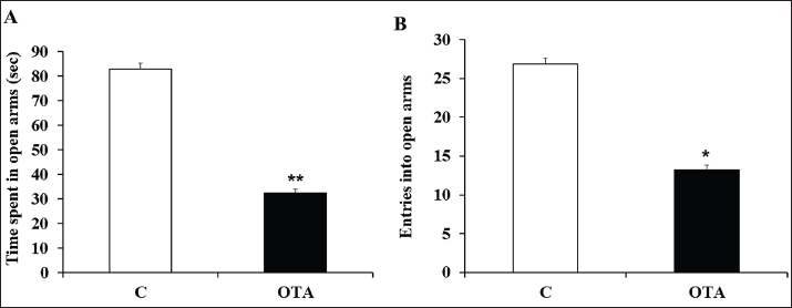
Fig. 1. Graphical representation of time spent in open arms (A) and number of entries into open arms (B) in the elevated plus maze for the different groups studied. C: control; OTA: mice prenatally treated with ochratoxin A. Data are presented as mean values ± SEM. ** p < 0.01 versus C. * p < 0.05 versus C.
Open field test
To confirm the previous findings of the EPM test, we evaluated anxiety via the OF test. Our results showed that OTA-exposed mice spent a significantly increased time in the corners (peripheral area) of the OF versus the control mice (C) (p=< 0.001) (Fig. 2).
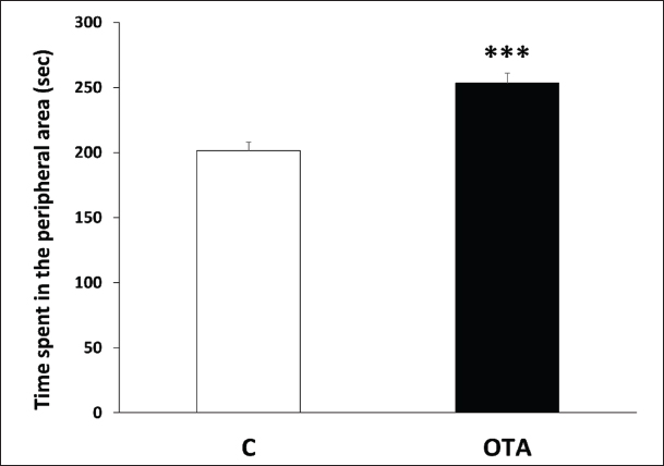
Fig. 2. Histogram showing the time spent in the peripheral area of the open field test for the different studied groups. C: control; OTA: mice prenatally treated with ochratoxin A. Data are presented as group mean values ± SEM. ***p < 0.001 versus C.
Forced swim test
We also studied depressive-like behaviors following prenatal OTA exposure in our mice using the forced swim test (FST). Our findings revealed a highly significant rise in immobility time, regarded as a depressive-like behavior, in OTA-treated mice versus controls (p=0.004) (Fig. 3).
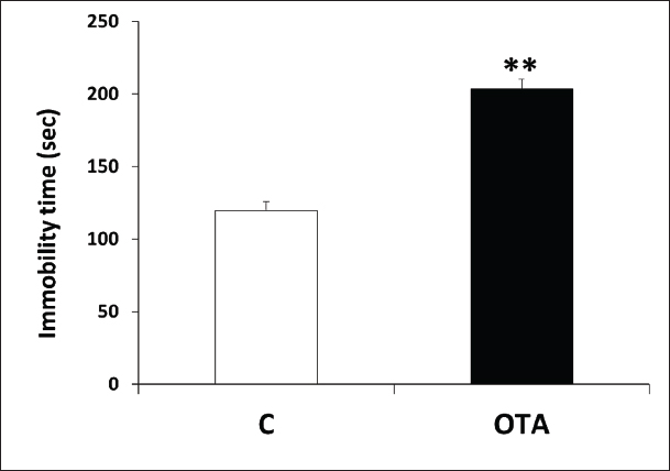
Fig. 3. Graph showing immobility time in the forced swimming test in the two groups studied. C: control; OTA: mice prenatally treated with ochratoxin A. Data are reported as mean values ± SEM. **p < 0.01 versus C.
Tail suspension test
We also used the tail suspension test (TST) to confirm and support the FST findings. The TST demonstrated a highly significant increase in the immobility time in mice prenatally exposed to OTA compared to the control group (p < 0.001), confirming the depressive-like behavior in OTA-treated mice (Fig. 4).
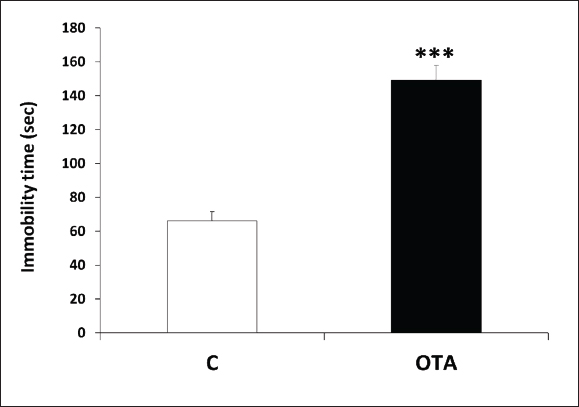
Fig. 4. Graphs representing immobility time in the tail suspension test in the two groups studied. C: control; OTA: mice given ochratoxin A. The data are presented as mean values ± SEM. ***p < 0.001 versus C.
Oxidative stress biomarkers measurement
Our results showed a highly significant increase in AChE and GST activities (p ≤ 0.001 and p ≤ 0.001, respectively), along with MDA levels (p ≤ 0.001) in OTA mice versus controls. While a trend toward a decreases in CAT activity was observed, it did not reach the significance level (p=1,000) in OTA-treated mice compared to controls (Fig. 5).
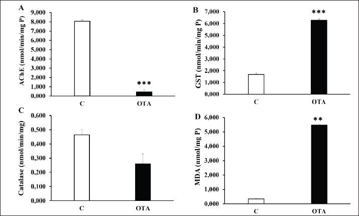
Fig. 5. Graphical representation of oxidative stress biomarker measurements in control and OTA-treated mice. (A): acetylcholinesterase (AChE). (B): glutathione S-transferase (GST). (C): catalase (CAT). (D): malondialdehyde (MDA). C: control (n=7); OTA: mice exposed prenatally to ochratoxin A (n=7). The data are presented as group mean values ± S.E.M. *** p < 0.001 versus C.
Discussion
Research on the neurotoxicity of ochratoxin A has expanded significantly in recent years (Doi and Uetsuka, 2011; Obafemi et al., 2023; García-Esparza et al., 2025). However, certain specific aspects, such as the exact mechanism of action and the role of OTA in human neurodegenerative diseases, remain insufficiently explored (Nourbakhsh and Tajbakhsh, 2021; Serrano-Civantos et al., 2025). Most of them do not provide strong evidence on the effect of prenatal OTA exposure during pregnancy on pups’ brain development, especially on the behavioral state, including anxiety control and depressive-like behaviors, are lacking. The current study clearly reveals that prenatal exposure to OTA induces long-lasting anxiety and depressive-like behaviors in mice, along with elevated brain oxidative stress. These results sustain the neurotoxic properties of OTA, extending current research beyond its already well-identified nephrotoxic and hepatotoxic aspects. Depressive-like behavior was particularly apparent in the FST and the TST, while anxiety was shown via the EPM and OF tests. This could be attributed to disrupted brain circuits and structures involved in regulating these behavioral aspects as a result of prenatal exposure to ochratoxin A, and these detrimental effects continued into adulthood.
In an earlier study in 1983, the administration of OTA to pregnant mice at doses of 1.25 and 2.25 mg/kg on gestational days 15 to 17 resulted in their offspring showing considerable delays in postnatal development. The offspring were assessed for swimming (days 6 to 20), surface righting (days 3 to 12), and pivoting (days 7, 9, and 11), with all three tests revealing statistically significant differences, which indicated impaired brain development (Poppe et al., 1983). These behavioral alterations could be a result of persistent brain oxidative stress, as our findings showed elevated oxidative stress marker activity in prenatally OTA-treated mice, which could affect brain circuits involved in this processing. In vitro studies further support this neurotoxicity, as OTA was found to be toxic to the Neuro-2a neuronal cell line. MTT and LDH assays demonstrated that OTA caused cell death in a dose-dependent manner in Neuro-2a cells, accompanied by elevated levels of reactive oxygen species (ROS) and MDA, as well as a dose-dependent loss of mitochondrial membrane potential (Bhat et al., 2018). Similarly, another study showed that exposure to OTA for 48 hours triggered oxidative stress in normal astrocytes, which is characterized by a reduction in the factors regulating the oxidative stress response (Von Tobel et al., 2014). OTA was also found to create oxidative stress across different parts of the mice's midbrain and hippocampus, leading to impaired brain development (Sava et al., 2006). Recently, OTA has been shown to alter behavior and neurochemical markers related to oxidative stress non-protein thiols (NPSH), glutathione peroxidase (GPx), glutathione-S-transferase (GST), and glutathione reductase (GR) in adult zebrafish, indicating its potential impact on the CNS at low doses (Valadas et al., 2021). Interestingly, OTA treatment was shown to induce apoptotic lesions in mouse blastocysts via the production of ROS, as well as stimulating mitochondria-dependent apoptotic signalling pathways that impair embryonic development thereafter (Hsuuw et al., 2013).
A study conducted by Mateo et al. also revealed that OTA could alter in vivo neurogenesis in the hippocampus and induce side reactions in the brain tissue cells of adult mice when given in accumulated doses (Mateo Jiménez et al., 2022), which could be partly linked to anxiety-like behaviors. On the other hand, depressive-like behavior was studied by Tanaka et al. (2016) who investigated the impact of exposure to OTA on GABAergic interneurons. They revealed that the malfunctioning of these GABAergic interneurons can result in a range of behavioral neuropsychiatric issues, notably anxiety and depression (Tanaka et al., 2016). Another study also demonstrated a link between the reduction in these interneurons and anxiety-like behaviors in mice (Bi et al., 2013). The anxiolytic effect of GABA is known to occur by blocking the hypothalamic–pituitary–adrenal axis through GABAergic neuronal transmission to the paraventricular nuclei in the hypothalamus, causing hyperpolarization and suppression of the target neurons (Zhu et al., 2019; Tafet and Nemeroff, 2020). Alternatively, monoamine neurotransmitters are well known to control mood state, and their dysregulation is associated with anxiety and depressive-like behaviors. Notably, OTA actually builds up in several brain regions, notably the hippocampus, cortex, gray matter, and striatum, where big quantities of receptors for dopamine are found. Importantly, OTA is able to cross the blood–brain barrier, which allows its access to the central nervous system and its accumulation in different brain regions (Belmadani et al., 1998; Belmadani et al., 1999). The neurotoxicity of OTA causes severe dopamine loss, leading to Parkinson-like symptoms, as well as low serotonin levels, causing further depression (Brahmi et al., 2025). Indeed, in a study by Zhang et al. (2009) the authors showed that OTA could contribute to the development of neurodegenerative diseases (e.g., Alzheimer's and Parkinson's diseases), in which apoptotic mechanisms play a central role (Zhang et al., 2009). In summary, our findings reinforce the necessity of reducing OTA exposure during pregnancy and the early stages of life, given that even minimal exposure can lead to serious enduring behavioral and neurochemical changes.
Conclusion
Prenatal exposure to OTA induces persistent anxiety-like and depressive-like behaviors in adult mice, as evidenced by concordant alterations observed in the elevated plus maze, open field, forced swim, and tail suspension tests. These neurobehavioral disturbances are accompanied by a significant increase in cerebral oxidative stress, characterized by increased lipid peroxidation and changes in antioxidant and detoxification enzyme activities. While the study presented some limitations, such as the use of one dose of OTA (based on our preliminary trials), our data strengthen the powerful neurotoxicity of mycotoxins on brain tissue functions and/or structure, with potent and permanent post-anal neurobehavioral disabilities. To complete the study, further investigations are needed to well define the neuro-glial mechanisms underlying these abnormalities, as well as a pharmacological approach to reverse or alleviate the OTA-induced brain injuries.
Acknowledgments
The authors express their deep thanks to all those who participated in the success of this work, particularly Prof. Rahmoun Miloud for English revision.
Funding
The study received funding from FSJ-UCD as a component of the annual budget allocated for research units.
Authors’ contributions
Conceptualization, O.E.; methodology, M.K.; FE-Z.S; software, M.K.; validation, O.E and A.B.; formal analysis, FE-Z.S.; A.Z.; and O.E.; investigation, FE-Z.S.; K.S and M.A.; resources, A.B.; A.Z.; and O.E.; data curation, M.M and FE-Z.S.; writing original draft preparation, FE-Z.S.; writing review and editing, K.S.; M.M.; O.E.; and B.E-M.; visualization, K.S.;B.E-M and O.E.; supervision, A.B and O.E.; project administration, O.E and A.B.; funding acquisition, O.E and A.B.;. All authors have read and agreed to the published version of the manuscript.
Conflicts of interest
The authors declare no conflict of interest.
Data availability
All data are available in the manuscript.
References
Abbaoui, A., El Hiba, O. and Gamrani, H. 2016. Copper poisoning induces neurobehavioral features of Parkinson’s disease in rat: alters dopaminergic system and locomotor performance. Parkinsonism. Rel. Disord. 22, 188.
Aebi, H. 1984. [13] Catalase in vitro. In Methods in enzymology. Academic press, Vol. 105, pp: 121–126.
Babayan, N., Tadevosyan, G., Khondkaryan, L., Grigoryan, R., Sarkisyan, N., Haroutiounian, R. and Stopper, H. 2020. Ochratoxin A induces global DNA hypomethylation and oxidative stress in neuronal cells in vitro. Mycotoxin. Res. 36, 73–81.
Bao, H., Yan, J., Huang, J., Deng, W., Zhang, C., Liu, C., Huang, A., Zhang, Q., Xiong, Y. and Wang, Q. 2023. Activation of endogenous retrovirus triggers microglial immuno-inflammation and contributes to negative emotional behaviors in mice with chronic stress. J. Neuroinflammation 20, 37.
Belmadani, A., Steyn, P.S., Tramu, G., Betbeder, A., -M.., Baudrimont, I. and Creppy, E.E. 1999. Selective toxicity of ochratoxin A in primary cultures from different brain regions. Arch. Toxicol. 73, 108–114.
Belmadani, A., Tramu, G., Betbeder, A.M., Steyn, P.S. and Creppy, E.E. 1998. Regional selectivity to ochratoxin A, distribution and cytotoxicity in rat brain. Arch. Toxicol. 72, 656–662.
Bhat, P.V., Anand, T., Mohan Manu, T. and Khanum, F. 2018. Restorative effect of l-Dopa treatment against Ochratoxin A induced neurotoxicity. Neurochem. Int. 118, 252–263.
Bi, L.L., Wang, J., Luo, Z.Y., Chen, S.P., Geng, F., Chen, Y.H., Li, S.J., Yuan, C.H., Lin, S. and Gao, T.M. 2013. Enhanced excitability in the infralimbic cortex produces anxiety-like behaviors. Neuropharmacology 72, 148–156.
Brahmi, M., Adli, D.E.H., Kaoudj, I., Alkholifi, F.K., Arabi, W., Sohbi, S., Ziani, K., Kahloula, K., Slimani, M. and Sweilam, S.H. 2025. Chemical composition, in vivo, and in silico molecular docking studies of the effect of Syzygium aromaticum (Clove) essential oil on Ochratoxin A-induced acute neurotoxicity. Plants 14, 130.
Brandão, A.A., Deus, D.L., Duarte-Filho, L.A., Menezes, P.M., Massaranduba, A.B., Silva, F.S. and Ribeiro, L.A. 2023. Nebulized and intraperitoneal ketamine have equivalent antidepressant-like effect in the forced swim and tail suspension tests in mice. Pharmacol. Biochem. Behav. 233, 173674.
Dahal, S., Lee, H.J., Gu, K. and Ryu, D. 2016. Heat stability of ochratoxin A in an aqueous buffered model system. J. Food Prot. 79, 1748–1752.
Doi, K. and Uetsuka, K. 2011. Mechanisms of mycotoxin-induced neurotoxicity through oxidative stress-associated pathways. Int. J. Mol. Sci. 12, 5213.
El Hiba, O., Gamrani, H. and Ahboucha, S. 2012. Increased Reissner’s fiber material in the subcommissural organ and ventricular area in bile duct ligated rats. Acta Histochem. 114, 673–681.
García-Esparza, M., Mateo, E.M., Robles, J.A., Capoferri, M., Jiménez, M. and Soria, J.M. 2025. Unveiling the neurotoxic effects of ochratoxin A and its impact on neuroinflammation. Toxins. (Basel). 17, 264.
Habig, W.H., Pabst, M.J. and Jakoby, W.B. 1974. Glutathione S-transferases: the first enzymatic step in mercapturic acid formation. J. Boil. Chem. 249(22), 7130–7139.
Hsuuw, Y.D., Chan, W.H. and Yu, J.S. 2013. Ochratoxin A inhibits mouse embryonic development by activating a mitochondrion-dependent apoptotic signaling pathway. Int. J. Mol. Sci. 14, 935–953.
Kruk-Slomka, M., Dzik, A. and Biala, G. 2025. The effects of indirect and direct modulation of endocannabinoid system function on anxiety-related behavior in mice assessed in the elevated plus maze test. Molecules 30, 867.
Kumar, P., Mahato, D.K., Sharma, B., Borah, R., Haque, S., Mahmud, M.M.C., Shah, A.K., Rawal, D., Bora, H. and Bui, S. 2020. Ochratoxins in food and feed: occurrence and its impact on human health and management strategies. Toxicon 187, 151–162.
Lamine, I., Elazzaoui, A., Ben-Haddad, M., Agnaou, M., Moukrim, A. and Ait Alla, A. 2023. Integrated biomarker responses and metal contamination survey in the wedge clam Donax trunculus from the Atlantic coast of Morocco. Environ. Sci. Pollut. Res. 30, 38465–38479.
Mateo Jiménez, E.M., Soria López, J.M., Tonino, R.P., Cantó Catalá, A., Monroy Noyola, A,. García Esparza, M.Á. and Miranda Sanz, M. 2022. The neurotoxic effect of ochratoxin-A on the hippocampal neurogenic niche of adult mouse brain. Toxins 14(9), 624.
Milani, J. and Heidari, S. 2017. Stability of ochratoxin A during bread making process. J. Food Saf. 37, e12283.
Montejo, U., Beldarrain, G., Olza, S., Alart, J.A., Chillida, M., García-Alonso, I., Alonso-Alconada, D., Alonso-Varona, A. and Herrero De La Parte, B. 2025. Microplate assay for the quantification of catalase activity in biological samples. Methods. 246, 186–94.
Nourbakhsh, F. and Tajbakhsh, E. 2021. Neurotoxicity mechanism of Ochratoxin A. Qual. Assur. Saf. Crop. Foods 13, 34–45.
Obafemi, B.A., Adedara, I.A. and Rocha, J.B.T. 2023. Neurotoxicity of ochratoxin A: molecular mechanisms and neurotherapeutic strategies. Toxicology 497, 153630.
Önel, T., Arıcıoğlu, F., Yıldırım, E., Zortul, H. and Yaba, A. 2023. The effect of maternal separation stress-induced depression on ovarian reserve in Sprague Dawley Rats: the possible role of imipramine and agmatine through a mTOR signal pathway. Physiol. Behav. 269, 114270.
Pleadin, J., Frece, J. and Markov, K. 2019. Mycotoxins in food and feed. Adv. Food. Nutr. Res. 89, 297–345.
Poppe, S.M., Stuckhardt, J.L. and Szczech, G.M. 1983. Postnatal behavioral effects of ochratoxin A in offspring of treated mice. Teratology 27, 293–300.
Reboux, G., 2006. Mycotoxins: health effects and relationship to other organic compounds.
Sava, V., Reunova, O., Velasquez, A., Harbison, R. and Sanchezramos, J. 2006. Acute neurotoxic effects of the fungal metabolite ochratoxin-A. Neurotoxicology 27, 82–92.
Seibenhener, M.L. and Wooten, M.C. 2015. Use of the open field maze to measure locomotor and anxiety-like behavior in mice. JoVE 96, e52434; doi: 10.3791/52434
Serrano-Civantos, M., Beraza, E., Alvarez-Erviti, L., De Cerain, A.L. and Vettorazzi, A. 2025. Potential role of ochratoxin A in Parkinson’s disease: a systematic review of current evidence. Arch. Toxicol. 99, 1769–1790.
Singh, L.K., Kumar, A., Siddiqi, N.J., Sharma, B., 2024. Heavy metals altered the xenobiotic metabolism of rats by targeting the GST enzyme: an in vitro and in silico study. Toxicology 509, 153946.
Smimih, K., El-Mansoury, B., Marghich, M., Azzouhri, C., Zouhairi, N., Agnaou, M., Guennouni, M., Fdil, N., Bitar, A. and Abulmeaty, M.M.A. 2026. Prenatal alcohol exposure induces anxiety and depressive-like behaviors with deficits in growth and food intake in mice. Front. Cell. Dev. Biol. 14, 1742806.
Smimih, K., El-Mansoury, B., Saad, F.E.Z., Khanouchi, M., El Amine, S., Aimrane, A., Zouhairi, N., Ferssiwi, A., Bitar, A., Merzouki, M. and El Hiba, O. 2023. Sensory motor function disturbances in mice prenatally exposed to low dose of ethanol: a neurobehavioral study in postnatal and adult stages. Neurol. Int. 15, 580–594.
Sowa-Kućma, M., Pańczyszyn-Trzewik, P., Gałka, N., Bobula, B. and Stachowicz, K. 2025. Vanadium shows no effect in stress-induced hyperthermia and the tail suspension test in healthy mice. J. Trace Elem. Med. Biol., 91, 127742.
Sunderman Jr, F.W., Marzouk, A.B.U.B.A.K.R., Hopfer, S.M., Zaharia, O. and Reid, M.C. 1985. Increased lipid peroxidation in tissues of nickel chloride-treated rats. Ann. Clin. Lab. Sci. 15(3), 229–236.
Tafet, G.E. and Nemeroff, C.B. 2020. Pharmacological treatment of anxiety disorders: the role of the HPA axis. Front. Psychiatry 11, 443.
Tanaka, T., Hasegawa-Baba, Y., Watanabe, Y., Mizukami, S., Kangawa, Y., Yoshida, T. and Shibutani, M. 2016. Maternal exposure to ochratoxin A targets intermediate progenitor cells of hippocampal neurogenesis in rat offspring via cholinergic signal downregulation and oxidative stress responses. Reprod. Toxicol. 65, 113–122.
Valadas, J., Sachett, A., Marcon, M., Bastos, L.M., Piato, A., 2021. Ochratoxin a induces behavioral and neurochemical changes in adult zebrafish. bioRxiv 2010–2021.
Von Tobel, J.S., Antinori, P., Zurich, M.G., Rosset, R., Aschner, M., Glück, F., Scherl, A. and Monnet-Tschudi, F. 2014. Repeated exposure to Ochratoxin A generates a neuroinflammatory response, characterized by neurodegenerative M1 microglial phenotype. Neurotoxicology 44, 61–70.
Zhang, X., Boesch-Saadatmandi, C., Lou, Y., Wolffram, S., Huebbe, P. and Rimbach, G. 2009. Ochratoxin A induces apoptosis in neuronal cells. Genes. Nutr. 4, 41–48.
Zhu, X., Yao, Y., Li, X., Dong, J. and Zhang, A. 2019. Alteration of GABAergic signaling is associated with anxiety-like behavior in temporal lobe epilepsy mice. Prog. Neuro-Psychopharmacology. Biol. Psychiatry. 93, 141–148.








