





| Research Article | ||
Open Vet. J.. 2026; 16(7): 4933-4942
Open Veterinary Journal, (2026), Vol. 16(7): 4933-4942 Research Article Pyramidal carapace deformity in a loggerhead sea turtle (Caretta caretta) in TunisiaSami Karaa1*, Arij Sadraoui2, Asma Chaabane2, Marwa Hrizi2, Nadya Tounsi3, Zeineb Mnif3 and Imed Jribi21National Institute of Marine Sciences and Technologies, Marine Biodiversity Laboratory, University of Carthage, Gabès, Tunisia 2Department of Biology, Faculty of Sciences of Sfax, Marine Biodiversity and Environment Laboratory, University of Sfax, Sfax, Tunisia 3Radiology Department, CHU Hedi Chaker, Sfax, Tunisia *Corresponding Author: Sami Karaa. National Institute of Marine Sciences and Technologies, Marine Biodiversity Laboratory, University of Carthage, Gabès, Tunisia. Email: sami.karaa [at] instm.rnrt.tn Submitted: 25/01/2026 Revised: 23/05/2026 Accepted: 07/06/2026 Published: 27/07/2026 © 2025 Open Veterinary Journal
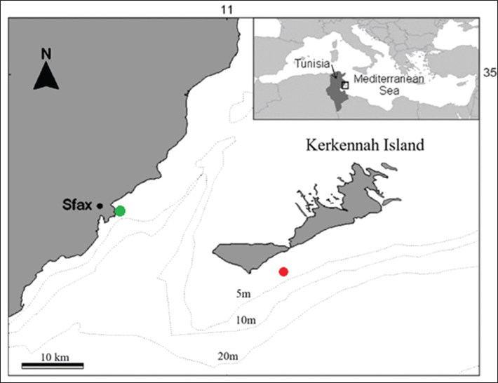
ABSTRACTBackground: Shell abnormalities in loggerhead sea turtles (Caretta caretta) remain insufficiently documented, although they may provide important clues about developmental disorders and environmental pressures impacting this species. Aim: This study aims to characterize and examine an atypical pyramidal carapace deformity identified in a loggerhead sea turtle incidentally captured in Tunisia. Methods: The transfer of a loggerhead sea turtle to the marine turtle first-aid center at the Faculty of Sciences of Sfax enabled a comprehensive clinical examination, which revealed a shell abnormality. Computed tomography (CT), performed using a CT scanner at the Radiology Department of Hedi Chaker University Hospital in Sfax, further characterized the deformity and allowed assessment of potential skeletal alterations. Results: Examination revealed a pronounced pyramidal carapace morphology accompanied by extensive epibiont colonization over the entire body except for the protruding portion of the carapace. Imaging confirmed the external deformity but showed no associated rib abnormalities, indicating the absence of traumatic skeletal injury. Conclusion: The observed shell deformity appears unlikely to be of traumatic origin. Instead, the findings suggest a congenital malformation or a developmental disturbance during ontogeny as the most plausible explanation for this unusual carapace anomaly. Keywords: Kerkennah Islands, Tunisia, Sea turtles, Shell abnormalities. IntroductionThe LIFE MedTURTLES project is a European initiative focused on the conservation of marine turtles in the Mediterranean Sea. It specifically targets two species designated as priority taxa under Annex II of the Habitats Directive: the loggerhead turtle (Caretta caretta) and the green sea turtle (Chelonia mydas). The project aims to enhance understanding of Mediterranean populations of these species, mitigate the anthropogenic pressures they face, and strengthen management measures throughout the Mediterranean basin, including its southern coast, where Tunisia plays a key role. Within the framework of this project, Tunisia established a marine turtle first-aid centre at the Faculty of Sciences of Sfax (FSS) in 2021, making it the second operational centre in the country and along the southern Mediterranean coast. The centre provides medical care for injured or debilitated turtles, conducts applied research, and participates in training and awareness programmes. Most admitted turtles are loggerheads, with fewer green turtles; both represent Mediterranean populations that are genetically and ecologically distinct from their Atlantic counterparts (Casale and Margaritoulis, 2010). In Tunisia, the green turtle (C. mydas) is regularly observed in coastal and marine waters, although nesting remains rare, with only a single nest reported in 2019 at Rejich (Mahdia) (Karaa et al., 2012; Ben Ismail et al., 2022). In contrast, the loggerhead turtle (C. caretta) is more widespread and nests frequently along the Tunisian coastline, particularly on the Kerkennah Islands and at Chebba (Bradai and Karaa, 2017; Jribi et al., 2023; Jribi, 2025). This species also interacts with various fishing gears and is frequently stranded in the Gulf of Gabès, a key area for overwintering and foraging in the Mediterranean (Jribi et al., 2007; Echwikhi et al., 2011; Karaa et al., 2016; Bradai et al., 2024). The marine turtle first-aid centre of Sfax, located in the northern Gulf of Gabès, plays a crucial role in rescuing marine turtles in the region and provides valuable data on their biology, ecology, and behaviour. Materials and MethodsOn 7 April 2024, following the report of a loggerhead turtle accidentally captured off the coast of the Kerkennah Islands (southeastern Tunisia, Fig. 1), a team from the FSS visited the site. The turtle, named “Zina,” had been trapped in gillnets less than one nautical mile from the coast in shallow waters (approximately 3 m deep) and was transferred the same day to the Sfax first-aid centre for marine turtles (Fig. 2). Upon arrival, she was examined, measured, weighed, and photographed, and epibionts attached to her body were removed.
Fig. 1. Location of the Kerkennah Island, indicating the capture site of the turtle “Zina” (red dot) and its release site (green dot).
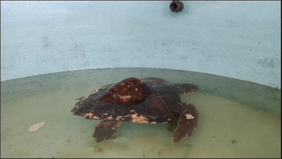
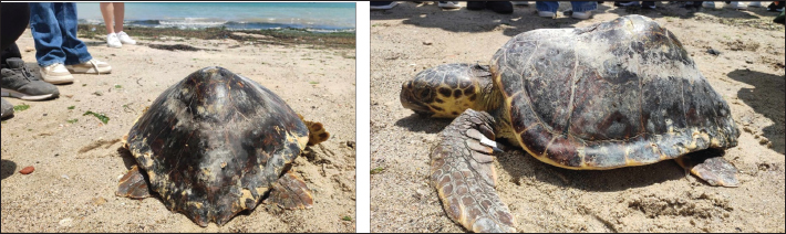
Fig. 2. Marine turtle first-aid center, FSS. The body condition index (BCI) of the turtle Zina was calculated from her standard curved carapace length (SCCL) and weight using the following equation (Tortosa et al., 2024): BCI=(P (kg)/SCCL (cm3)) × 10,000. where SCCL=standard curved carapace length (Bolten et al., 1999) and P=body mass. This index is used to assess the turtle’s general health, physical condition, and nutritional status based on its size and weight. The BCI can also be evaluated visually on a scale from 1 to 5, where 1 corresponds to an emaciated appearance characterized by sunken eyes and loss of musculature, and 5 indicates a healthy turtle (Norton and Wyneken, 2015). The turtle was thereafter placed in a tank, provided with fish and crab broth, and continuously monitored to assess her condition, including reactivity and feeding behavior (Fig. 3).
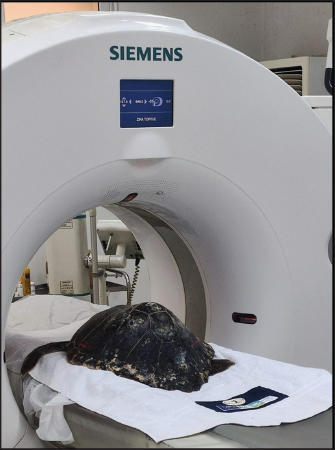
Fig. 3. Turtle “Zina” in a tank; the water level was reduced to facilitate feeding. A whole-body computed tomography (CT) examination was performed on 17, April 2024, at the radiology department of the Hedi Chaker University Hospital of Sfax using a Siemens Somatom Definition AS+ 64-slice CT. Magnetic resonance imaging was performed on 17 April 2024 by the Radiology Department of Hedi Chaker University Hospital, Sfax (CHU H. Chaker) (Fig. 4). CT imaging is considered a valuable non-invasive technique for the detailed evaluation of shell morphology and internal skeletal structures in marine turtles, allowing accurate characterization of congenital or acquired deformities. The acquisition protocol included an initial scout view encompassing the entire body of the turtle, followed by a helical scan without contrast medium administration. Image reconstructions were generated using standard and hard kernels with a reconstruction increment of 0.4 mm. The datasets were automatically transferred to the post-processing workstation Syngo.via CT, where multiplanar reconstructions (MPRs), curved planar reconstructions, and 3-D volume-rendered (VR) images were obtained to improve anatomical visualization and assessment of the shell deformity. Additional image manipulations, such as measurements and magnification, were performed when necessary. Image interpretation was performed by a senior radiologist. After several weeks of monitoring, the turtle was tagged and released on 17 May 2024 at Casino Beach in Sfax (Figs. 1 and 5).
Fig. 4. Turtle “Zina” positioned in the CT scanner at H. Chaker University Hospital.
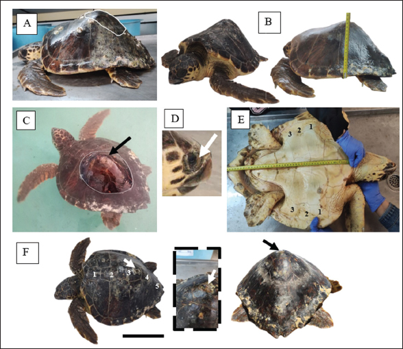
Fig. 5. Release of the turtle « “Zina” ». Ethical approvalNot needed for this study. ResultsExamination of the turtle “Zina” revealed (i) an unusual carapace deformation, with a pyramidal shape in the second vertebral third and a deep concavity at the apex; (ii) extensive epibiont coverage over the body, except for the prominent portion of the carapace; and (iii) localized trauma on the marginal scutes, mainly in the anterior region (Fig. 6). The animal exhibited buoyancy issues, with asymmetrical swimming and difficulty submerging.
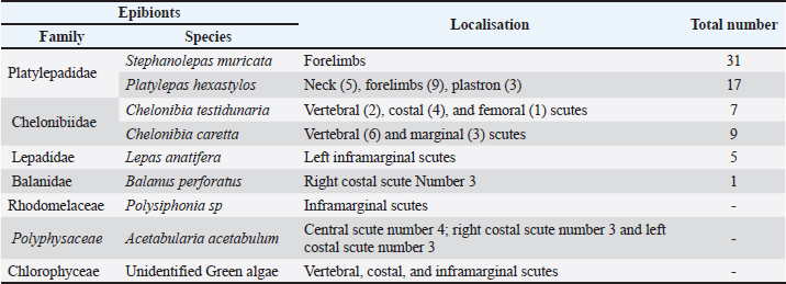
Fig. 6. Photos of turtle “Zina”: A, lateral view; B, pyramidal shape; C, white line indicating the bump area; D, white arrow indicating sunken eyes; E, plastron and inframarginal scutes; F, carapace and vertebral scutes; arrow indicates concave apex of the protuberance; scale bars 25 cm. The turtle “Zina” was treated with disinfectants, an antiparasitic, and tap water to manage parasites and buoyancy issues, likely caused by trauma at the apex of the carapace. The number and arrangement of the dorsal scutes were consistent with those of a loggerhead turtle, comprising five vertebral scutes, two pairs of five costal scutes bordering them, and 26 marginal scutes encircling both the costal and vertebral series. All scutes exhibited normal coloration and texture, displaying a light mahogany hue and typical firmness (Fig. 6). No disruption in the overall arrangement of the dorsal scutes was noted, although slight shape modifications were observed in vertebral scutes 2, 3, and 4 due to the pronounced protuberance: scute 3 was smaller and ovoid, whereas scutes 2 and 4 were more developed than usual. Vertebral scute 5 also showed a tendency to divide into two scutes (Fig. 6). The plastron appeared morphologically normal, displaying three pairs of inframarginal scutes and six pairs of symmetrical corneous scutes. However, the observed plastral concavity, reduced musculature, and sunken eyes suggest a poor nutritional status, corresponding to a visual body condition index (BCI) score of 1 (Norton and Wyneken, 2015) (Fig. 6). This assessment is further supported by the calculated BCI value (BCI=0.94), based on a SCCL of 56 cm and a body mass of 16.5 kg. Analysis of the epibionts identified six cirriped species and three macroalgal species (Table 1, Fig. 7). These organisms were predominantly distributed across the carapace, except in the area of the pronounced protuberance, which, being frequently emergent and prone to desiccation, showed no epibiont colonization. Table 1. Epibionts collected from turtle “Zina”.
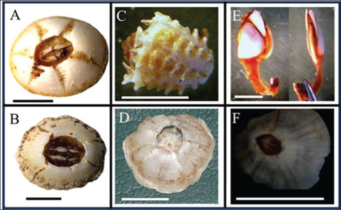
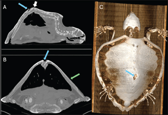
Fig. 7. Cirripedia collected from the turtle « “Zina” »; A: Chelonibia testidunaria: B: Chelonibia caretta; C: S. muricata; D: P. hexastylos; E: Lepas anatifera; F: Balanus perforates. Scale bars: 5 mm. Most epibionts were superficially attached, except for Platylepadidae cirripeds: Platylepas hexastylos, which was partially embedded in the skin, while Stephanolepas muricata penetrated deeply into the soft tissues. The turtle encapsulated these barnacles in fibrous connective tissue, allowing them to remain anchored and continue growing. The high abundance of Platylepadidae barnacles, which are epibionts specific to sea turtles, indicates that the animal exhibited low activity over an extended period. Furthermore, the presence of photophilic algae (Acetabularia and red algae of the genus Polysiphonia) and generalist epibionts, including Lepas anatifera and Balanus perforatus, which are typical of hard substrates like rocks or floating objects, further confirms that the turtle “Zina” was partially exposed out of the water for a considerable amount of time. CT performed on 17, April 2024, revealed an accentuated dorsal kyphosis associated with a soft-tissue discontinuity at the level of the dorsal protuberance, extending over approximately 22 mm, opposite an underlying thoracic vertebral fracture. Several ribs appeared slightly elongated compared with the normal morphology. No other abnormalities were identified at the thoracic or abdominopelvic levels, particularly with no evidence of deep lesions (Fig. 8). The deep wound observed at the apex of the fracture-associated dorsal protuberance may constitute a portal for pathogen entry, posing a significant health risk to the animal (Fig. 8). This lesion, together with those affecting the marginal scutes, is consistent with trauma inflicted by a single spear-shaped iron implement. The turtle “Zina” spent several weeks in the dedicated care tank. Initially, she was stressed, refused all food, and appeared to have difficulty submerging. Remnants of plastic and whole sea cucumbers, belonging to the genus Holothuria and measuring 3–6 cm, were found at the bottom of the tank, indicating that her digestive transit was functioning normally. A fish and crab broth, administered directly into the turtle’s mouth using a syringe, enabled her to feed and, after a few days, she exhibited encouraging responses such as increased movement and a renewed appetite.
Fig. 8. Whole-body CT of the turtle with MPRs in sagittal (A), axial (B), and 3-D VR technique (VRT) (C). The images demonstrate an accentuated dorsal kyphosis (white arrow) associated with a soft-tissue defect overlying the protuberance, corresponding to an underlying vertebral fracture (blue arrow), along with elongation of the ribs (green arrow). After 2 weeks, the turtle began feeding normally. She was provided with a varied and balanced diet, including crab, sardine, and occasional cuttlefish remains, to prevent osteofibrosis and maintain balanced calcium and phosphorus levels. Daily aeration ensured adequate light exposure, supporting vitamin D synthesis. By the end of the first month, live crabs were introduced into the tank to stimulate diving behavior. The turtle “Zina” regained strength and was released after spending 41 days at the first-aid centre. DiscussionThrough collaboration between Kerkennah fishermen and the marine turtle first-aid centre at the FSS, a turtle with an unusual carapace anomaly was rescued and monitored. The anomaly is indicative of kyphosis, characterized by an increased dorsal convexity. The plastron exhibited a concave shape, indicating a slight nutritional deficit, which was confirmed by the (BCI=0.94). BCI values for juvenile and adult loggerhead turtles generally range from 1.0 to 1.8, with Mediterranean juveniles often showing higher values (mean 1.78 ± 0.40; range 1.35–3.26) (Nishizawa and Joseph, 2022; Arienzo et al., 2023; Disclafani et al., 2024). Two factors provide insight into the turtle’s health. First, the presence of whole sea cucumber remains in its stomach indicates feeding on easily accessible and abundant prey in the coastal areas of the Gulf of Gabès (Azouz, 1971; Ben Othman, 1971; Sellem et al., 2017). Second, the high abundance of epibionts suggests limited locomotor activity (Frick and Pfaller, 2013); conversely, the absence of epibionts on the region of the carapace with the most pronounced protuberance indicates that this area is frequently exposed above water and subject to desiccation, potentially reflecting a particular swimming posture or buoyancy problem (Frick et al., 2000; Ten, 2024). Such behavior may increase interactions with fishing gear, contributing to the external traumas observed. CT identified external lesions affecting the marginal scutes and the apex of the carapace protuberance, which could constitute potential portals of entry for pathogenic agents. Nevertheless, the lack of scarring or deep rib deformities suggests that the observed kyphosis is unlikely to be trauma-related, with natural or nutritional factors being more probable causes (Doneley et al., 2017). Kyphoses are vertebral anomalies observed in chelonians, affecting both hatchlings and adults (Baron, 2014). Their origin, whether embryonic or postnatal, remains difficult to determine (Nguyen, 2013). Acquired deformities after hatching are primarily reported in captive individuals and are often associated with nutritional disorders (Nguyen, 2013). Congenital kyphoses have also been reported in chelonians, notably in softshell turtles (Trionichidae), likely caused by excess yolk compressing and deforming the spine during late embryonic development (Bellairs et al., 1981). The overall incidence of vertebral malformations in sea turtles is estimated at 0.11%, including 0.08% for kyphoses (Rhodin et al., 1984). Depending on the species and developmental stage, several anomalies have been observed at varying frequencies: 13.2% in aberrant olive ridley embryos from Nuevo Vallarta, Mexico, where the most common defects are compressed or irregular carapaces (Ibarra and Gasca, 2009); 1.4% in loggerhead turtle hatchlings from Florida, USA (Drennean, 1990); and 0.07% in adult leatherback turtles from French Guiana (Fretey, 1978). Congenital malformations in sea turtles result from complex interactions among genetic, epigenetic, and environmental factors during embryogenesis, frequently affecting the carapace and potentially leading to skeletal deformities (Martín-del -Campo In addition, a study on green turtle nests in the Mediterranean found correlations between nest temperature and the occurrence of malformations in hatchlings, including pigmentation abnormalities and craniofacial deformities (Sönmez and Sağol, 2024). A review of malformations in Mediterranean loggerhead sea turtles reports two cases on the eastern French coast. The most recent being a turtle that washed ashore at Canet-en-Roussillon on August 21, 2001; this turtle was severely deformed, with a pronounced dorsal hump (Oliver, 2014). The study of this specimen was limited to external observation, which revealed no deformation of the vertebral and costal plates other than their large size and the presence of two extra prefrontal scutes (Oliver, 2014). The specimen captured off Port-Vendres in August 1961 was a medium-sized male (42 cm carapace length, 15 kg) with a pyramidal carapace shape, altered dorsal scutes, a pronounced hump between vertebral scutes 4 and 5, converging costal scutes 3–5, and a modified plastron with four inframarginal scutes (Knoepffler, 1962). Kept in an aquarium, the turtle struggled to dive, refused food, and died; necropsy revealed a vertebral hump and slightly elongated ribs, with no evidence of injury, pointing to a congenital or developmental malformation (Knoepffler, 1962). The turtle captured off the Kerkennah Islands shows similarities with the case described by Knoepffler (1962): a pyramid-shaped bump was observed, with minor alterations in vertebral scutes 2, 3, and 4, and radiographs revealed no signs of internal trauma. In our case, the turtle was released in good condition, whereas the individual reported by Knoepffler (1962) died after several weeks in captivity, likely due to low temperatures and an inability to feed, ultimately succumbing to cachexia. ConclusionCases formerly considered isolated anomalies not requiring reporting, anatomical malformations in marine turtles should now receive increased attention in the pathology of these species. The carapace deformation observed in the turtle “Zina,” reported in this study, could negatively affect its survival in the wild. It may result in (i) reduced mobility, (ii) difficulties in foraging, or (iii) increased vulnerability to bycatch and predators. This hump is unlikely to be of traumatic origin but may result from disrupted embryonic development, potentially linked to epigenetic imbalances or environmental factors such as thermal stress or exposure to pollutants. Advances in scientific knowledge have improved our understanding of embryology and the origins of developmental anomalies. Although many uncertainties remain regarding the exact mechanisms, it is now well established that the embryo is strongly influenced by environmental conditions during incubation. It is therefore essential to continue research, systematically document observed anomalies, and expand this study through methodical surveys of malformations during nest monitoring at loggerhead turtle nesting sites on Tunisian beaches. AcknowledgmentsWe thank the fishermen who took part in the rescue of the turtle “Zina.” We are also grateful to all the fishermen who have helped rescue injured sea turtles, as well as the NGOs that facilitated their transfer to the centre. FundingThis work was carried out at the first-aid centre of the Faculty of Sciences of Sfax (FSS), established within the framework of the Life Medturtles Project, cofunded by the LIFE financial instrument of the European Union. Authors’ contributionsAll authors participated in data collection. Sami Karaa drafted the manuscript under the guidance of Imed Jribi, who oversaw the study’s conceptualization. Conflict of interestNot necessary for this manuscript. Data availabilityThe data that support the findings of this study are available from the corresponding author upon reasonable request. ReferencesArienzo, M., Toscanesi, M., Esposito, M., Iaccarino, D., Di Nocera, F., Canzanella, S., Ferrara, L., Di Natale, G. and Trifuoggi, M. 2023. Comparative study of polycyclic aromatic hydrocarbons (PAHs) in salt gland and liver of loggerhead turtle Caretta caretta (Linnaeus, Cheloniidae) stranded along the Mediterranean coast, Southern Italy. Ecotoxicol. Environ. Saf. 263, 115355. Azouz, A. 1971. Etude des biocénoses benthiques et de la faune ichtyologique des fonds chalutables de la Tunisie, région nord et sud-est, M. S. thesis, Caen Normandie Univ. Baron, S. 2014. Pathologie des tortues marines en Polynésie française : exemple du centre de soins de Moorea. M. S. thesis, Ecole Nationale Vétérinaire de Toulouse. Bellairs, A., Cooper, J.E. and Jackson, O.F. 1981. Congenital and developmental diseases. In Diseases of the Reptilia. Eds., Cooper, J.E. and Jackson Londres, UK: Academic Press, Vol. 14, pp: 469–85. Ben Ismail, M., Jribi, I., Kaska, Y., Ben Nakhla, L., Ben Fradj, A., Dibej, M., Souki, A., Dorai, S., Damak, M., Ghedira, A. and Bradai, M.N. 2022. The westernmost green turtle (Chelonia mydas) nest recorded in the Mediterranean from Tunisia. Med.Turtle. Bull. 1, 19–23. Ben Othman, S. 1971. Observations hydrologiques, dragages et chalutages dans le sud-est Tunisien. Bull. Inst. Natn. Scient. Tech. Océanogr. Pêche. Salammbô. 2(2), 103–120. Bolten, A.B., Eckert, K.L., Bjorndal, K.A., AbreuGrobois, F.A. and Donnelly, M. 1999. Techniques for measuring sea turtles. In Research and Management Techniques for the Conservation of Sea Turtles. Eds., Eckert, K.L., Bjorndal, K.A., Abreu-Grobois, F.A. and Donnelly Washington, DC, USA: IUCN/SSC Marine Turtle Specialist Group, pp: 110–4. Bradai, M.N., Jribi, I., Saidi, B., and Enajjar, S. 2024. Sea turtles in Tunisia: an overview on their status and conservation effort. 26 pp. London: IntechOpen Press. Bradai, M.N. and Karaa, S. 2017. Première mention de la nidification de la tortue caouanne Caretta caretta sur la plage Zouaraa (Nord de la Tunisie). Bull. Inst. Natn. Scien. Tech. Mer De Salammbô 44, 203–206. Casale, P. and Margaritoulis. D. 2010. Sea turtles in the Mediterranean: distribution, threats and conservation priorities. Gland : IUCN. 294 p. Disclafani, R., Galluzzo, P., Schirò, G., Vazzana, I., Lomonaco, C., Monteverde, V. and Dara, S. 2024. Evaluation of Biochemical Parameters in Caretta caretta Sea Turtles. Vet. Sci. 11, 571. Doneley, B., Monks, D., Johnson, R. and Carmel, B. 2017. Reptile medicine and surgery in clinical practice. Oxford, UK: Wiley-Blackwell Press, p: 978. Drennean, D.J. 1990. Occurrence of physical abnormalities in Caretta caretta at Hobe Sound National Wildlife Refuge, 1987 and 1988. Mar. Tur. Newslet. 48, 19–20. Echwikhi, K., Jribi, I., Bouain, A. and Bradai, M.N. 2011. Loggerhead Turtle Bycatch in the Gulf of Gabès, Tunisia: an Overview. Mar. Tur. Newslet. 131, 9–12. Ehsanpour, M., Afkhami, M., Khoshnood, R. and Reich, K.J. 2014. Determination and maternal transfer of heavy metals (Cd, Cu, Zn, Pb, and Hg) in the hawksbill sea turtle (Eretmochelys imbricata). Bull. Environ. Contam. Toxicol. 92(6), 667–673. Fretey, J. 1978. Mensurations de tortues luths femelles adultes, Dermochelys coriacea (Linné), en Guyane française. Bull. Soc. Zool. Fr. 103, 518–523. Frick, M.G. and Pfaller, J.B. 2013. Sea turtle epibiosis. In The biology of sea turtles. Eds., Wyneken, J., Lohmann, K.J. and Musick. Boca Raton, FL: CRC Press, pp: 399–426. Frick, M.G., Williams, K.L., Veljacic, D., Pierrard, L., Jackson, J.A. and Knight. S.E. 2000. Newly documented epibiont species from nesting loggerhead sea turtles (Caretta caretta) in Georgia, USA. Mar. Turtle Newsl. 88, 3–5. Guirlet, E., Das, K. and Girondot, M. 2008. Maternal transfer of trace elements in leatherback turtles (Dermochelys coriacea) from French Guiana. Aquat. Toxicol. 88(4), 267–276. Hopkins, B.C., Willson, J.D. and Hopkins, W.A. 2013. Mercury exposure is associated with negative effects on turtle reproduction. Environ. Sci. Technol. 47(5), 2416–2422. Ibarra, A.B. and Gasca, A.M. 2009. Malformations in embryos and neonates of Olive Ridley Sea turtle (Lepidochelys olivacea) in Nuevo Vallarta, Nayarit, Mexico. Vet. Mex. 40(4), 371–380. Jribi, I. 2025. Impact of light pollution on the success of loggerhead turtle nesting in Chebba, Tunisia. Open Vet. J. 15(10), 5346–5354. Jribi, I., Bradai, M.N. and Bouain, A. 2007. Impact of trawl fishery on marine turtles in the Gulf of Gabes, Tunisia. Herpetologica. J. 17, 110–114. Jribi, I., Hrizi, M. and Ben Nakhla, L. 2023. Uncovering hidden gems: exploring new marine turtle nesting sites and evaluating beach suitability along the Tunisian coast. Med. Turtle Bull. 4, 21–30. Karaa, S., Bradaï, M.N., Jribi, I. and Bouain.. 2012. The occurrence of the green sea turtle Chelonia mydas, in the Gulf of Gabes (Tunisia). Life Environ. 62(4), 1–6. Karaa, S., Maffucci, F., Jribi, I., Bologna, M.A., Borra, M., Biffali, E., Bradai, M.N. and Hochscheid, S. 2016. Connectivity and stock composition of loggerhead turtles foraging on the North African continental shelf (Central Mediterranean): implications for conservation and management. Mar. Ecol. 37, 1103–1115. Knoepffler, L.P. 1962. Une curieuse anomalie de la carapace chez Caretta caretta caretta (Linnaeus, 1758). Vie et Milieu. 13, 327–332. Martín-Del-Campo, R., Calderón-Campuzano, M.F., Rojas-Lleonart, I., Briseño-Dueñas, R. and García-Gasca, A. 2021. Congenital malformations in sea turtles: puzzling interplay between genes and environment. Animals 11(2), 444. Martín-Del-Campo, R., Sifuentes-Romero, I. and García-Gasca, A. 2019. Hox genes in reptile development, epigenetic regulation, and teratogenesis. Cytogenet. Genome. Res. 157(1–2), 34–45. Morão, I.F.C., Simões, T., Casado, R.B., Vieira, S., Ferreira-Airaud, B., Caliani, I., Di Noi, A., Casini, S., Fossi, M.C., Lemos, M.F.L. and Novais, S.C. 2024. Metal accumulation in female green sea turtles (Chelonia mydas) and potential implications for embryonic development. Sci. Total Environ. 931, 172710. Nguyen, L. 2013. Etude des malformations anatomiques chez les tortues marines, M. S. thesis École Nationale Vétérinaire, Agroalimentaire et de l’Alimentation Nantes Atlantique. Nishizawa, H. and Joseph, L. 2022. Morphometric condition indices in sea turtles: applications and limitations. Mar. Biol. Res. 18(3), 245–260. Norton, T., and Wyneken, J. 2015. Body condition scoring the Sea Turtle. Jekyll Island, GA: Georgia Sea Turtle Cente. Oliver, G. 2014. Variabilité et malformations de l’écaillure de la Caouanne, Caretta caretta (Linnaeus, 1758) (Reptilia cheloniidae), sur les côtes françaises de Méditerranée. Bull. Soc. Herp. Fr. 150, 9–23. Rhodin, A., Pritchard, P. and Mittermeier, R. 1984. The incidence of spinal deformities in marine turtles, with notes on the prevalence of kyphosis in Indonesian Chelonia mydas. Br. J. Herpetol. 6, 369–373. Sellem, F., Graja, S. and Brahmi, Z. 2017. Données biologiques et valeur nutritive de la paroi d’Holothuria polii (Delle Chiaje, 1823) (Holothuroidea aspidochirotida) des iles Kerkennah (golfe de Gabès, Tunisie). Bull. Inst. Natn. Scien. Tech. Mer de Salammbô. 44, 139–146. Sönmez, B. and Sağol, O. 2024. Congenital malformation in green turtle embryos and hatchlings. J. Exp. Zool. A. Ecol. Integr. Physiol. 341, 925–936. Ten, S.G. 2024. Epibionts as biological indicators of cetaceans and sea turtles: a multidisciplinary approach, M. S. thesis, Valencia University. Tortosa, M., Bellido, J.J. and Báez, J.C. 2024. Analysis of plastic ingestion by juvenile loggerhead sea turtles (Caretta caretta) Stranded from Tenerife, Canary Islands. Sustainability 16, 7147. | ||
| How to Cite this Article |
| Pubmed Style Karaa S, Sadraoui A, Chaabane A, Hrizi M, Tounsi N, Mnif Z, Jribi I. Pyramidal carapace deformity in a loggerhead sea turtle (Caretta caretta) in Tunisia. Open Vet. J.. 2026; 16(7): 4933-4942. doi:10.5455/OVJ.2026.v16.i7.70 Web Style Karaa S, Sadraoui A, Chaabane A, Hrizi M, Tounsi N, Mnif Z, Jribi I. Pyramidal carapace deformity in a loggerhead sea turtle (Caretta caretta) in Tunisia. https://www.openveterinaryjournal.com/?mno=308010 [Access: July 27, 2026]. doi:10.5455/OVJ.2026.v16.i7.70 AMA (American Medical Association) Style Karaa S, Sadraoui A, Chaabane A, Hrizi M, Tounsi N, Mnif Z, Jribi I. Pyramidal carapace deformity in a loggerhead sea turtle (Caretta caretta) in Tunisia. Open Vet. J.. 2026; 16(7): 4933-4942. doi:10.5455/OVJ.2026.v16.i7.70 Vancouver/ICMJE Style Karaa S, Sadraoui A, Chaabane A, Hrizi M, Tounsi N, Mnif Z, Jribi I. Pyramidal carapace deformity in a loggerhead sea turtle (Caretta caretta) in Tunisia. Open Vet. J.. (2026), [cited July 27, 2026]; 16(7): 4933-4942. doi:10.5455/OVJ.2026.v16.i7.70 Harvard Style Karaa, S., Sadraoui, . A., Chaabane, . A., Hrizi, . M., Tounsi, . N., Mnif, . Z. & Jribi, . I. (2026) Pyramidal carapace deformity in a loggerhead sea turtle (Caretta caretta) in Tunisia. Open Vet. J., 16 (7), 4933-4942. doi:10.5455/OVJ.2026.v16.i7.70 Turabian Style Karaa, Sami, Arij Sadraoui, Asma Chaabane, Marwa Hrizi, Nadya Tounsi, Zeineb Mnif, and Imed Jribi. 2026. Pyramidal carapace deformity in a loggerhead sea turtle (Caretta caretta) in Tunisia. Open Veterinary Journal, 16 (7), 4933-4942. doi:10.5455/OVJ.2026.v16.i7.70 Chicago Style Karaa, Sami, Arij Sadraoui, Asma Chaabane, Marwa Hrizi, Nadya Tounsi, Zeineb Mnif, and Imed Jribi. "Pyramidal carapace deformity in a loggerhead sea turtle (Caretta caretta) in Tunisia." Open Veterinary Journal 16 (2026), 4933-4942. doi:10.5455/OVJ.2026.v16.i7.70 MLA (The Modern Language Association) Style Karaa, Sami, Arij Sadraoui, Asma Chaabane, Marwa Hrizi, Nadya Tounsi, Zeineb Mnif, and Imed Jribi. "Pyramidal carapace deformity in a loggerhead sea turtle (Caretta caretta) in Tunisia." Open Veterinary Journal 16.7 (2026), 4933-4942. Print. doi:10.5455/OVJ.2026.v16.i7.70 APA (American Psychological Association) Style Karaa, S., Sadraoui, . A., Chaabane, . A., Hrizi, . M., Tounsi, . N., Mnif, . Z. & Jribi, . I. (2026) Pyramidal carapace deformity in a loggerhead sea turtle (Caretta caretta) in Tunisia. Open Veterinary Journal, 16 (7), 4933-4942. doi:10.5455/OVJ.2026.v16.i7.70 |


