





| Research Article | ||
Open Vet. J.. 2026; 16(7): 4915-4922
Open Veterinary Journal, (2026), Vol. 16(7): 4915-4922 Research Article In vitro anthelmintic activity of aqueous and ethanolic extracts of the wulung coconut husk (Cocos nucifera L. var. rubescens) against H. contortusMuhammad Kautsar1, Irkham Widiyono2*, Joko Prastowo3 and Zein Ahmad Baihaqi41Master of Veterinary Science Program, Faculty of Veterinary Medicine, Universitas Gadjah Mada, Yogyakarta, Indonesia 2Department of Internal Medicine, Faculty of Veterinary Medicine, Universitas Gadjah Mada, Yogyakarta, Indonesia 3Department of Parasitology, Faculty of Veterinary Medicine, Universitas Gadjah Mada, Yogyakarta, Indonesia 4Research Center for Animal Husbandry, National Research and Innovation Agency (BRIN), Bogor, Indonesia *Corresponding Author: Irkham Widiyono. Department of Internal Medicine, Faculty of Veterinary Medicine, Universitas Gadjah Mada, Yogyakarta, Indonesia. Email: irkhamwidiyono [at] ugm.ac.id Submitted: 27/01/2026 Revised: 25/05/2026 Accepted: 07/06/2026 Published: 27/07/2026 © 2025 Open Veterinary Journal
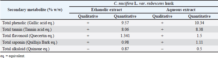
AbstractBackground: Haemonchosis remains a major constraint in small ruminant production due to reduced productivity. In tropical regions, it is estimated that haemonchosis can cause significant economic losses in livestock, with prevalence rates often exceeding 50% in some areas. Aim: This study aimed to assess and compare the in vitro anthelmintic activity of aqueous and ethanolic extracts of the wulung coconut husk against Haemonchus contortus by evaluating the phytochemical profile, adult worm mortality test, and surface morphological changes observed using a scanning electron microscope (SEM). Methods: Coconuts were collected from Kulon Progo Regency, Yogyakarta, Indonesia. Phytochemical screening was conducted to identify bioactive compounds. Each treatment group consisted of five adult female worms with four replicates, and adult worm mortality was evaluated at final concentrations of 1%, 4%, 7%, and 10%. Mortality was recorded at 0.5, 1, 2, 3, 4, 6, 8, 10, and 12 hours post-exposure and compared with both the negative and positive control groups. SEM was used to examine ultrastructural alterations, and statistical significance was set at p < 0.05. Results: Phytochemical analysis showed that the total phenol content was 10.34% in the aqueous extract and 9.57% in the ethanolic extract, while the total tannin content was 8.38% and 8.06%, respectively. In the in vitro assay, the aqueous extract at 10% induced 100% mortality within 10 hours. SEM examination revealed marked ultrastructural alterations, including surface aggregates and pronounced wrinkling in the buccal region, along the body surface, and around the genital area, suggesting substantial disruption of cuticular integrity following treatment, with these changes resembling those observed in worms treated with albendazole. Conclusion: Overall, these findings provide in vitro evidence that the aqueous extract has anthelmintic potential. A 10% aqueous extract achieved complete worm death within 10 hours, and SEM observations indicated that this effect may be related to the disruption of cuticular integrity. However, in vivo confirmation and toxicity assessment are needed before field application. Keywords: Haemonchus contortus, SEM, Cocos nucifera L. var. rubescens, Husk, Anthelmintic. IntroductionSmall ruminant farming is an essential component of smallholder livestock systems in many regions, providing diverse products such as meat, milk, wool, and hides. However, productivity in grazing-based systems is often constrained by gastrointestinal nematode infections, particularly Haemonchus contortus. As a haematophagous parasite, H. contortus can cause severe anemia, reduced performance, and even death under heavy infections (Liu et al., 2003; Roeber et al., 2013; Mavrot et al., 2015; Charlier et al., 2020). Haemonchosis is also widely recognized as a major cause of anemia in animals, especially in pasture-based systems under warm and humid conditions, making it a key challenge for herd health management (Flay et al., 2022). Control has traditionally relied heavily on anthelmintic drugs; however, the emergence and spread of anthelmintic resistance in nematodes have been reported as a major threat to sustainable production and animal welfare because they reduces the efficacy of previously reliable therapies (Roeber et al., 2013; Rose et al., 2015). In order to minimise these impacts and maintain productivity, it is essential to implement parasite control measures within the framework of integrated parasite management (IPM). IPM combines the strategic use of deworming drugs with good livestock management practices, such as pasture management, targeted selective treatment, improved nutrition, and the use of parasite-resistant livestock breeds. In this context, phytopharmaceutical agents can also serve as complementary tools in sustainable control programmes by helping to reduce reliance on conventional deworming drugs (Van Der Voort et al., 2013; Maurizio et al., 2023). Coconut has been extensively studied for its nutritional and medicinal properties. Various plant parts, including the husk, leaves, endosperm, coconut water, and haustorium, have been evaluated for their biological activities. These studies have reported a broad range of pharmacological effects, such as anthelmintic, antioxidant, antimicrobial, anti-inflammatory, antinociceptive, antitumor, and cardioprotective activities (Rinaldi et al., 2009; Lima et al., 2015; Tayler et al., 2019; Vatakkeel et al., 2024; Senthamaraikannan et al., 2025). Cocos nucifera L. var. rubescens is a coconut variety with a reddish husk that occurs in Indonesia, yet its husk is often underutilized. This is noteworthy because coconut husk may contain bioactive metabolite compounds, such as flavonoids, phenolics, and tannins, which have shown anthelmintic activity in other plant species (Lima et al., 2015; Vatakkeel et al., 2024). Therefore, the present study aimed to explore the bioactive constituents and to evaluate the in vitro anthelmintic activity of the aqueous and ethanolic extracts of C. nucifera L. var. rubescens husk against adult H. contortus. Materials and MethodsPlant extract preparation and phytochemical analysisCoconut husks were collected from a young coconut of a single coconut tree in Kulon Progo Regency, Yogyakarta, Indonesia. Plant identification was confirmed at the Plant Systematics Laboratory, Faculty of Biology, Universitas Gadjah Mada, under certificate number 00808/S.Tb./I/2025. Fresh husks were cut into small pieces and oven-dried at 45°C for 2–3 days. The dried material was then ground into powder and placed in an extraction container. The powder was macerated with aquadest (Onemed, Indonesia) at a 1:10 (w/v) ratio to ensure optimal extraction of bioactive compounds. Maceration was carried out at room temperature for 72 hours with periodic stirring to enhance solvent–material contact. The mixture was subsequently filtered using a filter cloth or Whatman paper to separate the filtrate from the residue. The filtrate was concentrated using a rotary evaporator at 40°C–50°C until the solvent was removed and a viscous extract was obtained. The same procedure was applied to prepare the ethanolic extract using ethanol 70% (Merck, Germany). Qualitative and quantitative phytochemical analyses were performed to determine the contents of total tannins, total flavonoids, total saponins, total alkaloids, and total phenolics. Quantitative analysis was performed using spectrophotometry with standard reference substances such as tannic acid, quercetin, quillaja bark, quinine, and gallic acid. All quantitative results are expressed as a percentage (% w/w) of the dry extract. (Baihaqi et al., 2020; Karseno et al., 2023; Sungpradit et al., 2025). Collection of adult H. contortusAdult H. contortus worms were collected from the abomasum of naturally infected sheep slaughtered at a local abattoir in Bantul Regency, Yogyakarta Province, Indonesia, following a previously published protocol with minor modifications (Indriati et al., 2025). All worms were sourced from a single batch of animals on the same day, and all tests were performed on that day under similar laboratory settings to ensure consistent results. The abomasum was transported to the laboratory and opened along the greater curvature, after which adult worms were recovered by carefully examining and washing the abomasal contents and mucosal surface. Worms were identified based on morphological characteristics. The collected worms were immediately transferred into containers containing 0.9% physiological NaCl solution, maintained at room temperature, and used within 30 minutes after slaughter for the in vitro adult worm mortality assay. In vitro adult worm mortality testThe adult worm mortality assay was performed with minor modifications following Indriati et al. (2025). Treatment groups received extracts at final concentrations of 1%, 4%, 7%, and 10%. Albendazole (2 mg/ml) was used as the positive control. A 0.9% NaCl solution was used as the negative control. For each group, five adult Haemonchus worms were placed in a Petri dish containing 10 ml of the extract solution, with four replicates per treatment, resulting in a total of 20 worms per treatment. Mortality was recorded at 0.5, 1, 2, 3, 4, 6, 8, and 10, and 12 hours post-exposure by noting the time of death. Worms were considered dead when no movement was observed after gentle mechanical stimulation with a stirring rod and agitation of the medium. Worms should be transferred to fresh saline solution to ensure that the lack of movement is not due to temporary paralysis. Scanning electron microscopy (SEM)SEM was performed on adult female Haemonchus worms selected from the extract-treated groups. Following fixation in 2.5% glutaraldehyde for 48 hours, the worms were rinsed with phosphate-buffered saline, and then dehydration was carried out using graded ethanol (50%, 75%, 90%, 95%, and 100%), and dried using critical point drying to maintain structural integrity. The samples were subsequently sputter-coated with gold for 15 minutes using an automatic fine coater (JEOL JEC-3000FC, Tokyo, Japan). The specimens were examined using a scanning electron microscope (JEOL JSM6510LA, Tokyo, Japan) operated in secondary electron detector (SED) mode at an accelerating voltage of 15 kV. Statistical analysisMortality data are presented as percentage of mortality and were visualized using GraphPad Prism 10. Differences between treatment groups were assessed using the generalized linear mixed model (GLMM), with statistical significance set at p < 0.05. When significant differences were found, post hoc analysis was performed to identify pairwise differences. Quantitative phytochemical data were expressed as percentages. All statistical analyses were conducted using SPSS version 27. SEM findings are presented descriptively. Ethical approvalThis research was approved by the Research Ethics Committee of the Faculty of Veterinary Medicine, Universitas Gadjah Mada, under document number 118/EC-FKH-int./2025. ResultsPhytochemical profiles analysisThe qualitative and quantitative phytochemical profiles of C. nucifera L. var. rubescens (CNR) extracts are presented in Table 1. Qualitatively, both the aqueous and ethanolic extracts tested positive for major secondary metabolites, including tannins, saponins, flavonoids, and phenolics, indicating that both solvents were able to extract bioactive constituents that may contribute to biological activity. Quantitatively, the aqueous extract tended to yield numerically higher levels for several parameters, with total phenolics of 10.34% w/w, total tannins of 8.38% w/w, total flavonoids of 1.50% w/w, total saponins of 1.11% w/w, and total alkaloids of 0.50% w/w. In contrast, the ethanolic extract contained total phenolics of 9.57% w/w and total tannins of 8.06% w/w, along with total flavonoids of 1.34% w/w, total alkaloids of 0.98% w/w, and total saponins of 0.87% w/w. Table 1. Qualitative and quantitative phytochemical analyses.
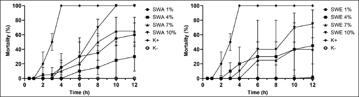
In vitro adult worm mortality testThe adult H. contortus mortality assay showed a descriptive time- and concentration-dependent response among the treatment groups, as shown in Figure 1. The positive control (K+) reached 100% mortality at approximately 4 hours, whereas the negative control (K−) remained at 0% throughout the 12-hours observation period, confirming the validity of the assay conditions. The positive control consisted of a commercial albendazole formulation dissolved in distilled water at a concentration of 2 mg/ml, selected based on previous studies demonstrating effective anthelmintic activity. In the aqueous extract group (SWA), increasing the concentration accelerated the onset of mortality and increased its magnitude. The 10% concentration showed the highest mortality response, with mortality increasing after 6 hours, reaching 70% at 8 hours and 100% at 10 hours. SWA 7% produced high but incomplete mortality 65% at 12 hours. Lower concentrations produced only partial effects, with SWA 4% increasing gradually to about 30% at 12 hours and SWA 1% resulting in low mortality (15%). A similar pattern was observed for the ethanolic extract (SWE), but the effect was generally more moderate; SWE 10% achieved high mortality but remained lower than SWA 10% (75% at 12 hours), while SWE 7% and SWE 4% showed intermediate mortality levels (45% and 40% at 12 hours, respectively). SWE 1% produced only limited mortality, reaching approximately 30% by the end of the experiment. However, the GLMM analysis showed no statistically significant differences among treatment groups (p > 0.05). The plant extract still exhibits potential anthelmintic activity, although slower, which may be due to its nature as a crude mixture of bioactive compounds.
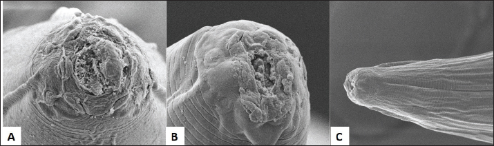
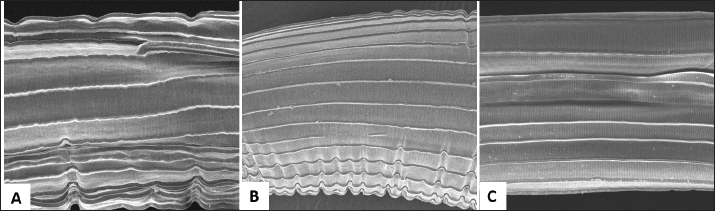
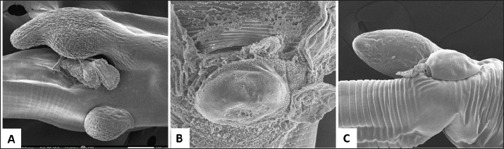
Fig. 1. In vitro mortality of H. contortus after exposure to extracts of wulung coconut husk: (a) aqueous extract (SWA) and (b) ethanolic extract (SWE) at concentrations 1, 4, 7, and 10%. K+: the positive control (albendazole 2 mg/ml), and K−: negative control (0.9% NaCl). The data are provided for the observation period, revealing no statistically significant differences between the groups (p > 0.05). SEM analysisSEM observations revealed marked ultrastructural changes on the surface of adult female worms after 12 hours of exposure to the 10% aqueous extract of C. nucifera L. var. rubescens, as shown in Figures 2–4. A total of n=5 worms were imaged per treatment group, and the observed changes were consistently detected across all specimens. In the buccal region, treated specimens exhibited surface aggregates accompanied by pronounced cuticular wrinkling (Fig. 2A), indicating disruption of the normal cuticular surface architecture. Comparable alterations were also evident in the positive control, which showed aggregate deposition and surface structural changes in the same region (Fig. 2B), whereas the negative control (NaCl 0.9%) preserved a smooth and intact cuticular surface without observable damage (Fig. 2C) with minimal, uniform wrinkling caused by the fixation and dehydration process, clearly distinguishable from the more prominent and irregular lesions observed in the positive control and treatment groups. Similar patterns were observed along the body surface extract-treated worms displayed extensive cuticular wrinkling and irregular folding (Fig. 3A), and the positive control likewise showed cuticular wrinkling (Fig. 3B), while the negative control maintained a regular, undisturbed cuticle (Fig. 3C). In addition, the area surrounding the vulva demonstrated wrinkling and surface irregularities after extract exposure (Fig. 4A), which were also present in the positive control (Fig. 4B) but absent in the negative control (Fig. 4C).
Fig. 2. SEM micrographs of the buccal region of H. contortus. (A) 10% aqueous extract of wulung coconut husk showed surface aggregates and marked cuticular shrinkage; (B) The positive control also exhibited surface aggregates and visible cuticular alterations; (C) The negative control showed no observable cuticular changes. n=5 worms per treatment. Mag.: ×3,500.
Fig. 3. SEM micrographs of the body surface of H. contortus. (A) 10% aqueous extract of wulung coconut husk showed cuticular shrinkage; (B) The positive control also exhibited cuticular shrinkage; (C) The negative control showed no observable cuticular changes. n=5 worms per treatment. Mag.: ×3,500.
Fig. 4. SEM micrographs of the vulvar region of H. contortus. (A) Worms treated with 10% aqueous extract of black coconut (C. nucifera) husk showed cuticular shrinkage; (B) The positive control also exhibited cuticular shrinkage; (C) The negative control showed no observable cuticular changes. n=5 worms per treatment. Mag.: ×3,500. DiscussionThe increasing prevalence of anthelmintic resistance in gastrointestinal nematodes has intensified the need for alternative control strategies to manage parasitic infections, including the use of plants rich in bioactive compounds. Numerous in vitro studies have explored the potential of plant extracts as antiparasitic agents, as secondary metabolites such as tannins, phenolics, flavonoids, saponins, and alkaloids are known to impair motility, damage surface structures, and induce physiological dysfunction that ultimately leads to parasite death (Davuluri et al., 2020; Sungpradit et al., 2025). In line with this, our study demonstrated that both the aqueous and ethanolic extracts contained phenolics, tannins, flavonoids, alkaloids, and saponins, which are a class of metabolites commonly associated with in vitro anthelmintic activity. These findings are consistent with Widiarso et al. (2021) who reported that an aqueous extract of Artocarpus heterophyllus caused high mortality in H. contortus, suggesting that polar, water-based fractions can effectively carry certain active constituents. Additional support has been reported for aqueous seed extracts of Zanthoxylum armatum, which showed lethal effects against H. contortus (Singh et al., 2016), and for aqueous seed extracts of Carica pubescens, which are rich in phenolics and tannins and have been shown to suppress the viability of adult H. contortus (Baihaqi et al., 2023). However, the specific mechanisms underlying this effect remain hypothetical and are inferred from previous research, as no fractionation or targeted mechanistic testing was conducted in this study. Multiple in vitro studies have consistently shown that plant extracts with high levels of phenolic compounds and tannins tend to exhibit significant anthelmintic activity, reinforcing the role of polyphenols, particularly tannins, as promising active candidates for nematode control. For example, extracts of Neolamarckia cadamba rich in phenolics and tannins have been reported to exert superior anthelmintic effects against several helminths, including Ascaridia galli and Railietina spiralis (Kumar et al., 2023), suggesting that polyphenol dominance may correlate with strong inhibition of parasite viability. A similar pattern has been observed for Mimosa caesalpinifolia, which is known for its high content of condensed tannins and has been shown to effectively inhibit egg hatching and larval exsheathment in H. contortus and Trichostrongylus colubriformis (Brito et al., 2017). Furthermore, the anthelmintic activity of tannin-containing forage plants such as Onobrychis viciifolia, Lotus pedunculatus, and Lotus corniculatus has been confirmed using larval inhibition and larval exsheathment assays, underscoring the key contribution of condensed tannins in suppressing nematode development and maintaining parasite viability (Novobilský et al., 2011). Tannins are thought to interact with surface proteins and cuticular structures, thereby compromising cuticular integrity and protective function and ultimately triggering physiological dysfunction that can lead to parasite death. A previous study reported that the ethyl acetate extract of green coconut husk liquid (C. nucifera L.) exhibited ovicidal and larvicidal activity against H. contortus (Oliveira et al., 2009), suggesting the presence of bioactive compounds with anthelmintic potential, although differences in susceptibility at various life stages of the parasite need to be considered. Based on these findings, we evaluated the in vitro potential of aqueous and ethanolic extracts of C. nucifera L. var. rubescens against adult female H. contortus. Notably, the 10% aqueous extract induced 100% mortality within 10 hours of exposure, indicating that C. nucifera L. var. rubescens aqueous husk extract possesses measurable antiparasitic activity. However, despite these promising in vitro findings, potential in vivo limitations such as reduced feed intake, antinutritional effects, or toxicity at high tannin levels must be taken into consideration, highlighting the need for further evaluation of safety and efficacy under in vivo conditions. Our study showed cuticular damage characterized by an irregular surface appearance, the presence of aggregates around the buccal region, and marked wrinkling observed in the buccal area, along the body surface, and around the genital region. This damage pattern indicates that the extract treatment may compromise cuticular integrity as the worm’s primary protective barrier, thereby reducing the parasite’s ability to maintain homeostasis and contributing to increased mortality. Additional mechanisms, such as neuromuscular function disorders or energy metabolism disorders, may also be involved, as reported for various plant compounds, and these possibilities require further investigation. These findings are consistent with other reports showing that exposure to a combined extract of Areca catechu L. and Piper betle L. caused cuticular damage and wrinkling in the buccal region, along the body, and in the vulvar area, and that such destructive morphological changes likely contributed to worm death (Sungpradit et al., 2025). Similarly, observations have shown that H. contortus treated with Paraserianthes falcataria husk extract exhibited distinct morphological alterations, particularly damage in the buccal area, along with wrinkling of the annular cuticle, indicating disruption of the cuticular surface structure following extract exposure (Baihaqi et al., 2020). In this study, the aqueous extract from wulung coconut husk caused similar cuticle modifications in H. contortus. This effect may be caused by the high total tannin content in the extract, found to be 8.38% w/w. The high total tannin content could be the main factor in this change, supporting previous findings that indicate a relationship between tannin content and the extent to which plant extracts affect parasitic nematodes. Supporting this interpretation, SEM-based evidence has shown aggregation around the buccal capsule together with transverse and longitudinal thickening and wrinkling of the cuticle after tannin exposure (Martínez-Ortíz-de-Montellano et al., 2013). Furthermore, a recent study in Caenorhabditis elegans reported that condensed tannins can induce cuticle stiffening (Greiffer et al., 2022), providing a plausible mechanistic explanation for tannin-rich extracts causing structural disruption and functional failure of the nematode cuticle. Other studies have also reported morphological alterations of the vulvar flap in female H. contortus, as well as the appearance of bubbles and surface wrinkling of the cuticle after exposure to carvacryl acetate, indicating structural disruption of the worm’s external surface layer (Andre et al., 2016). It should be noted that these changes were observed at relatively lower concentrations, indicating greater potency compared to the currently used extract, which requires a 10% concentration to induce comparable ultrastructural changes. Cuticular wrinkling following exposure to putative anthelmintic compounds has likewise been repeatedly documented in SEM studies, including after treatment with tannin-rich plants, suggesting that such changes can be regarded as a common indicator of cuticular damage in H. contortus (Martínez-Ortíz-de-Montellano et al., 2013; Cavalcante et al., 2020; Komáromyová et al., 2022). This is relevant because the nematode cuticle is the primary protective barrier against the external environment, contributes to locomotion through its functional linkage with the musculature, and participates in exchanges with the parasite’s surroundings (Page and Johnstone, 2007; Widiarso et al., 2018). In addition, cuticular surface components are implicated in interactions with the host immune system. Therefore, surface damage may further compromise the worm’s ability to maintain homeostasis (Martínez-Ortíz-de-Montellano et al., 2013; Ribeiro et al., 2017). This damage may elevate the exposure of parasite antigens to the host immune system, thereby augmenting immune identification and clearance in vivo. Nonetheless, this potential has yet to be formally validated for C. nucifera L. var. rubescens husk extracts. ConclusionOur study directly shows a 10% aqueous extract of C. nucifera L. var. rubescens exhibits in vitro anthelmintic activity against adult H. contortus. The extract induced parasite death and was associated with characteristic cuticular damage, indicating disruption of the nematode’s primary protective barrier as a likely contributor to mortality. Leveraging its phytochemical richness, this plant-based approach may offer a novel and potentially sustainable alternative to conventional synthetic anthelmintics. Nonetheless, these results are confined to in vitro conditions; hence, they cannot currently be directly extrapolated to practical efficacy. Additional in vivo investigations are required to assess their safety, efficacy, and practical uses. AcknowledgmentsWe would like to express our sincere gratitude to the Ministry of Higher Education, Science, and Technology of Indonesia for its support. We also thank the Department of Internal Medicine and the Department of Parasitology, Faculty of Veterinary Medicine, Universitas Gadjah Mada, as well as the National Research and Innovation Agency (BRIN), for providing laboratory facilities. FundingThe authors would like to express their sincere gratitude to the Ministry of Higher Education, Science, and Technology of the Republic of Indonesia for providing financial support through the PMDSU Scholarship program (Grant No. 067/C3/DT.05.00/PL/2025; 2520/UN1/DITLIT/Dit-Lit/PT.01.03/2025). Authors’ contributionsMK contributed to conceptualization, methodology, investigation, formal analysis, and writing the original draft. IW and JP contributed to supervision, conceptualization, methodology, and writing, review and editing. ZAB contributed to formal analysis, data curation, visualization, and writing, review and editing. All authors read and approved the final article. Conflict of interestThe authors declare that there is no conflict of interest. Data availabilityAll data were presented in the manuscript. ReferencesAndre, W.P.P., Ribeiro, W.L.C., Cavalcante, G.S., Santos, J.M.L.D., Macedo, I.T.F., Paula, H.C.B.D., De Freitas, R.M., De Morais, S.M., Melo, J.V.D. and Bevilaqua, C.M.L. 2016. Comparative efficacy and toxic effects of carvacryl acetate and carvacrol on sheep gastrointestinal nematodes and mice. Vet. Parasitology. 218, 52–58; doi:10.1016/j.vetpar.2016.01.001 Baihaqi, Z.A., Widiyono, I. and Nurcahyo, W. 2020. In vitro anthelmintic activity of aqueous and ethanol extracts of Paraserianthes falcataria bark waste against Haemonchus contortus obtained from a local slaughterhouse in Indonesia. Vet. World. 13(8), 1549. Baihaqi, Z.A., Widiyono, I., Angeles, A.A., Suwignyo, B. and Nurcahyo, W. 2023. Anthelmintic activity of Carica pubescens aqueous seed extract and its effects on rumen fermentation and methane reduction in Indonesian thin-tailed sheep: an in vitro study. Vet. World. 1421, 1421–1428; doi:10.14202/vetworld.2023.1421-1428 Brito, D.R.B., Costa Júnior, L.M., Garcia, J.L., Lopes, S.G., Santos, G.C. de O. and Sousa, J.V. da S. 2017. In vitro action of Mimosa caesalpinifolia ketone extract on Haemonchus contortus and Trichostrongylus colubriformis. Semina: Ciências Agrárias, 38(4), 1963–1972; doi:10.5433/1679-0359.2017v38n4p1963 Cavalcante, G.S., Morais, S.M.D., André, W.P.P., Araújo-Filho, J.V.D., Muniz, C.R., Rocha, L.O.D., Ribeiro, W.L.C., Rodrigues, A.L.M., Oliveira, L.M.B.D., Bevilaqua, C.M.L. and Ramos, M.V. 2020. Chemical constituents of Calotropis procera latex and ultrastructural effects on Haemonchus contortus. Revista Brasileira De Parasitologia Veterinária 29(2), 1320; doi:10.1590/s1984-29612020045 Charlier, J., Rinaldi, L., Musella, V., Ploeger, H.W., Chartier, C., Vineer, H.R., Hinney, B., Von Samson-himmelstjerna, G., Băcescu, B., Mickiewicz, M., Mateus, T.L., Martinez-Valladares, M., Quealy, S., Azaizeh, H., Sekovska, B., Akkari, H., Petkevicius, S., Hektoen, L., Höglund, J., Morgan, E.R., Bartley, D.J. and Claerebout, E. 2020. Initial assessment of the economic burden of major parasitic helminth infections to the ruminant livestock industry in Europe. Prev. Vet. Med. 182, 105103; doi:10.1016/j.prevetmed.2020.105103 Davuluri, T., Chennuru, S., Pathipati, M., Krovvidi, S. and Rao, G.S. 2020. In vitro anthelmintic activity of three tropical plant extracts on Haemonchus contortus. Acta. Parasitologica. 65(1), 11–18; doi:10.2478/s11686-019-00116-x Flay, K.J., Hill, F.I. and Muguiro, D.H. 2022. A review: Haemonchus contortus infection in pasture-based sheep production systems, with a focus on the pathogenesis of anaemia and changes in haematological parameters. Animals 12(10), 1238. Greiffer, L., Liebau, E., Herrmann, F.C. and Spiegler, V. 2022. Condensed tannins act as anthelmintics by increasing the rigidity of the nematode cuticle. Scientific. Rep. 12(1), 18850. Haryanti, K. and Poetri, R.K. 2023. Phytochemical characteristic and antimicrobial activity of coconut coir extract on various solvents. In Proceedings of the 3rd International Conference on Sustainable Agriculture for Rural Development (ICSARD 2022) (Vol. 30), Atlantis Press International BV, 30, pp 169–182. doi:10.2991/978-94-6463-128-9_19 Indriati, D., Erista, Y.N., Widiyono, I., Baihaqi, Z.A. and Yanuartono, Y. 2025. In vitro anthelmintic activity of Tectona grandis Linn. leaves nanoemulsion. J. Anim. Health. Prod. 13(2), 346–354; doi:10.17582/journal.jahp/2025/13.2.346.354 Karseno, Haryanti, P. and Poetri, R.K. 2023. Phytochemical characteristic and antimicrobial activity of coconut coir extract on various solvents. In Proceedings of the 3rd International Conference on Sustainable Agriculture for Rural Development (ICSARD 2022). Atlantis Press, pp. 169–182; doi:10.2991/978-94-6463-128-9_19 Komáromyová, M., Barčák, D., Königová, A., Urda Dolinská, M. and Várady, M. 2022. Does in vitro and in vivo exposure to medicinal herbs cause structural cuticular changes in Haemonchus contortus ?. Helminthologia 59(3), 265–274; doi:10.2478/helm-2022-0023 Kumar, P., Tripathi, L. and Verma, A. 2023. Phytochemical analysis and in vitro anthelmintic evaluation of Wild Cinchona (Neolamarckia cadamba) fruits. Mongolian J. Chem. 24(50), 24. Lima, E.B.C., Sousa, C.N.S., Meneses, L.N., Ximenes, N.C., Santos Júnior, M.A., Vasconcelos, G.S., Lima, N.B.C., Patrocínio, M.C.A., Macedo, D. and Vasconcelos, S.M.M. 2015. Cocos nucifera (L.)(Arecaceae): a phytochemical and pharmacological review. Braz. J. Med. Biol. Res. 48, 953–964. Liu, S.M., Masters, D.G. and Adams, N.R. 2003. Potential impact of nematode parasitism on nutrient partitioning for wool production, growth and reproduction in sheep. Austral. J. Exp. Agriculture. 43(12), 1409–1417. Martínez-Ortíz-De-Montellano, C., Arroyo-López, C., Fourquaux, I., Torres-Acosta, J.F.J., Sandoval-Castro, C.A. and Hoste, H. 2013. Scanning electron microscopy of Haemonchus contortus exposed to tannin-rich plants under in vivo and in vitro conditions. Exp. Parasitol. 133(3), 281–286; doi:10.1016/j.exppara.2012.11.024 Maurizio, A., Perrucci, S., Tamponi, C., Scala, A., Cassini, R., Rinaldi, L. and Bosco, A. 2023. Control of gastrointestinal helminths in small ruminants to prevent anthelmintic resistance: the Italian experience. Parasitology 150(12), 1105–1118. Mavrot, F., Hertzberg, H. and Torgerson, P. 2015. Effect of gastro-intestinal nematode infection on sheep performance: a systematic review and meta-analysis. Parasites & Vectors 8(1), 557. Novobilský, A., Mueller-Harvey, I. and Thamsborg, S.M. 2011. Condensed tannins act against cattle nematodes. Vet. Parasitology. 182(2–4), 213–220; doi:10.1016/j.vetpar.2011.06.003 Oliveira, L.M.B., Bevilaqua, C.M.L., Costa, C.T.C., Macedo, I.T.F., Barros, R.S., Rodrigues, A.C.M., Camurça-Vasconcelos, A.L.F., Morais, S.M., Lima, Y.C., Vieira, L.S. and Navarro, A.M.C. 2009. Anthelmintic activity of Cocos nucifera L. against sheep gastrointestinal nematodes. Vet. Parasitology 159(1), 55–59. Page, A., and Johnstone, I. 2007. WormBook: the online review of C. elegans biology [Internet]. Pasadena (CA): WormBook. Ribeiro, W.L.C., Andre, W.P.P., Cavalcante, G.S., De Araújo-filho, J.V., Santos, J.M.L., Macedo, I.T.F., De Melo, J.V., De Morais, S.M. and Bevilaqua, C.M.L. 2017. Effects of Spigelia anthelmia decoction on sheep gastrointestinal nematodes. Small. Ruminant. Res. 153, 146–152; doi:10.1016/j.smallrumres.2017.06.001 Rinaldi, S., Silva, D.O., Bello, F., Alviano, C.S., Alviano, D.S., Matheus, M.E. and Fernandes, P.D. 2009. Characterization of the antinociceptive and anti-inflammatory activities from Cocos nucifera L. (Palmae). J. Ethnopharmacol 122(3), 541–546; doi:10.1016/j.jep.2009.01.024 Roeber, F., Jex, A.R. and Gasser, R.B. 2013. Impact of gastrointestinal parasitic nematodes of sheep, and the role of advanced molecular tools for exploring epidemiology and drug resistance—An Australian perspective. Parasites Vectors. 6(1), 153; doi:10.1186/1756-3305-6-153 Rose, H., Rinaldi, L., Bosco, A., Mavrot, F., De Waal, T., Skuce, P., Charlier, J., Torgerson, P.R., Hertzberg, H., Hendrickx, G., Vercruysse, J. and Morgan, E.R. 2015. Widespread anthelmintic resistance in European farmed ruminants: a systematic review. Vet. Rec. 176(21), 546. Senthamaraikannan, Y., Sundaram, V. and Shanmugam, R. 2025. Bio-efficacy of polyethylene glycol-coated selenium nanocomposite synthesised using Cocos nucifera haustorium against HepG2 cell line. Int. Res. J. Multidiscip. Technovation 7(2), 165–183; doi:10.54392/irjmt25213 Singh, G., Singh, R., Verma, P.K., Singh, R. and Anand, A. 2016. Anthelmintic efficacy of aqueous extract of Zanthoxylum armatum DC. seeds against Haemonchus contortus of small ruminants. J. Parasitic Dis. 40(2), 528–532; doi:10.1007/s12639-014-0540-5 Sungpradit, S., Leesombun, A., Chanakarn, C., Nakthong, C. and Boonmasawai, S. 2025. Anthelmintic effects of Areca catechu L. (Arecaceae) and Piper betle L.(Piperaceae) combination on adult Haemonchus spp.: a scanning electron microscopy study. BMC. Vet. Res. 21(1), 491. Tayler, N.M., Boya, C.A., Herrera, L., Moy, J., Ng, M., Pineda, L., Almanza, A., Rosero, S., Coronado, L.M., Correa, R., Santamaría, R., Caballero, Z., Durant-Archibold, A.A., Tidgewell, K.J., Balunas, M.J., Gerwick, W.H., Spadafora, A., Gutiérrez, M. and Spadafora, C. 2019. Analysis of the antiparasitic and anticancer activity of the coconut palm (Cocos nucifera L. Arecaceae) from the natural reserve of Punta Patiño, Darién. PLos One. 14(4), 214193. Van Der Voort, M., Charlier, J., Lauwers, L., Vercruysse, J., Van Huylenbroeck, G. and Van Meensel, J. 2013. Conceptual framework for analysing farm-specific economic effects of helminth infections in ruminants and control strategies. Prev. Vet. Med. 109(3–4), 228–235; doi:10.1016/j.prevetmed.2012.10.017 Vatakkeel, B., Mythreyi, R. and Siju, E.N. 2024. Cocos nucifera Linn.: a promising candidate for drug development. J. Nat. Remedies 24(1), 27–35; doi: 10.18311/jnr/2024/34866 Widiarso, B.P., Dewi, D.A., Sarwendah, K. and Pratiwi, D.E. 2021. In vitro potency of a crude aqueous extract of Artocarpus heterophyllus leaves as an anthelmintic against Haemonchus contortus in Jawarandu goats. Adv. Anim. Vet. Sci. 9(9), 1498–1503; doi:10.17582/journal.aavs/2021/9.9.1498.1503 Widiarso, B.P., Kurniasih, K., Prastowo, J. and Nurcahyo, W. 2018. Morphology and morphometry of Haemonchus contortus exposed to Gigantochloa apus crude aqueous extract. Vet. World 11(7), 921–925; doi:10.14202/vetworld.2018.921-925 | ||
| How to Cite this Article |
| Pubmed Style Kautsar M, Widiyono I, Prastowo J, Baihaqi ZA. In vitro anthelmintic activity of aqueous and ethanolic extracts of the wulung coconut husk (Cocos nucifera L. var. rubescens) against H. contortus. Open Vet. J.. 2026; 16(7): 4915-4922. doi:10.5455/OVJ.2026.v16.i7.68 Web Style Kautsar M, Widiyono I, Prastowo J, Baihaqi ZA. In vitro anthelmintic activity of aqueous and ethanolic extracts of the wulung coconut husk (Cocos nucifera L. var. rubescens) against H. contortus. https://www.openveterinaryjournal.com/?mno=308250 [Access: July 27, 2026]. doi:10.5455/OVJ.2026.v16.i7.68 AMA (American Medical Association) Style Kautsar M, Widiyono I, Prastowo J, Baihaqi ZA. In vitro anthelmintic activity of aqueous and ethanolic extracts of the wulung coconut husk (Cocos nucifera L. var. rubescens) against H. contortus. Open Vet. J.. 2026; 16(7): 4915-4922. doi:10.5455/OVJ.2026.v16.i7.68 Vancouver/ICMJE Style Kautsar M, Widiyono I, Prastowo J, Baihaqi ZA. In vitro anthelmintic activity of aqueous and ethanolic extracts of the wulung coconut husk (Cocos nucifera L. var. rubescens) against H. contortus. Open Vet. J.. (2026), [cited July 27, 2026]; 16(7): 4915-4922. doi:10.5455/OVJ.2026.v16.i7.68 Harvard Style Kautsar, M., Widiyono, . I., Prastowo, . J. & Baihaqi, . Z. A. (2026) In vitro anthelmintic activity of aqueous and ethanolic extracts of the wulung coconut husk (Cocos nucifera L. var. rubescens) against H. contortus. Open Vet. J., 16 (7), 4915-4922. doi:10.5455/OVJ.2026.v16.i7.68 Turabian Style Kautsar, Muhammad, Irkham Widiyono, Joko Prastowo, and Zein Ahmad Baihaqi. 2026. In vitro anthelmintic activity of aqueous and ethanolic extracts of the wulung coconut husk (Cocos nucifera L. var. rubescens) against H. contortus. Open Veterinary Journal, 16 (7), 4915-4922. doi:10.5455/OVJ.2026.v16.i7.68 Chicago Style Kautsar, Muhammad, Irkham Widiyono, Joko Prastowo, and Zein Ahmad Baihaqi. "In vitro anthelmintic activity of aqueous and ethanolic extracts of the wulung coconut husk (Cocos nucifera L. var. rubescens) against H. contortus." Open Veterinary Journal 16 (2026), 4915-4922. doi:10.5455/OVJ.2026.v16.i7.68 MLA (The Modern Language Association) Style Kautsar, Muhammad, Irkham Widiyono, Joko Prastowo, and Zein Ahmad Baihaqi. "In vitro anthelmintic activity of aqueous and ethanolic extracts of the wulung coconut husk (Cocos nucifera L. var. rubescens) against H. contortus." Open Veterinary Journal 16.7 (2026), 4915-4922. Print. doi:10.5455/OVJ.2026.v16.i7.68 APA (American Psychological Association) Style Kautsar, M., Widiyono, . I., Prastowo, . J. & Baihaqi, . Z. A. (2026) In vitro anthelmintic activity of aqueous and ethanolic extracts of the wulung coconut husk (Cocos nucifera L. var. rubescens) against H. contortus. Open Veterinary Journal, 16 (7), 4915-4922. doi:10.5455/OVJ.2026.v16.i7.68 |


