Open Veterinary Journal, (2026), Vol. 16(7): 4796-4809
Research Article
10.5455/OVJ.2026.v16.i7.58
Antioxidant, anti-inflammatory, and hypouricemic properties of methanolic fruit extract of Tribulus terrestris in potassium oxonate-induced hyperuricemic rats
Taysser Khalid Ibrahim1, Ahmed Saeed Kabbashi2,3,4, Ahmed Ali Mustafa5*, Doaa Rafaat Zahran6,
Salwa Mohamed Elbashir7 and Shadia Abdoelatti Omer1
1Department of Basic Science, College of Veterinary Medicine and Animal Production, Sudan University of Science and Technology, Khartoum, Sudan
2Department of Biomedical Science, Faculty of Pharmacy, Omar Al-Mukhtar University, Al-Bayda, Libya
3Department of Microbiology and Parasitology, Medicinal and Aromatic Plants and Traditional Medicine Research Institute, National Centre for Research, Khartoum, Sudan
4Department of Microbiology, Faculty of Medical Laboratory Sciences, International University of Africa, Khartoum, Sudan
5Department of Botany and Microbiology, Faculty of Science, Gezira University, Wed Madani, Sudan
6Department of Botany and Microbiology, Faculty of Science, University of Cairo, Cairo, Egypt
7Department of Biochemistry, Veterinary Research Institute, Khartoum, Sudan
*Corresponding Author: Ahmed Ali Mustafa. Department of Botany and Microbiology, Faculty of Science, Gezira University, Wed Madani, Sudan. Email: ahmad.ali11526 [at] gmail.com
Submitted: 27/01/2026 Revised: 06/06/2026 Accepted: 20/06/2026 Published: 20/07/2026
© 2025 Open Veterinary Journal
This is an Open Access article distributed under the terms of the Creative Commons Attribution-Non Commercial-No Derivatives License (http://creativecommons.org/licenses/by-nc-nd/4.0/), which permits non-commercial re-use, distribution, and reproduction in any medium, provided the original work is properly cited, and is not altered, transformed, or built upon in any way.
ABSTRACT
Background: Hyperuricemia is a key risk factor for gout and other metabolic disorders. Conventional xanthine oxidase (XO) inhibitors, like allopurinol, can cause adverse effects, prompting the search for natural alternatives. Tribulus terrestris is a traditional medicinal plant with diuretic and anti-inflammatory properties.
Aim: This study aimed to evaluate the in vitro antioxidant and anti-inflammatory activities, and in vivo hypouricemic and XO inhibitory effects of a methanolic extract of T. terrestris fruit (TTFME) in a rat model of hyperuricemia.
Methods: Hyperuricemia was induced in Wistar rats (n=6/group) using yeast extract (20 g/kg, p.o.) and potassium oxonate (250 mg/kg, intraperitoneally). The rats were treated with TTFME (200 and 400 mg/kg) or allopurinol (5 mg/kg) for 40 days. In vitro assays were used to assess 2,2-diphenyl-1-picrylhydrazyl (DPPH) radical scavenging and albumin denaturation inhibition. Serum uric acid, XO activity, liver and kidney function markers, and globulin levels were measured. IC50 values were calculated using nonlinear regression analysis.
Results: TTFME showed modest in vitro antioxidant activity (16.7% DPPH scavenging at 100 µg/ml), consistent with its low phenolic content (2.69 mg GAE/g). However, it demonstrated potent dose-dependent anti-inflammatory activity (84.4% inhibition of protein denaturation at 300 µg/mL; IC50=100 µg/ml). In hyperuricemic rats, both TTFME doses significantly reduced serum uric acid levels to normal levels, comparable to allopurinol. Notably, 200 mg/kg TTFME showed numerically higher XO inhibition (77.5%) than allopurinol (72.2%), but this difference was not statistically significant (p > 0.05). A nonlinear dose–response was observed, with 400 mg/kg XO inhibition (47.0%) being lower. TTFME also significantly lowered elevated serum globulin and creatinine levels, indicating anti-inflammatory and nephroprotective effects. However, dose-dependent elevations in aspartate aminotransferase and alkaline phosphatase levels were observed, warranting further toxicological evaluation.
Conclusion: This preclinical study demonstrated that TTFME has hypouricemic, XO inhibitory, and anti-inflammatory properties in a rat model of hyperuricemia, providing preclinical evidence supporting its potential as a natural therapeutic candidate. However, the nonlinear dose–response and elevated hepatic enzymes warrant further toxicological investigation before clinical translation can be considered.
Keywords: Anti-inflammatory, Antioxidant, Hyperuricemia, Tribulus terrestris, Xanthine Oxidase.
Introduction
Hyperuricemia, characterized by serum uric acid levels exceeding 7.0 and 6.0 mg/dl in men and 6.0 mg/dl in women, is a prevalent metabolic disorder with a rising global burden (Fathallah-Shaykh and Cramer, 2014; Skoczyńska et al., 2020). Hyperuricemia pathogenesis involves either uric acid overproduction or, more frequently, impaired renal excretion. Key contributing factors include high purine diets, genetic predisposition, and existing kidney dysfunction (Perez-Ruiz et al., 2015). Persistently elevated uric acid is the primary etiological agent of gout, a painful inflammatory arthritis resulting from deposition of monosodium urate crystals in the joints (Dalbeth et al., 2021). The clinical significance of hyperuricemia extends beyond gout, as it has been established as an independent risk factor for a spectrum of systemic conditions, including hypertension, chronic kidney disease, cardiovascular ailments, and metabolic syndrome (Perez-Ruiz et al., 2015; Parvin and Tartte, 2025).
First-line pharmacotherapy for hyperuricemia involves xanthine oxidase inhibitors, such as allopurinol and febuxostat, which reduce uric acid synthesis (Pacher et al., 2006; Strilchuk et al., 2019). Despite their efficacy, their use is often limited by adverse effects, including severe cutaneous reactions, hepatotoxicity, and nephropathy (Mackenzie et al., 2020; Vickneson and George, 2021). This safety profile underscores the need for safer and naturally derived alternatives.
Given this need, medicinal plants remain a vital source of novel drugs. Tribulus terrestris L. (Zygophyllaceae), a plant with a long history of use in traditional Ayurvedic, Chinese, and Unani medicine, is widely used for its diuretic, anti-inflammatory, and tonic properties (Chhatre et al., 2014). Modern phytochemical investigations have identified the major bioactive constituents, including steroidal saponins (e.g., protodioscin), flavonoids, and phenolic compounds, which are associated with a spectrum of pharmacological activities, including antioxidant, anti-inflammatory, and nephroprotective effects (Ștefănescu et al., 2020; Gunarathne et al., 2023).
The T. terrestris fruit was specifically selected for this investigation based on ethnopharmacological evidence indicated that the fruit is the most commonly used part in traditional formulations for urinary disorders and inflammatory conditions (Chhatre et al., 2014). In addition, phytochemical studies have demonstrated that steroidal saponins, the primary bioactive constituents implicated in xanthine oxidase (XO) inhibition, are predominantly concentrated in the fruit compared with other aerial parts (Ștefănescu et al., 2020; Gunarathne et al., 2023).
Preliminary evidence indicates that T. terrestris extracts and their isolated compounds possess significant XO inhibitory activity, providing a direct mechanistic basis for their potential use in managing hyperuricemia (Abu-Gharbieh et al., 2018; Ashiq et al., 2021; Ajala et al., 2022).
Despite these promising preliminary findings, critical knowledge gaps remain. Previous in vivo studies of T. terrestris for hyperuricemia have been limited by the following: (1) short treatment durations (typically 7–14 days) that do not assess sustained efficacy; (2) lack of simultaneous evaluation of both serum uric acid reduction and direct XO enzyme inhibition; (3) insufficient assessment of organ-protective effects; and (4) failure to mechanistically link observed in vivo effects to the phytochemical profile and in vitro bioactivities of the extract. Therefore, a comprehensive investigation that systematically integrates these elements in a validated long-term animal model remains lacking.
This study was designed to address these gaps with the following specific objectives: (1) characterizing the phytochemical profile of TTFME; (2) evaluating its in vitro antioxidant and anti-inflammatory activities; and (3) validating its in vivo hypouricemic efficacy, XO inhibitory activity, and effects on renal and hepatic function in a 40-day yeast extract/potassium oxonate-induced hyperuricemic rat model.
Materials and Methods
Chemicals and reagents used
We purchased potassium oxonate, xanthine, 2,2-diphenyl-1-picrylhydrazyl (DPPH), and other analytical-grade chemicals from Sigma-Aldrich (St. Louis, MO, USA). The yeast extract was obtained from Oxoid (Basingstoke, UK). Allopurinol tablets were provided by EPICO Pharmaceuticals (Cairo, Egypt). All biochemical assay diagnostic kits were purchased from Biosystem S.A. (Barcelona, Spain). Analytical-grade solvents were used for extraction.
Plant material and extract preparation
Mature T. terrestris fruits were purchased from a certified herbal market in Omdurman, Sudan, in October 2022. Following botanical authentication, a voucher specimen (TT-OMD-1022) was deposited at the herbarium of the Medicinal and Aromatic Plants and Traditional Medicine Research Institute, National Center for Research, Khartoum, Sudan. Air-dried fruits (1,000 g) were coarsely ground using an electric mill. The powdered material was subjected to cold maceration with 4 L of 80% aqueous methanol (v/v) at room temperature (25°C ± 2°C) for 72 hours with occasional shaking, following the methods described by Harborne (1998) and (Kumar et al., 2011). The process was repeated twice with fresh solvent. The combined filtrates were filtered through Whatman No. 1 filter paper and concentrated using a rotary evaporator under reduced pressure at 40°C (Heidolph, Germany). The resulting crude extract was dried in a vacuum desiccator over anhydrous calcium chloride to obtain a constant weight. The final yield of the dry TTFME was 12.8% (w/w), and it was stored at −20°C in airtight amber glass vials until use.
Phytochemical analysis
Preliminary qualitative screening
To detect the presence of major classes of secondary metabolites, phytochemical screening was performed using standard qualitative procedures as described by Harborne (1998). Specific tests included alkaloids (Mayer’s, Wagner’s, and Dragendorff’s reagents), flavonoids (Shinoda test), saponins (foam test), tannins and phenolics (ferric chloride test), steroids and terpenoids (Liebermann–Burchard test), glycosides (Legal’s and Keller–Killiani tests), carbohydrates (Molisch’s test), and amino acids (ninhydrin test).
Quantitative determination
Quantitative analysis was performed using established colorimetric methods. Total phenolic content (TPC) was determined using the Folin–Ciocalteu method (Vl, 1999) and expressed as milligrams of gallic acid equivalent per gram of dry extract (mg GAE/g). Total flavonoid content (TFC) was estimated using the aluminum chloride colorimetric method and expressed as milligrams of quercetin equivalent per gram (mg QUE/g). Total tannin content was measured after adsorption onto insoluble polyvinylpyrrolidone and expressed as mg of tannic acid equivalent per gram (mg TAE/g).
In vitro biological activities
Antioxidant activity (DPPH radical scavenging assay)
The free radical scavenging capacity of TTFME was assessed against the stable DPPH radical using the method described by Shimada et al. (1992). Briefly, 2 ml of various TTFME concentrations (25–300 µg/ml in methanol) was mixed with 2 ml of 0.1 mM methanolic DPPH solution. The mixture was then vortexed and incubated for 30 minutes in the dark at room temperature. The absorbance of the resulting solution was measured at 517 nm using a UV–Vis spectrophotometer (Shimadzu, Japan). A control was prepared by mixing methanol with a DPPH solution. Propyl gallate was used as the standard antioxidant reference.
A solvent control (methanol without extraction) was included in all assays to correct for any background absorbance. All in vitro assays were performed in triplicate (n=3), and the results are expressed as the mean ± SD of three independent experiments. The percentage of DPPH radical scavenging activity was calculated as follows:
% Scavenging Activity=[(A. control – A. sample) / A. control] × 100
where A. control is the absorbance of the control, and A. sample is the absorbance of the test sample/standard.
Anti-inflammatory activity (protein denaturation inhibition assay)
In vitro anti-inflammatory activity was evaluated using an egg albumin (EA) denaturation assay, following the method described by Sakat et al. (2010). This assay is based on the principle that protein denaturation, which is the loss of tertiary structure, is a well-established marker of inflammatory processes. When proteins, such as EA, are exposed to heat (51°C), they undergo denaturation, which can be measured spectrophotometrically as increased turbidity (absorbance at 660 nm). Agents that inhibit heat-induced denaturation are considered to have potential anti-inflammatory activity, as protein structure stabilization is one mechanism by which non-steroidal anti-inflammatory drugs exert their effects.
The reaction mixture (5 ml) consisted of 0.5 ml of the test sample (TTFME at 100, 200, and 300 µg/ml in distilled water) and 4.5 ml of 1% aqueous solution of EA (pH 6.3). The volume of the test sample (0.5 ml) was selected to maintain a final concentration range of 100–300 µg/ml in the total reaction mixture, based on preliminary experiments establishing this range as optimal for detecting inhibition without exceeding the linear range of the spectrophotometric assay. This volume ratio (1:10, sample to total volume) is consistent with the established protocol (Sakat et al., 2010). The mixture was incubated at 37°C for 20 minutes and then heated to 51°C in a water bath for 20 minutes to induce denaturation. After cooling, turbidity was measured spectrophotometrically at 660 nm. Diclofenac sodium (100–300 µg/ml) was used as a standard anti-inflammatory drug. A control test (representing 0% inhibition) was performed using distilled water instead of the extract. The percentage inhibition of protein denaturation was calculated as follows:
% Inhibition=[1 – (A. sample/A. control)] × 100
where A. sample and A. control are the absorbance values of the sample and control, respectively.
In vivo hypouricemic study
Animals
A total of 30 adult male Wistar albino rats (160–180 g) were obtained from the Central Animal Breeding Facility of the College of Veterinary Medicine, University of Khartoum. The animals were housed in polypropylene cages (three rats per cage) under standard laboratory conditions: temperature, 25°C ± 2°C; relative humidity, 55% ± 5%; and a 12-hour light/dark cycle. They were provided with a standard commercial pellet diet (purchased from the Sudan Veterinary Council Feed Unit) and water ad libitum. All animals were acclimatized to laboratory conditions for 1 week before the experiment.
The sample size (n=6 animals per group) was determined based on a power analysis assuming 80% power and a significance level of α=0.05 to detect a clinically meaningful difference in serum uric acid levels. This sample size is also consistent with previous studies using similar hyperuricemic rat models (Yong et al., 2018; Bai et al., 2024).
Experimental design and induction of hyperuricemia
After acclimatization, the rats were randomly divided into five groups of six animals each (n=6). Hyperuricemia was induced using a modified yeast extract and potassium oxonate (YE/PO) method, as described by Xilifu et al. (2014). The modification involved orally administering YE 12 hours before potassium oxonate injection, as detailed below.
Group I (Normal Control): Received vehicle (distilled water) daily for 40 days.
Group II (Hyperuricemic Control): YE (20 g/kg, p.o.) was administered once daily. Potassium oxonate (PO; 250 mg/kg body weight, intraperitoneally) was injected 12 hours after YE administration. The induction protocol was followed throughout the 40-day experimental period.
Group III (Standard Drug Control): Induced with YE/PO, as in Group II, and treated with allopurinol (5 mg/kg body weight, p.o.) daily, 1 hours before YE administration.
Group IV (TTFME Low Dose): YE/PO was induced and treated with TTFME (200 mg/kg body weight, p.o.) daily, 1 hours before YE.
Group V (TTFME High Dose): YE/PO was induced and treated with TTFME (400 mg/kg body weight, p.o.) daily, 1 hours before YE.
All treatments were administered via oral gavage for 40 consecutive days.
A 40-day treatment duration was selected to evaluate sustained efficacy and potential adaptive responses, as most previous studies using the YE/PO model have employed shorter durations (7–14 days), which may not adequately reflect the chronic nature of hyperuricemia management in clinical settings (Yong et al., 2018; Bai et al., 2024).
The doses of TTFME (200 and 400 mg/kg) were selected based on the following: (1) pilot studies conducted in our laboratory indicating significant urate-lowering effects at these doses without overt toxicity; (2) previously reported effective doses of T. terrestris extracts in rodent models of metabolic disorders (150–500 mg/kg) (Al-Ali et al., 2003; Amin et al., 2006); and (3) conversion of traditional human doses (approximately 5–10 g of dried fruit/day) to rat-equivalent doses using standard body surface area normalization (Reagan-Shaw et al., 2008). Although these doses showed urate-lowering efficacy in pilot studies, we acknowledge that the subsequent observation of dose-dependent elevations in hepatic enzymes [aspartate aminotransferase (AST) and alkaline phosphatase (ALP) at 400 mg/kg in the present study indicates a potentially narrow therapeutic window. Therefore, we have moderated our conclusions and explicitly recommend histopathological evaluation before clinical translation. Based on hepatic enzyme profiles, the 200 mg/kg dose appeared to be better tolerated and is suggested as the preferred dose for future studies.
Sample collection and biochemical analysis
Blood samples (approximately 1.0 ml) were collected from the retro-orbital plexus at 10-day intervals (0, 10, 20, 30, and 40 days) under light isoflurane anesthesia to minimize pain and distress. Collections were performed at 10-day intervals (rather than more frequent intervals) to reduce stress from repeated sampling, and animals were monitored daily for signs of distress, including weight loss, piloerection, and behavioral changes. No significant adverse effects were observed. Blood was allowed to clot at room temperature, and the serum was separated by centrifugation at 3,000 rpm for 15 minutes. The serum was aliquoted and stored at −80°C until further analysis.
All animals were euthanized by cervical dislocation under deep isoflurane anesthesia at the end of the 40-day experimental period (day 40) before final blood collection and tissue harvesting. This method is consistent with the American Veterinary Medical Association guidelines for euthanasia of laboratory animals.
Serum levels of uric acid, urea, creatinine, total protein, albumin, alanine aminotransferase (ALT), AST, and ALP were determined using commercial enzymatic colorimetric diagnostic kits (Biosystem S.A., Spain) on a semi-automated biochemical analyzer (Mindray BA-88A, Shenzhen, China) according to the manufacturer’s instructions. The Uric Acid PAP Kit (Biosystem S.A., Spain) was used for uric acid specifically, based on the uricase–peroxidase method with detection at 520 nm. Serum globulin concentration was calculated by subtracting the albumin value from the total protein value (Globulin=Total Protein − Albumin).
Xanthine oxidase activity assay
Xanthine oxidase activity was measured in serum samples collected at the study endpoint (day 40) using a chemical assay based on the method described by Prajda and Weber (1975). This assay measures UA formation from xanthine by monitoring the increase in absorbance at 290 nm. One unit of enzyme activity was defined as the amount of enzyme required to produce 1 µmol uric acid per minute at 37°C. The percentage inhibition was calculated relative to the control group.
The absorbance was measured at 290 nm using a UV–Vis spectrophotometer (Shimadzu UV-1800, Kyoto, Japan). The reaction mixture contained xanthine (50 µM) in phosphate buffer (pH 7.4).
Statistical analysis
All data are presented as the mean ± standard deviation for six animals per group (n=6). Statistical analysis was performed using IBM SPSS Statistics (version 26.0; IBM Corp., Armonk, NY, USA) and GraphPad Prism (version 10.0; GraphPad Software Inc., San Diego, CA, USA).
Before performing parametric tests, the assumptions of normality and homogeneity of variance were verified using the Shapiro–Wilk test and Levene’s test, respectively. These assumptions were met in all cases (p > 0.05).
For single-time-point comparisons (XO activity, renal function, hepatic enzymes, and globulin), one-way analysis of variance was performed, followed by Tukey’s honestly significant difference post-hoc test for multiple comparisons. Exact p-values are reported in the text where appropriate, with significance levels denoted as *p < 0.05, **p < 0.01, and ***p < 0.001 in the figures.
For time-course data (serum uric acid over 40 days), two-way repeated-measures analysis of variance was performed with treatment and time as factors, followed by Bonferroni’s post-hoc test for multiple comparisons at each time point. This approach accounts for repeated measurements on the same animals and corrects for multiple comparisons across time points. All assays were performed in triplicate, and the results are expressed as the mean ± SD of three independent experiments. IC50 values (concentration required to inhibit 50% of activity) were calculated by nonlinear regression analysis using the GraphPad Prism software. Statistical significance was set at p < 0.05.
Ethical approval
The ethical approval for this study was granted on March 15, 2023 (Approval No. UK.028. B.22.16) by the Institutional Animal Ethics Committee of the Faculty of Veterinary Medicine, University of Khartoum. All procedures were conducted in strict accordance with the relevant national and institutional guidelines for the care and use of laboratory animals and ARRIVE guidelines.
Results
Phytochemical screening of TTFME
Qualitative phytochemical screening of the methanolic extract of T. terrestris fruit confirmed the presence of several classes of secondary metabolites. High saponin concentrations and moderate flavonoid levels were detected. Low concentrations of amino acids, carbohydrates, phenols, and tannins were also detected. Alkaloids, glycosides, sterols, and terpenoids were not present. Table 1 shows the qualitative profile.
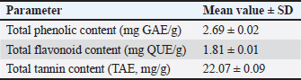
Notably, despite the relatively low phenolic and flavonoid contents, the high tannin content (22.07 mg TAE/g) and abundance of saponins (Table 2) suggest that non-phenolic constituents may be primarily responsible for the observed bioactivities.
Table 1. Qualitative phytochemical profile of TTFME.
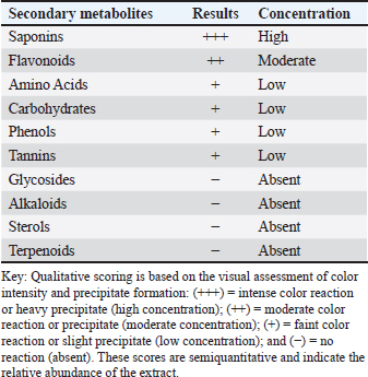
Table 2. Quantitative analysis of the major phytoconstituents in the TTFME.
w
The apparent discrepancy between the qualitative detection of flavonoids as «moderate» (Table 1) and the low quantitative total flavonoid content (1.81 mg QUE/g, Table 2) can be explained by methodological differences. The Shinoda test (Table 1) is highly sensitive to specific flavonoid subclasses (e.g., flavones and flavonols) and can produce a positive reaction even at low concentrations. In contrast, the aluminum chloride colorimetric method (Table 2) specifically quantifies flavonoids, primarily quercetin and its derivatives, that form stable complexes with aluminum ions. Therefore, the low TFC value (1.81 mg QUE/g) does not necessarily indicate an absolute deficiency of all flavonoid types but rather reflects the low abundance of quercetin-equivalent flavonoids. Other flavonoid subclasses that do not react strongly with aluminum chloride may still be present and contribute to the positive qualitative result.
In vitro antioxidant and anti-inflammatory activities
Antioxidant activity
The antioxidant capacity of TTFME in vitro was evaluated using a DPPH radical scavenging assay. At a concentration of 100 µg/ml, TTFME exhibited modest radical-scavenging activity, achieving 16.7% ± 2.1% inhibition. In contrast, the standard antioxidant, propyl gallate (100 µg/ml), exhibited 92.3% ± 1.5% inhibition (Fig. 1).
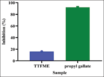
Fig. 1. In vitro antioxidant activity of TTFME extract compared with that of the standard compound (propyl gallate).
The IC50 value for DPPH radical scavenging could not be accurately determined because the maximum inhibition at the highest tested concentration (300 µg/ml) was only 21.3% ± 2.4%, which is below the 50% threshold. In contrast, the IC50 for inhibition of protein denaturation was calculated to be 156.7 ± 8.3 µg/ml for TTFME, compared to 48.2 ± 3.1 µg/ml for diclofenac sodium.
Anti-inflammatory activity
The anti-inflammatory potential of TTFME was evaluated using an EA denaturation inhibition assay. TTFME significantly and dose-dependently inhibited protein denaturation. At a concentration of 300 µg/ml, TTFME achieved an inhibition rate of 84.3% ± 3.8%. Diclofenac sodium, the standard drug, exhibited 97.8% ± 0.9% inhibition at the same concentration (Fig. 2).
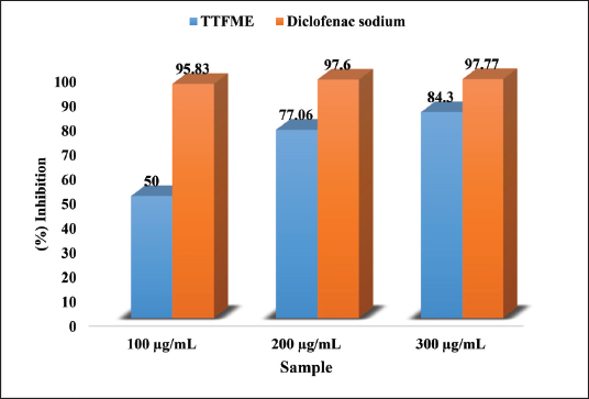
Fig. 2. Anti-inflammatory activity of TTFME extract compared with that of the standard compound (diclofenac sodium).
In vivo hypouricemic and inhibitory effects of xanthine oxidase
Validation of the hyperuricemic model
The administration of YE/PO successfully induced a stable hyperuricemic state. The hyperuricemic control group (Group II) maintained significantly elevated serum uric acid levels above those of the normal control group (Group I) throughout the 40-day experimental period (p < 0.001 at all measured time points).
Effect on serum uric acid levels
Treatment with both TTFME and allopurinol resulted in a significant time-dependent reduction in serum uric acid levels compared with those in the hyperuricemic control (Fig. 3). By day 40, the serum uric acid level in the 200 mg/kg TTFME group was reduced to 1.33 ± 0.9 mg/dl. This level was statistically equivalent (p > 0.05) to the levels in the allopurinol-treated (1.09 ± 0.47 mg/dl) and normal control (1.11 ± 0.14 mg/dl) groups. The 400 mg/kg TTFME group also showed a significant reduction (1.23 ± 0.5 mg/dl).
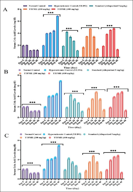
Fig. 3. Time-course effect of TTFME extract on serum uric acid levels in a yeast extract/potassium oxonate (YE/PO)-induced hyperuricemic rat model over 40 days. Values represent mean ± SD (n=6). Statistical significance (two-way repeated-measures ANOVA with Bonferroni post-hoc test) compared with the hyperuricemic control group at each time point is denoted as follows: *p < 0.05, **p < 0.01, ***p < 0.001. NS=not significant (p > 0.05). (A) Normal control group, (B) hyperuricemic control (YE/PO), and (C) standard treatment (allopurinol, 5 mg/kg).
Effect on XO activity
Liver XO activity was measured at the study endpoint (day 40) (Fig. 4). The hyperuricemic control group exhibited significantly elevated XO activity (36.7 ± 5.8 U/L). All treatment groups exhibited marked inhibition. TTFME at 200 mg/kg demonstrated potent inhibition, reducing XO activity to 8.2 ± 6.3 U/L, representing 77.5% inhibition relative to the hyperuricemic control. This inhibition was numerically higher than that achieved by allopurinol 5 mg/kg (10.5 ± 6.1 U/L; 72.2% inhibition), although this difference was not statistically significant (p > 0.05). The 400 mg/kg TTFME group showed 47.0% inhibition (19.4 ± 3.2 U/L), demonstrating a nonlinear dose–response relationship.
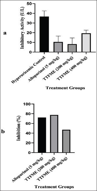
Fig. 4. Effect of TTFME extract and allopurinol on XO activity and percentage inhibition in a hyperuricemic rat model at day 40. (4A) Xanthine oxidase (XO) inhibitory activity (U/L) at day 40. (4B) Percentage inhibition of XO activity for each treatment group relative to the control group. Values represent mean ± SD (n=6).
Effects on renal function parameters
Induction of hyperuricemia led to significant renal impairment, as evidenced by marked increases in serum urea and creatinine levels in the hyperuricemic control group (Fig. 5). TTFME treatment, particularly 400 mg/kg, significantly attenuated this increase. The urea and creatinine levels in the high-dose TTFME group were significantly lower than those in the hyperuricemic control group (p < 0.01) but were not significantly different from those in the normal control group (p > 0.05).
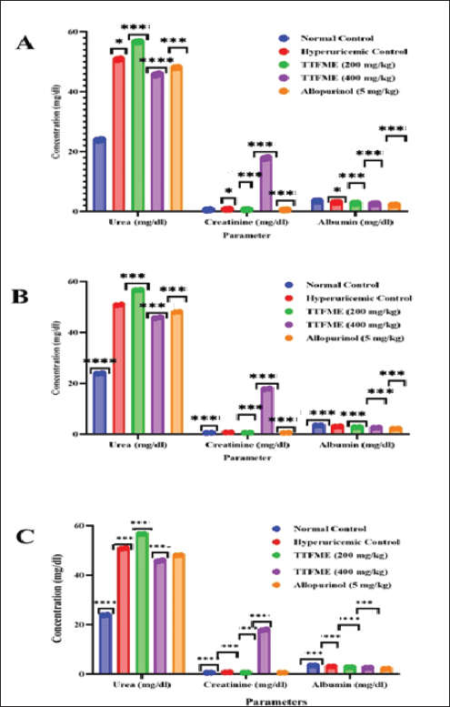
Fig. 5. Effects of TTFME extract and allopurinol on renal function parameters (urea, creatinine, and albumin) in the serum of hyperuricemic rats at day 40. Statistical significance compared with the hyperuricemic control group is denoted with asterisks (*p < 0.05, **p < 0.01, ***p < 0.001) for comparisons with the (A) normal control group, (B) hyperuricemic control group (YE/PO), and (C) standard treatment group (allopurinol, 5 mg/kg).
Effects on serum globulin and hepatic enzymes
Hyperuricemia induced a significant pro-inflammatory response, as evidenced by markedly elevated serum globulin levels in the hyperuricemic control group (14.36 ± 0.14 g/L) compared with the normal control (2.76 ± 0.24 g/L) (p < 0.001). All treatments significantly attenuated hyperglobulinemia. Treatment with TTFME (200 mg/kg) was particularly effective, reducing serum globulin to 7.00 ± 0.66 g/L (Fig. 7).
Hyperuricemia was associated with significant alterations in the hepatic enzyme profiles. Serum levels of ALT, AST, and ALP were elevated in the control group. Notably, the administration of TTFME was associated with dose-dependent elevations in AST and ALP. At 400 mg/kg, AST levels (168.3 ± 15.7 U/L) were significantly higher than those in the normal control (98.4 ± 10.2 U/L; p < 0.01) and hyperuricemic control (132.5 ± 12.8 U/L; p < 0.05). Similarly, ALP levels at 400 mg/kg (245.6 ± 22.3 U/L) exceeded those in the normal control (156.7 ± 14.5 U/L; p < 0.001). In the absence of corresponding increases in ALT, these elevations suggest possible cholestatic effects rather than hepatocellular injury, although histopathological confirmation is needed. The effects on hepatic enzymes are shown in Figure 6.
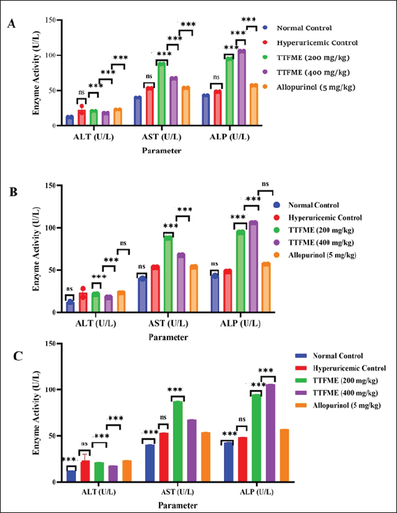
Fig. 6. Effects of TTFME extract and allopurinol on hepatic enzyme levels (ALT, AST, and ALP) on day 40. Statistical significance compared with the hyperuricemic control group is denoted with asterisks (*p < 0.05, **p < 0.01, ***p < 0.001) for comparisons with the (A) normal control group, (B) the hyperuricemic control group (YE/PO), and (C) the standard treatment group (allopurinol, 5 mg/kg).
Discussion
The findings of this study provide a comprehensive validation of the anti-hyperuricemic potential of T. terrestris fruit, linking its distinctive phytochemical profile to its demonstrable in vivo efficacy through a multi-targeted mechanistic approach. The core discovery was the potent hypouricemic action of the extract, which was equivalent to that of the standard drug allopurinol, and its numerically higher in vivo XO inhibitory activity at a dose of 200 mg/kg, although this difference was not statistically significant. This primary effect was synergistically supported by significant anti-inflammatory and nephroprotective outcomes.
Phytochemical analysis revealed a composition dominated by saponins and tannins, with low levels of phenolics and flavonoids. An apparent discrepancy exists between the low total phenolic (2.69 mg GAE/g) and flavonoid (1.81 mg QUE/g) contents and the observed potent in vivo bioactivity. This finding suggests that the therapeutic effects of TTFME may be mediated primarily by non-phenolic constituents, particularly steroidal saponins, which are present in high concentrations (as indicated by the strong positive foam test, Table 1). Saponins from T. terrestris possess diuretic, anti-inflammatory, and enzyme-modulatory activities (Ștefănescu et al., 2020; Yang et al., 2022). Furthermore, the high tannin content (22.07 mg TAE/g) may contribute to XO inhibition, as tannins are recognized as natural XO inhibitors (Ling and Bochu, 2014). This phytochemical profile, dominated by saponins and tannins rather than phenolics, explains the modest DPPH scavenging activity while supporting the observed enzyme inhibition and anti-inflammatory effects.
Furthermore, the Folin–Ciocalteu method for total phenolics can overestimate phenolic content by reacting with non-phenolic reducing compounds (e.g., ascorbic acid and reducing sugars) that may be present in the extract (Prior et al., 2005). This methodological limitation may partially explain the apparent disconnect between TPC values and antioxidant activity. The modest DPPH scavenging activity suggests that the phenolic compounds present may be predominantly in glycosylated form, which has a reduced radical-scavenging capacity due to the presence of substituted hydroxyl groups (Akhlaghi and Foshati, 2017).
The most significant finding was the potent, dose-dependent reduction in serum uric acid levels and direct inhibition of XO. However, an intriguing finding was the non-linear dose–response relationship, in which the lower dose (200 mg/kg) demonstrated numerically higher XO inhibition (77.5%) than the higher dose (400 mg/kg; 47.0% inhibition). Although unexpected, this inverted U-shaped dose–response has been previously reported for plant extracts containing multiple bioactive constituents (Calabrese, 2018). The numerically higher inhibition observed with 200 mg/kg TTFME (77.5%) compared to allopurinol (72.2%) is noteworthy, although the statistical comparison did not reach significance (p > 0.05). This trend may reflect potential synergistic interactions between the extract constituents, but no claim of superiority can be made based on these data.
Several mechanisms may explain this observation: (1) certain constituents may self-associate or form complexes that reduce bioavailability at higher concentrations; (2) high-dose saponins may induce efflux transporters (e.g., P-glycoprotein) in the gut, thereby reducing absorption (Ștefănescu et al., 2020); (3) competitive inhibition among multiple compounds at the XO active site may exhibit bell-shaped kinetics; and (4) compensatory metabolic pathways that increase uric acid production may be activated by high-dose constituents.
This phenomenon underscores the importance of dose optimization in botanical drug development and suggests that the therapeutic window for TTFME may be narrower than initially anticipated. Future pharmacokinetic studies are needed to elucidate the absorption and metabolism of TTFME constituents at different doses.
The numerically higher inhibition observed with 200 mg/kg TTFME (77.5%) than with allopurinol (72.2%) is noteworthy, although the statistical comparison did not reach significance. This trend may reflect potential synergistic interactions between the extract constituents. Steroidal saponins, a hallmark of T. terrestris, have been independently linked to diuretic and renoprotective effects, which can enhance uric acid excretion (Al-Ali et al., 2003). Similar to tannins, polyphenolic compounds are natural XO inhibitors (Ling and Bochu, 2014). Recent molecular docking studies have further elucidated that specific T. terrestris saponins and flavonoids can competitively bind to the active site of XO, thereby blocking substrate access and providing a structural basis for this inhibition (Patel et al., 2021; Ajala et al., 2022; Bouabdallah et al., 2024).
The significant reduction in serum globulin levels suggests systemic anti-inflammatory effects, which may be particularly relevant to gouty inflammation. Although the NLRP3 inflammasome and Interleukin-1 beta (IL-1β) pathway are well-established mediators of urate crystal-induced inflammation (So and Martinon, 2017), these mediators were not directly measured. The observed reduction in globulin levels provides preliminary evidence that further investigation of specific cytokine profiles [e.g., IL-1β, tumor necrosis factor-alpha (TNF-α), and Interleukin-6 (IL-6)] is warranted to explain the anti-inflammatory mechanism.
The dose-dependent elevation of hepatic enzymes (AST and ALP) in the TTFME-treated groups is a critical finding that requires careful interpretation, which appears to contradict the nephroprotective narrative and raises potential safety concerns. Several explanations, not mutually exclusive, may account for this observation: First, these elevations may represent adaptive hepatocellular responses rather than true hepatotoxicity. Steroidal saponins, abundant in T. terrestris, are known inducers of hepatic cytochrome P450 enzymes (particularly CYP3A4), which can lead to hepatocellular hypertrophy and transient, non-pathological increases in enzyme levels without tissue damage (Ștefănescu et al., 2020). This interpretation is supported by the absence of concomitant increases in bilirubin or decreases in albumin, indicating synthetic dysfunction. Second, the observed pattern of increased AST and ALP without significant ALT elevation suggests possible cholestatic or biliary effects rather than hepatocellular injury, as ALP is a bile duct function marker. This may be related to saponin-mediated effects on bile acid metabolism or excretion. Third, administration of high-dose polyphenols has been associated with pro-oxidant effects in hepatic tissues under certain conditions, potentially inducing adaptive oxidative stress responses (Andrés et al., 2023).
These findings highlight a critical safety signal that cannot be ignored. The nonlinear dose–response (lower enzyme elevations at 200 mg/kg compared to 400 mg/kg) suggests a potential therapeutic window; however, histopathological examination is urgently needed to distinguish between adaptive changes and genuine hepatotoxicity. The clinical significance of these enzyme elevations remains uncertain without histological correlation. Future studies must include comprehensive liver histopathology, assessment of synthetic function (albumin, prothrombin time), and biliary markers (γ-GT, bilirubin) to fully characterize the hepatic effects of TTFME. Until such data are available, hepatoprotective claims for T. terrestris should be viewed with caution.
This study has several limitations that should be acknowledged. First, in vitro antioxidant assessment was limited to a single assay (DPPH radical scavenging). Although DPPH is widely used, it measures only one aspect of antioxidant capacity. Future studies should employ a battery of assays ABTS (2,2’-azino-bis(3-ethylbenzothiazoline-6-sulfonic acid)), FRAP (Ferric Reducing Antioxidant Power), ORAC (Oxygen Radical Absorbance Capacity) to fully characterize the antioxidant potential of TTFME, particularly given the observed high tannin content, which may exhibit different antioxidant mechanisms. Second, while qualitative phytochemical screening identified major classes of compounds, the absence of High-Performance Liquid Chromatography or Liquid Chromatography-Tandem Mass Spectrometry profiling limits the reproducibility and mechanistic insights of this study. The observed bioactivities cannot be definitively attributed to specific constituents, and the batch-to-batch consistency of the extract cannot be guaranteed without chromatographic fingerprinting. Future studies should include comprehensive metabolomic profiling to identify and quantify the specific saponins, flavonoids, and tannins responsible for the observed effects. Third, while biochemical markers indicated alterations in renal and hepatic function, histopathological examination of kidney and liver tissues was not performed. Such an examination is essential to distinguish between adaptive physiological responses and true tissue damage, particularly given the observed elevations in AST and ALP in the TTFME-treated groups. Future studies should include a comprehensive histopathological evaluation to confirm the nephroprotective effects of creatinine and urea normalization and to assess potential hepatotoxicity. Fourth, this study evaluated only the methanolic extract. Given that traditional use often involves aqueous decoctions, comparative studies of methanolic versus aqueous extracts would be valuable in determining which preparation optimally extracts the bioactive constituents responsible for XO inhibition and anti-inflammatory effects. Fifth, the in vivo anti-inflammatory mechanism was inferred from globulin reduction; measuring specific cytokines (e.g., TNF-α, IL-1β, IL-6, and Interleukin-18) and inflammasome activity would provide deeper insights. Finally, to establish a genuine therapeutic window, the observed effects on liver markers necessitate detailed sub-chronic toxicology studies.
Despite these limitations, the present study provides valuable preclinical evidence supporting the potential of T. terrestris fruit extract as a multi-targeted therapeutic approach for hyperuricemia. Future investigations should focus on the following: (1) bioassay-guided fractionation to identify the specific compounds responsible for XO inhibition; (2) comprehensive toxicological evaluation, including histopathology; (3) pharmacokinetic studies to understand the absorption and metabolism of active constituents; and (4) exploration of potential synergistic combinations with low-dose conventional drugs to enhance efficacy while minimizing adverse effects.
Conclusion
In conclusion, this preclinical study demonstrates that the methanolic extract of TTFME exhibits multi-targeted efficacy in a yeast extract/potassium oxonate-induced hyperuricemic rat model. The primary mechanism appears to involve the inhibition of XO, with the 200 mg/kg dose achieving XO inhibition comparable to that of allopurinol (77.5% vs. 72.2%; p > 0.05) and effectively normalizing serum uric acid levels. Concurrently, TTFME showed significant anti-inflammatory effects, as evidenced by reduced serum globulin levels and inhibition of protein denaturation, as well as nephroprotective effects, as indicated by normalization of creatinine and urea levels. However, the observed elevations in hepatic enzymes (AST and ALP) at higher doses warrant careful toxicological evaluation in future studies. This finding represents a critical safety signal that requires histopathological confirmation and cannot be dismissed. These findings provide compelling preclinical evidence supporting the potential of T. terrestris as a natural therapeutic approach for hyperuricemia and gout. However, further studies, including bioassay-guided fractionation to identify active compounds, histopathological examination, and sub-chronic toxicity assessments, are necessary before clinical translation can be considered. The nonlinear dose–response observed in this study underscores the importance of dose optimization and suggests that the therapeutic window may be narrower than initially anticipated.
Acknowledgments
The authors would like to thank the technicians and staff of the Department of Basic Science at the College of Veterinary Medicine and Animal Production, Sudan University of Science and Technology, and the Veterinary Research Institute, Khartoum, for their invaluable technical support and assistance with animal care and laboratory analyses. We are also grateful to the Medicinal and Aromatic Plants and Traditional Medicine Research Institute (MAPTMRI), National Center for Research, Khartoum, for their assistance with authentication of the plant material.
Conflict of interest
The authors declare no conflict of interest.
Funding
This study was not supported by any external funding source.
Authors› contributions
Taysser K. Ibrahim: Conceptualization, methodology, resources, project administration, and writing original draft. Ahmed S. Kabbashi: Validation, resources, supervision, writing—review and editing. Ahmed A. Mustafa: Investigation, formal analysis, and data curation. Doaa R. Zahran: Methodology, investigation, and resources. Salwa M. Elbashir: Investigation, validation, and data curation. Shadia A. Omer: Investigation, methodology, and visualization. All authors have read and approved the final version of the manuscript.
Data availability
The data supporting the findings of this study are available from the corresponding author upon reasonable request.
References
Abu-Gharbieh, E., Shehab, N.G., Almasri, I.M. and Bustanji, Y. 2018. Antihyperuricemic and xanthine oxidase inhibitory activities of Tribulus arabicus and its isolated compound, ursolic acid: in vitro and in vivo investigation and docking simulations. PLoS One 13(8), e0202572; doi: 10.1371/journal.pone.0202572
Ajala, O.S., Ayeleso, A.O., Owolabi, M., Akinleye, M.O. and Ukpo, G. 2022. Xanthine oxidase inhibitory potentials of flavonoid aglycones of Tribulus terrestris in vivo, in silico and in vitro studies. Future J. Pharm. Sci. 8, 58.
Akhlaghi, M. and Foshati, S. 2017. Bioavailability and metabolism of flavonoids: a review. Int. J. Nutr. Sci. 2, 180–184.
Al-Ali, M., Wahbi, S., Twaij, H. and Al-Badr, A. 2003. Tribulus terrestris: preliminary study of its diuretic and contractile effects and comparison with Zea mays. J. Ethnopharmacol. 85, 257–260.
Amin, A., Lotfy, M., Shafiullah, M. and Adeghate, E. 2006. The protective effect of Tribulus terrestris in diabetes. Ann. New York Acad. Sci. 1084, 391–401.
Andrés, C.M.C., Pérez De La Lastra, J.M., Juan, C.A., Plou, F.J. and Pérez-Lebeña, E. 2023. Polyphenols as antioxidant/pro-oxidant compounds and donors of reducing species: relationship with human antioxidant metabolism. Processes 11, 2771.
Ashiq, K., Hussain, K., Islam, M., Shehzadi, N., Ali, E. and Ashiq, S. 2021. Medicinal plants of Pakistan and their xanthine oxidase inhibition activity to treat gout: a systematic review. Turkish J. Botany 45, 723–738.
Bai, H., Zhang, Z., Zhu, M., Sun, Y., Wang, Y., Li, B., Wang, Q. and Kuang, H. 2024. Research progress of treating hyperuricemia in rats and mice with traditional Chinese medicine. Front. Pharmacol. 15, 1428558.
Bouabdallah, S., Ibrahim, M.H., Brinza, I., Boiangiu, R.S., Honceriu, I., Amin, A., Ben-Attia, M. and Hritcu, L. 2024. Anxiolytic and antidepressant effects of Tribulus terrestris ethanolic extract in scopolamine-induced amnesia in zebrafish: supported by molecular docking investigation targeting monoamine oxidase A. Pharmaceuticals 17, 1208.
Calabrese, E.J. 2018. Hormesis: path and progression to significance. Int. J. Mol. Sci. 19, 2871.
Chhatre, S., Nesari, T., Kanchan, D., Somani, G. and Sathaye, S. 2014. Phytopharmacological overview of Tribulus terrestris. Pharmacognosy Rev. 8, 45.
Dalbeth, N., Gosling, A., Gaffo, A. and Abhishek, A. 2021. Gout. Lancet 397(10287), 1843–1855.
Fathallah-Shaykh, S.A. and Cramer, M.T. 2014. Uric acid and the kidney. Pediatric Nephrology 29, 999–1008.
Gunarathne, R., Nadeeshani, H., Lu, A., Li, J., Zhang, B., Ying, T. and Lu, J. 2023. Potential nutraceutical use of Tribulus terrestris L. in human health. Food. Rev. Int. 39, 5326–5355.
Harborne, A. 1998. Phytochemical methods a guide to modern techniques of plant analysis, springer science & business media.
Kumar, M., Soni, P. and Singh, S.S., 2011. A phytochemical review on Tribulus terrestris Linn. Res. J. Pharmacol. Pharmacodyn. 3(5), 211–214.
Ling, X. and Bochu, W. 2014. A review of phytotherapy of gout: perspective of new pharmacological treatments. Die Pharmazie. Int. J. Pharm. Sci. 69, 243–256.
Mackenzie, I.S., Ford, I., Nuki, G., Hallas, J., Hawkey, C.J., Webster, J., Ralston, S.H., Walters, M., Robertson, M., De Caterina, R., Findlay, E., Perez-Ruiz, F., Mcmurray, J.J.V., Macdonald, T.M., Aziz, J., Dobson, G., Doney, A.S.F., Flynn, R.W.V., Furnace, J., Grieve, J.W.K., Guthrie, G., Jamieson, D., Jennings, C.G., Kean, S., Lund, L.C., Mcconnachie, A., Pigazzani, F., Riches, P.L., Rix Hanson, M., Rogers, A., Rooke, E.D.M., Thomson, J., Warren, M., Wetherall, K., Wilson, R., Hall, C.P., Maseri, A., Bird, H.A., Murray, G., Dear, J.W., Petrie, M., Macdonald, M., Jhund, P.S., Connolly, E., Murphy, D.J., Paul, N., Olsson, A., Koskinen, P.T., Fuat, A., Foster, A., Saywood, W., Barr, R.J., Mcconnachie, L., Wilson, L.F., Larsen Rasmussen, L., Mcginnis, A.R., Birrell, H., Keiller, M., Bremner, I.S., Forbes, G.J., Dumbleton, J.S., Rhodes, J. and Waller, T. 2020. Long-term cardiovascular safety of febuxostat compared with allopurinol in patients with gout (FAST): a multicentre, prospective, randomised, open-label, non-inferiority trial. Lancet 396, 1745–1757.
Pacher, P., Nivorozhkin, A. and Szabó, C. 2006. Therapeutic effects of xanthine oxidase inhibitors: renaissance half a century after the discovery of allopurinol. Pharmacol. Rev. 58, 87–114.
Parvin, J.S. and Tartte, V. 2025. Bioinoculants impact on growth and bioactive compound production in genetically diverse Tribulus terrestris. In Advances in medicinal plant research. Ed., Yakkaldevi, A. Solapur: Laxmi Book Publication, pp. 145–168.
Patel, A., Bhatt, M., Soni, A. and Sharma, P. 2021. Identification of steroidal saponins from Tribulus terrestris and their in silico docking studies. J. Cell. Biochem. 122, 1665–1685.
Perez-Ruiz, F., Dalbeth, N. and Bardin, T. 2015. A review of uric acid, crystal deposition disease, and gout. Adv. Therapy 32, 31–41.
Prajda, N. and Weber, G. 1975. Malignant transformation-linked imbalance: decreased xanthine oxidase activity in hepatomas. FEBS. Lett. 59, 245–249.
Prior, R.L., Wu, X. and Schaich, K. 2005. Standardized methods for the determination of antioxidant capacity and phenolics in foods and dietary supplements. J. Agricult. Food. Chem. 53, 4290–4302.
Reagan-Shaw, S., Nihal, M. and Ahmad, N. 2008. Dose translation from animal to human studies revisited. FASEB. J. 22, 659–661.
Sakat, S., Juvekar, A.R. and Gambhire, M.N. 2010. In vitro antioxidant and anti-inflammatory activity of methanol extract of Oxalis corniculata Linn. Int. J. Pharm. Pharm. Sci. 2, 146–155.
Shimada, K., Fujikawa, K., Yahara, K. and Nakamura, T. 1992. Antioxidative properties of xanthan on the autoxidation of soybean oil in cyclodextrin emulsion. J. Agric. Food Chem. 40, 945–948.
Skoczyńska, M., Chowaniec, M., Szymczak, A., Langner-Hetmańczuk, A., Maciążek-Chyra, B. and Wiland, P. 2020. Pathophysiology of hyperuricemia and its clinical significance–a narrative review. Reumatologia/Rheumatology 58, 312–323.
So, A.K. and Martinon, F. 2017. Inflammation in gout: mechanisms and therapeutic targets. Nature Rev. Rheumatol. 13, 639–647.
Ștefănescu, R., Tero-Vescan, A., Negroiu, A., Aurică, E. and Vari, C.E. 2020. A comprehensive review of the phytochemical, pharmacological, and toxicological properties of Tribulus terrestris L. Biomolecules 10, 752.
Strilchuk, L., Fogacci, F. and Cicero, A.F. 2019. Safety and tolerability of available urate-lowering drugs: a critical review. Expert. Opin. Drug. Saf. 18, 261–271.
Vickneson, K. and George, J. 2020. Xanthine oxidoreductase inhibitors. In Reactive Oxygen Species. Handbook of Experimental Pharmacology. Eds., Schmidt, H.H.H.W., Ghezzi, P., Cuadrado, A. Cham, Springer, Vol. 264; doi: 10.1007/164_2020_383
Vl, S. 1999. Analysis of total phenols and other oxidation substrates and antioxidants by means of Folin-Ciocalteu reagent. Methods Enzymol. 299, 152–178.
Xilifu, D., Abudula, A., Rehemu, N., Zhao, L., Zhou, X. and Zhang, X. 2014. Effect of rosuvastatin on hyperuricemic rats and the protective effect on endothelial dysfunction. Exp. Therapeutic. Med. 8, 1683–1688.
Yang, M., Oppong, M.B., Di, J., Yuan, Q., Chang, Y., Jiang, M., Cao, S., Dong, P., Li, L., Xie, Y., Luan, X., Hu, L., Chai, L. and Zhang, P. 2022. Steroidal saponins with anti-inflammatory activity from Tribulus terrestris L. Acupuncture. Herbal. Med. 2, 41–48.
Yong, T., Chen, S., Xie, Y., Chen, D., Su, J., Shuai, O., Hu, H., Zuo, D. and Liang, D. 2018. Hypouricemic effects of Armillaria mellea on hyperuricemic mice regulated through OAT1 and CNT2. Am. J. Chin. Med. 46, 585–599.
Key: Qualitative scoring is based on the visual assessment of color intensity and precipitate formation: (+++)=intense color reaction or heavy precipitate (high concentration); (++)=moderate color reaction or precipitate (moderate concentration); (+)=faint color reaction or slight precipitate (low concentration); and (−)=no reaction (absent). These scores are semiquantitative and indicate the relative abundance of the extract.








