





| Research Article | ||
Open Vet. J.. 2026; 16(7): 4491-4502
Open Veterinary Journal, (2026), Vol. 16(7): 4491-4502 Research Article Clinical skin scoring and pathogenic dermatophytes detection in cats in a shelter setting in Bogor, IndonesiaNicole Ting Qian Wen1, Novericko Ginger Budiono2*, Agustin Indrawati2, Titiek Sunartatie2, Dwi Utari Rahmiati3 and Agus Wijaya41Study Program of Veterinary Professional Education, School of Veterinary Medicine and Biomedical Sciences, IPB University, Bogor, Indonesia 2Division of Medical Microbiology, School of Veterinary Medicine and Biomedical Sciences, IPB University, Bogor, Indonesia 3Division of Surgery and Radiology, School of Veterinary Medicine and Biomedical Sciences, IPB University, Bogor, Indonesia 4Division of Internal Medicine and Clinical Pathology, School of Veterinary Medicine and Biomedical Sciences, IPB University, Bogor, Indonesia *Corresponding Author: Novericko Ginger Budiono. Division of Medical Microbiology, School of Veterinary Medicine and Biomedical Sciences, IPB University, Bogor, Indonesia. Email: novericko-gi [at] apps.ipb.ac.id Submitted: 30/01/2026 Revised: 06/06/2026 Accepted: 15/06/2026 Published: 11/07/2026 © 2025 Open Veterinary Journal
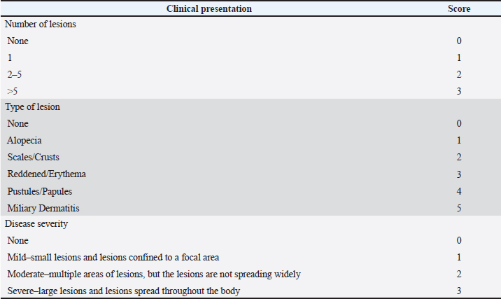
AbstractBackground: Dermatophytosis is a zoonotic fungal infection of concern in both human and veterinary medicine. It typically manifests as alopecia, scaling, and erythematous skin lesions. Cats, particularly those in shelters, may act as reservoirs; however, data from Indonesia are scarce. Aim: This study aimed to determine the point prevalence of dermatophytosis among cats in a shelter in Bogor, Indonesia, using culture-based confirmation. Methods: Samples were collected from 20 cats from a cat shelter in Bogor, Indonesia, using the MacKenzie toothbrush technique, supplemented with skin scrapings and hair plucks for direct microscopy and fungal culture. The lesions were scored, and Wood’s lamp examination was used as a screening tool. Fungal cultures on Sabouraud Dextrose Agar and Potato Dextrose Agar with chloramphenicol and cycloheximide provided a definitive diagnosis. Results: Of the 20 cats, 14 showed clinical signs, and lesion scores ranged from 0 to 8. Wood’s lamp testing was positive in 11 cats (55%), and microscopy revealed fungal elements (not a specific dermatophyte) in all skin scrapings and hair plucks. Culture confirmed Microsporum canis (1/20; 5%) and Trichophyton verrucosum (1/20; 5%), yielding an overall prevalence of 10%. Both positive cats were symptomatic, underscoring the risk of transmission. Conclusion: Dermatophytosis was present in shelter cats. Preventive measures should include early detection, isolation, environmental disinfection, and routine screening of shelter populations. Keywords: Animal shelter, Cats, Feline dermatophytosis, Microsporum canis, Trichophyton verrucosum. IntroductionCats are companion animals that provide emotional support and encourage physical activity. Other cats live as strays or in shelters. Animal shelters provide medical care, adoption options, short-term housing, and public health promotion to reduce the number of free-roaming cats and stop the spread of disease (Vojtkovská et al., 2020; Munir et al., 2023). However, the shelter condition—stressful and crowded—can predispose cats to diseases, highlighting the significance of effective management strategies and welfare (Vojtkovská et al., 2020). Cats may have infectious or noninfectious diseases, with some contagious diseases being transmissible to humans (Gordon et al., 2020). Dermatophytosis is the most common integumentary disease in shelter cats (Newbury and Moriello, 2014). The prevalence of feline dermatophytosis ranges from 0.48% in Canada (Gordon et al., 2020) and 1.80% in the USA (DeTar et al., 2019) to much higher rates (5.3%–65.6%) in Chile, India, Iraq, Nigeria, and Malaysia (Nweze, 2011; Debnath et al., 2016; Ridzuan et al., 2021; Hussein et al., 2025; Núñez et al., 2025). Clinical signs include hyperpigmentation, crusts, scales, papules, erythema, ring-shaped lesions, and nail deformities (Fauziyyah et al., 2024), although these are non-specific and may resemble other dermatoses. Asymptomatic carriers can transmit infection to humans and animals, complicating control efforts (Debnath et al., 2016; Chupia et al., 2022; Moskaluk and Vandewoude, 2022; Lúcia Sousa Resende et al., 2024; Núñez et al., 2025). The causative agents of dermatophytosis are dermatophytes, the fungi that infect keratinized tissues, such as skin, hair, and nails, in both animals and humans (Fauziyyah et al., 2024). Despite being one of the most common dermatological disorders globally, the true incidence of the disease is often undervalued (Afonso et al., 2024). The nine genera of dermatophytes are Guarromyces, Lophophyton, Paraphyton, Arthroderma, Ctenomyces, Nannizzia, Trichophyton, Epidermophyton, and Microsporum, with Microsporum being the most commonly found genus in felines (Gordon et al., 2020; Chupia et al., 2022; Afonso et al., 2024; Budiono et al., 2026). Dermatophytes are divided into zoophilic (animals as hosts), anthropophilic (humans as hosts), and geophilic (soil as habitat) based on their habitat (Jamin et al., 2020; Ridzuan et al., 2021). Despite the growing prevalence, previous research on Indonesian feline dermatophytosis has focused only on clinics, households, and stray cats and has not included data from shelter settings (Indarjulianto et al., 2017; Husna et al., 2020; Endrawati and Kusumaningtyas, 2021; Paryuni et al., 2023; Fauziyyah et al., 2024; Budiono et al., 2026). Shelters present distinct challenges—communal housing, frequent intake, and limited resources—making dermatophytosis endemic and difficult to control (Frymus et al., 2013; Newbury and Moriello, 2014). Diagnostic tools, such as Wood’s lamp, microscopy, culture, and molecular techniques, have inherent limitations, and treatment may last up to 7 weeks (Moskaluk and VandeWoude, 2022; DeTar et al., 2025; Zineldar et al., 2025). Therefore, this study aimed to investigate skin lesions and the point prevalence of dermatophytosis among cats at an animal shelter in Parung, Bogor, West Java, Indonesia. Materials and MethodsTime and place of the studyThis study was conducted from the end of 2024 to early 2025. Samples were collected from an animal shelter located in Bogor, Indonesia. Laboratory examinations were conducted at the Bacteriology-Mycology Laboratory of the Medical Microbiology Division, School of Veterinary Medicine and Biomedical Sciences, IPB University. Cats, anamnesis, identity record, lesion scoring, and Wood’s lamp testWin Episcope 2.0 software was used to determine the minimum detectable point prevalence with 95% confidence to assess the statistical strength of the sampling for a known shelter population size of 150 cats. The software analysis revealed that the sample size of 20 animals is sufficient to detect a prevalence of at least 13.33%. The samples were collected with convenience sampling. Medical records, including anamnesis, signalment, sex, age, breed, and coat color, were recorded. Lesions were scored according to established criteria with modifications (Moriello et al., 2004; Bexton and Nelson, 2016; Puls et al., 2018; Zineldar et al., 2025) (Table 1). The final lesion score for each cat was obtained by summing three independently assessed components (Table 1): (1) lesion distribution (number of affected anatomical sites), (2) lesion type (e.g., alopecia, scale/crust skin, erythema, etc.), and lesion severity. Each component was scored according to predefined criteria from previously published scoring systems, and the total score represented the cumulative burden of skin lesions in an individual animal. A maximum score of 11 represents the most severe combination of all 3 components. However, none of the cats in the present study exhibited the full combination of lesion number, type, and severity required to reach this maximum score (11). A score of 0 indicated the absence of visible skin lesions, whereas higher scores reflected increased lesion severity and extent. The Wood’s lamp was examined during scoring. All cats were photographed and systematically documented for lesions. Table 1. Dermatophytosis lesion scoring in cats was modified from references (maximum score: 11).
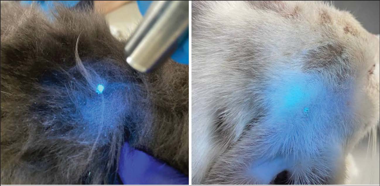
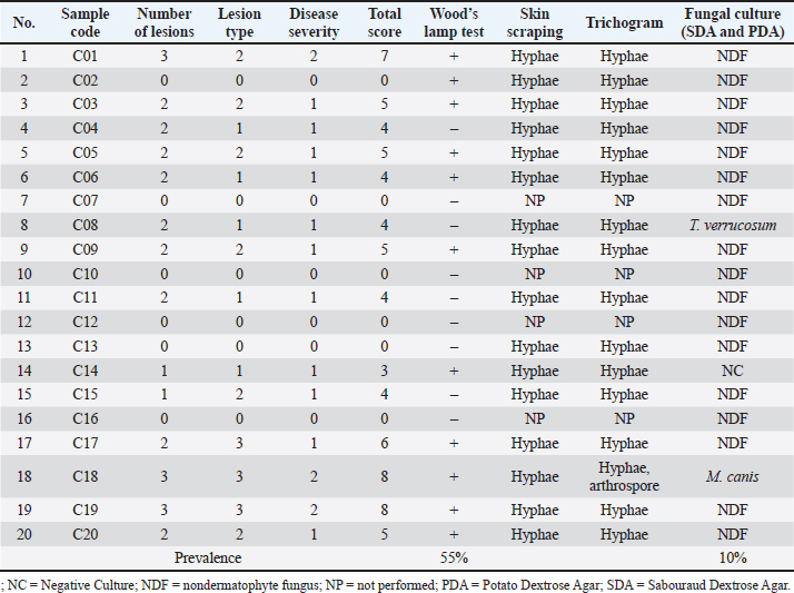
Samples were collected from 20 cats, 14 of which had skin lesions, using the MacKenzie toothbrush technique (Code B). In non-lesioned cats, brushing was performed over the entire body for ≥3 minutes. In lesioned cats, two and a half minutes were spent on unaffected areas and 30 seconds on lesions. More samples were obtained via skin scraping with sterile blunt scalpels (Code A) or hair plucking with sterile forceps (Code C), whenever possible. The lesioned area was disinfected with 70% alcohol before skin scraping. Each sample was individually stored in sterile plastic bags at room temperature. Direct microscopy and fungal cultureSkin scrapings and/or hair plucks were used for direct microscopy analysis. Skin scrapings or hair plucks were examined by direct microscopy after treatment with 10% potassium hydroxide (KOH). Two to three drops of 10% KOH were placed on a clean, grease-free slide, the sample was added, and the slide was covered with a slip to avoid air bubbles. After 10 minutes in 10% KOH, fungal elements were observed in the material. Every slide was carefully inspected for hyphae, macroconidia, microconidia, and/or arthroconidia at low (10 × 10) and high (40 × 10) magnification under the microscope. Hyphae fragments were observed internally (endothrix) or externally (ectothrix) around the hair shaft. Fungal culture was performed on samples collected using the MacKenzie technique, skin scrapings, and hair plucks. These samples were inoculated into a Petri dish containing Sabouraud Dextrose Agar (SDA) (HiMedia, India). The SDA media were supplemented with 0.0125% chloramphenicol (to inhibit the growth of a variety of Gram-negative and Gram-positive bacteria) and 0.04% cycloheximide (to inhibit the growth of saprophytic fungi). Cultures were incubated for 21 days, and fungal growth was examined in the Petri dishes 2–3 times a week. The incubation period was extended to 28 days to confirm the absence of growth, as recommended by previous studies (Stuntebeck and Moriello, 2020; Hussein et al., 2025; Rahmadani et al., 2025; Budiono et al., 2026). For further sporulation, other subcultures from suspected fungal colonies grown on SDA were transferred to Potato Dextrose Agar (PDA) (HiMedia, India) supplemented with chloramphenicol (0.0125%) and cycloheximide (0.04%), whenever needed. The identification of dermatophyte isolates began by examining microscopic characteristics and macroscopic colony morphology on SDA and/or PDA. Macroscopic observations of fungal colonies included growth rate, color, topography, and texture. Microscopic morphology was stained with lactophenol cotton blue and then examined at 40 × 10 and 10 × 10 magnification. The observed microscopic characteristics include the shapes, sizes, and arrangements of macroconidia and microconidia, as well as the hyphal shape and arrangement (Chupia et al., 2022). Fungal colony identification was carried out according to previous studies (Campbell et al., 2013; Walsh et al., 2018; Westblade et al., 2023). Data analysisThe data were presented in a table and described in a descriptive manner. No inferential statistical tests were performed, and results are presented as frequencies and proportions to provide a preliminary overview of lesion scores, Wood's lamp findings, and fungal culture outcomes. Ethical approvalThe procedure of sample collection of the study has been approved by the Animal Ethics Committee of the School of Veterinary Medicine and Biomedical Sciences, IPB University (250/KEH/SKE/IX/2024). Informed consent for animal sample collection was obtained from the veterinarian in charge of the shelter. ResultsThis study described the scoring of skin lesions and detected the point prevalence of feline dermatophytosis in one shelter setting in Bogor, Indonesia. Sampled animals included 20 shelter cats (7 males and 13 females), and of these, 1 was a kitten. The breeds were 4 Persians and 16 domestic shorthairs. Skin lesion scores in the sampled cats ranged from 0 to 8, with some showing none (maximum score: 11). All 14 lesioned cats had alopecia; 57.1% had scales/crusts; and 21.4% had erythema, including two with combined redness, crusts, and alopecia on the body and face. Lesions were mostly mild (78.6%), with 21.4% being moderate and multiarea. Of the 14 lesioned cats, 11 (55%) tested positive for Wood’s lamp (Table 2, Fig. 1).
Fig. 1. Cats sampled that showed Wood’s lamp positive from C06 (left) on the dorsal area and around the ear pinna from cat C08 (right). Table 2. Lesion scoring of the sampled cats and results of the Wood’s lamp test.
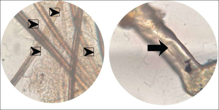
The health history of the sampled cats revealed that 14 (70%) had the flu, 4 (20%) had swollen lymph nodes, and 4 (20%) had a previous dermatophytosis diagnosis. Other medical records included neurological issues, gingivitis, stud tail, tooth decay, diarrhea, and eye enucleation. Among the 4 cats with a history of dermatophytosis, half were male, and half showed skin lesions. Before this study, cats with dermatophytosis with a single lesion were treated with topical ketoconazole and weekly grooming with Malaseb shampoo, as recommended by the veterinarian in charge of the shelter. If the lesions were multifocal, additional oral itraconazole or griseofulvin was added. Microscopy and trichoscopy revealed hyphae and/or arthrospores in hair plucks and skin scraping samples from cats with lesions (Table 2, Fig. 2).
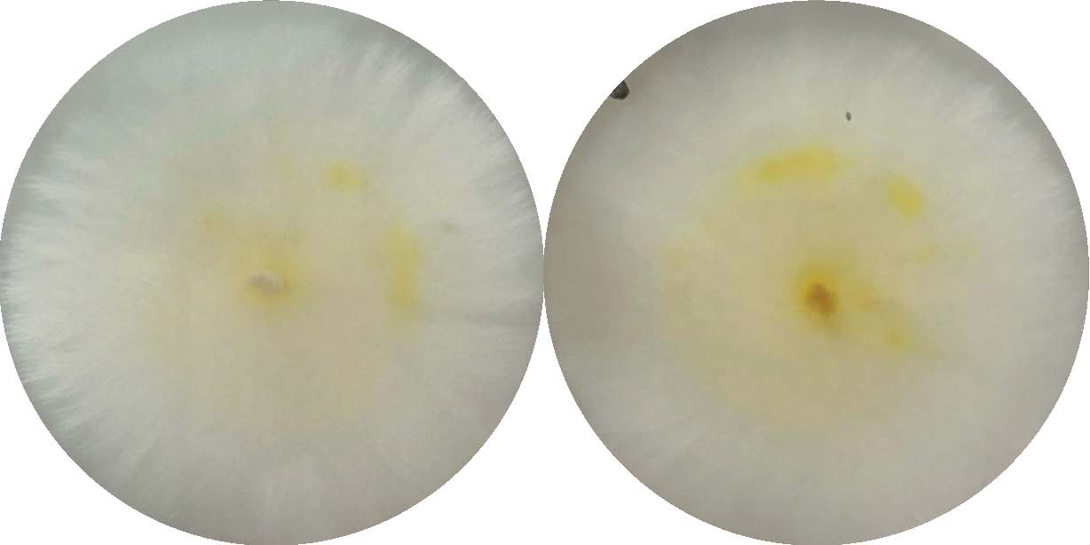
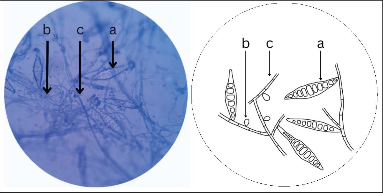
Fig. 2. Fungal elements found in direct examination with 10% KOH at 40 × 10 magnification. The hyphal structures (black arrowheads) were found in the skin scraping sample C08A of cat number C08 (left). Arthroconidia (black arrows) structures were found in the hair-pluck sample C18A of cat number C18 (right). Two dermatophyte species were isolated and identified by macroscopic and microscopic observations: Microsporum canis from cat C18 and T. verrucosum from cat C08, indicating a 10% prevalence (2/20) of dermatophytosis in this cat shelter. Colony diameters for the C18B2 isolation cultures (from cat C18) on SDA were 18.27 mm on day 7, 37.58 mm on day 14, and 51.03 mm on day 21. On day 7, the colony's surface was white, with a brown background and a white border. On days 14 and 21, the reverse turned yellow and eventually brownish yellow with age, while the surface remained white, with a flat topography and cottony texture (Fig. 3). Microscopically, C18B2 produced 6 to 12-celled macroconidia, club-shaped microconidia, and septate hyphae (Fig. 4), consistent with typical M. canis (Campbell et al., 2013; Walsh et al., 2018; Westblade et al., 2023).
Fig. 3. Macroscopic morphology of M. canis isolate (C18B2) grown on Potato Dextrose Agar on the 14th day of culture. The diameter was 53 mm, and the surface was white (left), with a yellow back and a white margin (right). The texture is flat with a cottony texture.
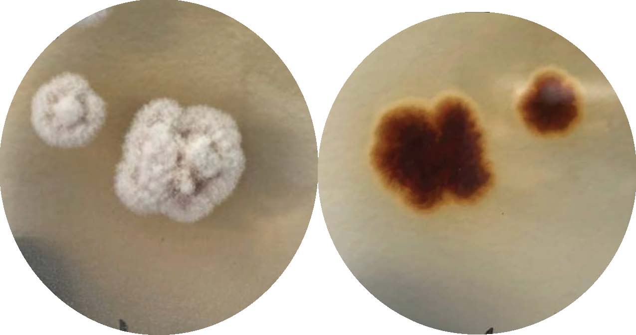
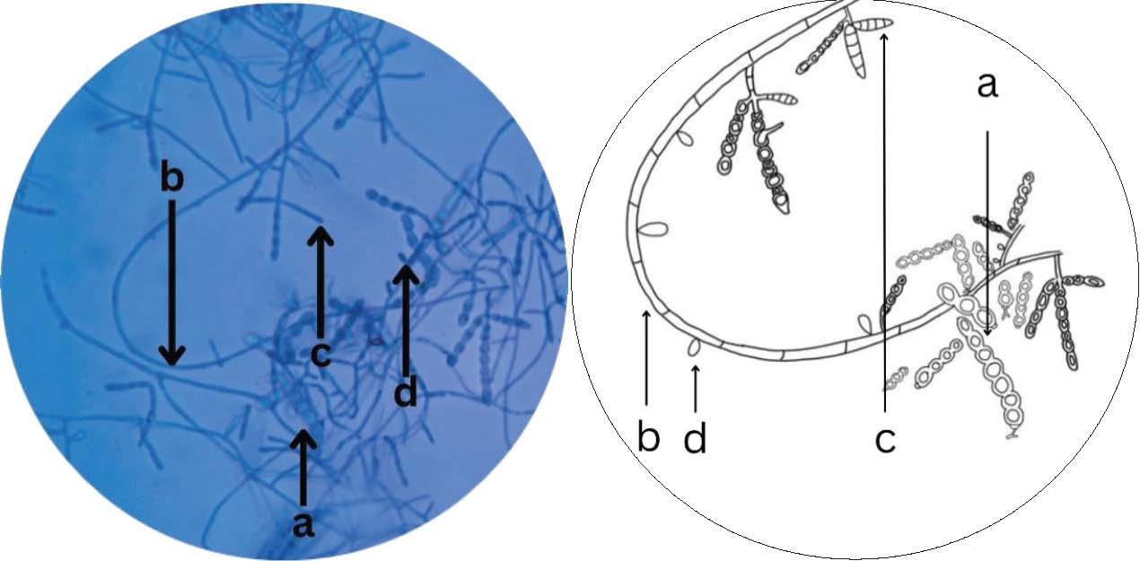
Fig. 4. Microsporum canis isolate C18B2 (left) stained with lactophenol cotton blue and 40 × 10 magnification with spindle-shaped 6–12-celled macroconidia (a), microconidia (b), and septate hyphae (c). The first author (Nicole Ting Qian Wen) illustrated the schematic (right). The colony growth of isolate C08B1 (cat C08) on Sabouraud Dextrose Agar (SDA) was slow, reaching 4.84 on day 14 and 12.77 mm on day 29. Its subculture on PDA measured 8.71 on day 10, and it became 20 mm on day 40 (Fig. 5). Colonies on both media were small, button-shaped, with white surfaces, brown reverses, domed topography, and glabrous texture. SDA and PDA microscopy revealed septate hyphae, terminal, and abundant chain-forming thick-walled chlamydospores, along with rat tail macroconidia and tear-shaped microconidia. These macroscopic and microscopic features confirmed that the fungal colony was Trichophyton verrucosum (Fig. 6) (Campbell et al., 2013; Walsh et al., 2018; Westblade et al., 2023).
Fig. 5. Macroscopic morphology of T. verrucosum isolate (C08B1) grown on Potato Dextrose Agar on the 40th day of culture. The colonies were small (20 mm in diameter), button-shaped, with white surfaces (left), brown reverses (right), domed topography, and a glabrous texture.
Fig. 6. Microscopy of T. verrucosum on PDA with lactophenol cotton blue stain and observed in 40 × 10 magnification showed chlamydospores in chains (a), septate hyphae (b), rat-tail-shaped macroconidia (c), and tear-shaped microconidia (d). The first author (Nicole Ting Qian Wen) illustrated the schematic (right). DiscussionDermatophytosis is a zoonotic skin infection transmitted from cats to humans, which is more common in tropical and subtropical regions. Since dermatophytes are not normal skin flora, infected animals can spread the disease to other animals, humans, and the environment (Łagowski et al., 2019; Lúcia Sousa Resende et al., 2024). Feline dermatophytosis prevalence varies by season, geography, and living conditions, with shelters showing particularly high rates (Afonso et al., 2024). Diagnosis is frequently presumptive, leaving regional differences poorly defined (Moskaluk and VandeWoude, 2022). Although typically mild in healthy pets, the disease can persist and be difficult to manage in crowded shelters (Gordon et al., 2020), and no reports have yet emerged from Indonesian shelters. This study was designed as a preliminary, descriptive investigation; therefore, no inferential statistical analyses were conducted, and any apparent associations between lesion severity, Wood's lamp positivity, and culture results should be interpreted descriptively rather than as statistically supported relationships. Fourteen of 20 cats exhibited dermatological lesions. The lesion scores ranged from 0 (no lesion) in 6 cats to 8 (2 cats). The most common symptoms among the cats with lesions were erythema (21.4%), redness, crusts, and alopecia on the face and body (14.3%), and scales and crusts (57.1%). Three cats had moderate multi-area involvement, whereas most lesions (78.6%) were mild. Two dermatophyte species, namely M. canis and T. verrucosum, were found in C18 and C08 cats, respectively. Both infected cats had skin lesions. Although several studies have reported that dermatophytes can be isolated from non-lesioned cats (Debnath et al., 2016; Chupia et al., 2022; Moskaluk and Vandewoude, 2022; Lúcia Sousa Resende et al., 2024; Núñez et al., 2025), this study isolated dermatophytes only from lesioned cats. In this study, Wood's lamp screening identified dermatophytosis in 11 of 20 cats (55%). Wood’s lamp is a low-cost diagnostic tool in resource-limited settings that uses UV light (320–400 nm) to detect fluorescence in Microsporum spp., showing bright-green and T. schoenleinii pale blue signals (Newbury and Moriello, 2014; Mojeski et al., 2020; Moskaluk and VandeWoude, 2022). On the contrary, T. tonsurans and T. verrucosum lack fluorescence, which limits sensitivity but still makes the lamp useful for identifying infection reservoirs (Potekaev et al., 2020). Of the 20 individuals evaluated, 11 cats were positive with the Wood’s lamp test, but only 1 was confirmed by culture, resulting in frequent false positives. These findings support earlier reports that examiner skill, protocols, equipment, animal behavior, and external factors (such as the presence of sebum or topical antifungal therapy) can influence Wood’s lamp accuracy (Dyer and Foy, 2022; Mrazkova et al., 2023). In this study, three cats with a history of confirmed dermatophyte infection and prior topical antifungal therapy may have led to negative fungal culture results despite positive Wood’s lamp results. Wood’s lamp examination showed a low positive predictive value in this dataset, with only 1 of 11 fluorescing cats yielding culture-confirmed M. canis. Wood’s lamp fluorescence reflects the presence of fluorescing hair shafts but does not necessarily indicate a viable dermatophyte infection. False-positive fluorescence may occur because of keratinous debris, topical residues, environmental contamination, or nonviable fungal elements remaining after prior treatment or spontaneous resolution. In such cases, fluorescence can persist even in the absence of fungal growth in culture. Similar discrepancies between Wood’s lamp positivity and culture-confirmed dermatophytosis have been reported in shelter and clinical studies, where Wood’s lamp examination demonstrated moderate sensitivity but low positive predictive value, supporting its role as a screening rather than a diagnostic tool (DeTar et al., 2019; Sattasathuchana et al., 2020). Fungal culture remains essential for confirming active infection and avoiding overestimation of dermatophytosis prevalence in shelter populations (Moriello et al., 2017). The experts recommended that further tests, such as microscopy, fungal culture, or molecular-based testing, be performed after the Wood’s lamp test; however, these tests require skilled personnel. Shelters usually use accessible screening techniques during entrance examinations, exposure history, and perceived risk factors, and follow-up diagnostic confirmation to try to identify dermatophyte-infected cats upon admission (Gordon et al., 2020; Afonso et al., 2024). Although the Wood’s lamp examination has a relatively high predictive value for a particular species (M. canis), the results should always be verified by culture or microscopy (Mrazkova et al., 2023). This study employed fungal culture, Wood’s lamp examination, and direct microscopy to diagnose dermatophytosis. Veterinarians have demonstrated the effectiveness of the application of the Wood’s lamp in animal shelters. Fungal culture was used as the gold-standard diagnostic because it can distinguish infected cats from fomite carriers and confirm mycological cure (Newbury and Moriello, 2014; Newbury et al., 2015; DeTar et al., 2019). Viable growth in fungal culture and clinical correlation are necessary to identify active dermatophytosis, even though microscopy can reveal nonviable or accidental fungal formations (Moriello et al., 2017; Moskaluk and VandeWoude, 2022). In this study, fungal culture remains an essential accompanying test to confirm active infection and differentiate between pathogenic activity and initial detection. Although 10% KOH microscopy is a useful screening tool for detecting fungal elements, it should not be interpreted as definitive evidence of dermatophytosis without culture confirmation (Moriello et al., 2017; Mendes et al., 2024). Other than dermatophytes, positive 10% KOH microscopy findings may reflect non-dermatophyte fungi or artifacts, and culture remains the gold standard for establishing a definitive diagnosis (Moriello et al., 2017; Moskaluk and VandeWoude, 2022). In addition, although polymerase chain reaction (PCR) offers greater specificity, sensitivity, and accuracy, fungal culture remains the gold standard for diagnosing active infection that PCR cannot (Mendes et al., 2024; Zineldar et al., 2025). Ten percent (2/20) were culture-positive for dermatophytosis. One of them (C08) was infected by T. verrucosum, and the other (C18) was infected by M. canis. Cat C18 had a lesion score of 8 (more extensive clinical signs) and was positive for M. canis and Wood’s lamp test. Cat C18 also had a history of dermatophytosis; thus, the positive fungal culture result confirms that this cat has not fully recovered from the disease. Microsporum canis is the most common dermatophyte isolated from cats (Indarjulianto et al., 2017; Budiono et al., 2026). On the contrary, three other cats (C17, C19, and C20) had a history of dermatophytosis and tested positive for the Wood’s lamp test but negative for dermatophyte fungal culture. These other negative fungal cultures resulted from Wood’s lamp-positive animals, possibly because they had received treatment for the disease according to medical history. Meanwhile, cat C08 had a lower score of 4 and did not fluoresce under the Wood’s lamp, consistent with T. verrucosum, a species that typically lacks fluorescence in cats. Trichophyton verrucosum is often spread zoonotically, particularly to people who regularly manage ruminants, such as farmers and livestock handlers (Moosavi et al., 2019). A study in Iran reported a dermatophytosis prevalence of 14.5% (15/103), with T. verrucosum as the most frequent (13 of 15 dermatophytes) causative agent (Moosavi et al., 2019). Other studies have also reported cases of dermatophytosis due to T. verrucosum infection in felines (de Melo et al., 2022). Feline dermatophytosis spreads via contact, fomites, or airborne routes, with wandering cats at higher risk (Moosavi et al., 2019; Gnat et al., 2022). The presence of livestock farming nearby suggests that the T. verrucosum infection in the shelter cat likely resulted from prior exposure before it was admitted to the shelter or from indirect transmission via contaminated workers/environment (Moosavi et al., 2019). The presence of a culture-positive patient with mild lesions (C08) and a total score of 8 indicates that clinical severity does not accurately predict infection status, although this could imply a relationship between lesion severity and infection. These results highlight the importance of laboratory confirmation in addition to clinical examinations. Indonesian publications on feline dermatophytosis are limited. Earlier reports of feline dermatophytosis prevalence in Indonesia varied due to differences in sampling locations or regions, sample size, subjects (household cats, stray cats, or cats attending veterinary clinics), and environmental settings (rural vs. urban). The prevalence of feline dermatophytosis differs between studies in the country, ranging from 0.03% to 100% (Indarjulianto et al., 2017; Yen et al., 2018; Husna et al., 2020; Paryuni et al., 2023; Fauziyyah et al., 2024; Budiono et al., 2026). In addition, other cases have been reported, including pseudomycetoma dermatophytosis (Indarjulianto et al., 2020; Hafizsha et al., 2025). These results highlight the importance of the disease despite the limited data available. This study also reported a point prevalence of 10% (2/20) for feline dermatophytosis in one shelter setting in Indonesia’s Bogor District in 2024. To the authors’ knowledge, this study is Indonesia's first publication on the point prevalence of dermatophytosis in felines in a shelter setting. The prevalence of dermatophytosis varies among nations when studies on shelter cats are considered alone. In this study, the prevalence of feline dermatophytosis was 10% (2/20), which is lower than the prevalence of 42.34%–64.29% in shelter settings in the Czech Republic (Načeradská et al., 2021; Mrazkova et al., 2023), 21.95% (Marques et al., 2023), and 82% in Russia (Ovchinnikov et al., 2020). However, the point prevalence of this study’s result is higher than that reported in Portugal (0%) (Afonso et al., 2024), Canada (0.48%) (Gordon et al., 2020), and the USA (1.80%–5.5%) (Boyanowski et al., 2000; DeTar et al., 2019). The sample sizes of these studies vary from 17 to 50,599 cats. Dermatophytosis diagnosis in shelter cats is crucial, as infected cats can transmit the disease to humans and other animals and contaminate the environment. A study conducted in a Russian shelter reported that 82% (14/17) of lesioned cats had dermatophytosis, and the environmental samples (cat houses, mats, cage pads, and hammocks) were also positive for M. canis (Ovchinnikov et al., 2020). Similarly, other researchers have reported evidence of environmental contamination from infected cats (Mancianti et al., 2003; Yen et al., 2018). The mitigation of dermatophytosis in animal shelters can be achieved by prioritizing the rapid recovery of infected animals, achieving a high cure rate, minimizing transmission risk to other animals and humans, and reducing environmental contamination (DeTar et al., 2025). Achieving these objectives requires antifungal therapy (a combination of systemic and topical agents). Lime sulfur has demonstrated a 100% cure in cats with dermatophytosis; hydrogen peroxide could be a substitute (DeTar et al., 2025). Other approaches to eradicate dermatophytosis include the application of oral itraconazole and lime sulfur cleanses (Newbury et al., 2007; Newbury et al., 2015) and strict admission control, pulsed itraconazole, and topical enilconazole. Effective management also necessitates mechanical cleaning and environmental disinfection, such as weekly enilconazole or daily bleach (1:20, 10 minutes), to lower the spore burden (Carlotti et al., 2010). Although these procedures have been effective overseas, further research is required to validate their use in Indonesian cat shelters. It is necessary to consider the study’s shortcomings. First, the small sample size of 20 cats, including 2 positive fungal culture results and 11 positive Wood’s lamp tests, limits the ability to draw clear conclusions about the prevalence and risk factors for dermatophyte infection in cats. Thus, Win Episcope 2.0 was used to determine the sample size. Emphasizing the need for more research with larger sample sizes to better understand these elements. This study included only one cat shelter; therefore, it can be interpreted as a point prevalence in that shelter. Variations in population dynamics, environmental conditions, and management strategies may cause dermatophytosis to manifest differently across shelters. Therefore, further studies from other shelter environments are advisable. In addition, it was challenging to determine whether the cats’ prior antifungal therapy affected dermatophyte identification. Without this information, it is still unknown whether cats were administered medications that inhibited fungal growth, leading to false-negative results. Another limitation of this study is that T. verrucosum was identified solely by phenotypic methods, without molecular confirmation. Therefore, misidentification of closely related species, such as T. rubrum, cannot be excluded. However, slow-growing heaped colonies, abundant chains of intercalary chlamydoconidia, and rare macroconidia resembling rats' tails were the most distinctive features that differentiated T. verrucosum from other Trichophyton species (Westblade et al., 2023). ConclusionThis study reported a point prevalence of dermatophyte parasitic fungal infections in one cat shelter setting in Bogor, Indonesia. Skin lesions were scored from 0 to 8, and 14 of 20 cats had lesions. A total of 55% (11/20) of the sampled cats were positive with the Wood’s lamp test, while only two out of 20 sampled cats (10%) were positive for fungal culture, one of which was M. canis (5%), while the other was T. verrucosum (5%). Rapid screening and accurate diagnosis of dermatophytosis in a shelter can help improve cure rates, quick recovery, reduced contamination, and lower transmission risks, which are key priorities in shelter treatment plans for dermatophytosis outbreaks. AcknowledgmentsThe authors would like to thank IPB University’s Direktorat Riset dan Inovasi for sponsoring this work through the “Penelitian Dosen Muda 2024 Program (23434/IT3/PT.01.03/P/B/2024)”. The authors thank the Animal Ethics Committee for approving the study protocol. The authors would like to thank the shelter veterinarian for allowing the research, the shelter personnel who assisted with sample collection, and the laboratory personnel for media preparation. FundingThis study was funded by “the Direktorat Riset dan Inovasi of IPB University” through the “Penelitian Dosen Muda 2024 Program (23434/IT3/PT.01.03/P/B/2024)”. Authors’ contributionsEach author contributed to this research. All authors reviewed and approved the final manuscript. Conflict of interestThe authors declare no conflict of interest. Data availabilityAll data supporting this study’s findings are available within the manuscript. ReferencesAfonso, P., Quintas, H., Vieira, A., Pinto, E., Matos, M., Soares, A., Cardoso, L. and Coelho, A.C. 2024. Furry hosts and fungal guests: investigating dermatophyte carriage in shelter and clinic cats and dogs of northern Portugal. Veterinarski. Glasnik. 78, 28–46; doi:10.2298/VETGL240130006A Bexton, S. and Nelson, H. 2016. Comparison of two systemic antifungal agents, itraconazole and terbinafine, for the treatment of dermatophytosis in European hedgehogs (Erinaceus europaeus). Vet. Dermatology. 27, 500; doi:10.1111/vde.12378 Boyanowski, K.J., Ihrke, P.J., Moriello, K.A. and Kass, P.H. 2000. Isolation of fungal flora from the hair coats of shelter cats in the Pacific coastal USA. Vet. Dermatology. 11, 143–150; doi:10.1046/j.1365-3164.2000.00161.x Budiono, N.G., Indrawati, A., Sunartatie, T. and Rahmiati, D.U. 2026. The prevalence of Microsporum canis dermatophytosis in symptomatic stray cats in Bogor, Indonesia. Veterinarski. Arhiv. 96, 41–49; doi:10.24099/vet.arhiv.2873 Campbell, C.K., Johnson, E.M. and Warnock, D.W. 2013. Identification of Pathogenic Fungi. Carlotti, D.N., Guinot, P., Meissonnier, E. and Germain, P.A. 2010. Eradication of feline dermatophytosis in a shelter: a field study. Vet. Dermatology 21, 259–266; doi:10.1111/j.1365-3164.2009.00789.x Chupia, V., Ninsuwon, J., Piyarungsri, K., Sodarat, C., Prachasilchai, W., Suriyasathaporn, W. and Pikulkaew, S. 2022. Prevalence of Microsporum canis from pet cats in small animal hospitals, Chiang Mai, Thailand. Vet. Sci. 9, 21; doi:10.3390/vetsci9010021 de Melo, D.R., dos Santos, J.N., Rocha, E.C.D.F., de Oliveira, A.P., dos Santos, J.P., de Souza, V.F.M., Rodrigues, V.T.D.S., Vieira, L.C.A.D.S., 2022. Dermatophytosis by Trichophyton verrucosum in domestic feline – case report. Res. Soc. Dev. 11, e43211225773; doi:10.33448/rsd-v11i2.25773 Debnath, C., Mitra, T., Kumar, A. and Samanta, I. 2016. Detection of dermatophytes in healthy companion dogs and cats in eastern India. Iranian J. Vet. Res. 17, 20–24. Detar, L., Janke, K.J. and Jacobson, L. 2025. Randomized, non-inferiority clinical trial of three topical dermatophytosis treatments in shelter cats. J. Feline Med. Surg. 27(1), 1–8; doi:10.1177/1098612X251360611 Detar, L.G., Dubrovsky, V. and Scarlett, J.M. 2019. Descriptive epidemiology and test characteristics of cats diagnosed with Microsporum canis dermatophytosis in a Northwestern US animal shelter. J. Feline Med. Surg. 21, 1198–1205; doi:10.1177/1098612X19825519 Dyer, J.M. and Foy, V.M. 2022. Revealing the unseen: a review of Wood's lamp in dermatology. J. Clin. Aesthetic Dermatology 15, 25–30. Endrawati, D. and Kusumaningtyas, E. 2021. Molecular profile of Trichophyton mentagrophytes and Microsporum canis based on PCR-RFLP of Internal Transcribed Spacer. Jurnal Ilmu Ternak Dan Veteriner 26, 10–21; doi:10.14334/jitv.v26i1.2546 Fauziyyah, S.A., Dewi, I.M.W. and Khairani, S. 2024. Incidence and characteristics of dermatophytosis lesions in cats (Felis catus) at Go Pet Care Animal Clinic (2020-2022). J. Parasite. Sci. 8(2), 59–62; doi:10.20473/jops.v8i2.61621 Frymus, T., Gruffydd-Jones, T., Pennisi, M.G., Addie, D., Belák, S., Boucraut-Baralon, C., Egberink, H., Hartmann, K., Hosie, M.J., Lloret, A., Lutz, H., Marsilio, F., Möstl, K., Radford, A.D., Thiry, E., Truyen, U. and Horzinek, M.C. 2013. Dermatophytosis in cats: aBCD guidelines on prevention and management. J. Feline. Med. Surg. 15, 598–604; doi:10.1177/1098612X13489222 Gnat, S., Łagowski, D., Dyląg, M., Osińska, M. and Nowakiewicz, A. 2022. Airborne dermatophyte propagules concentration in cowsheds as an underestimated reservoir of potential zoonoses. J. Appl. Microbiol. 133, 544–554; doi:10.1111/jam.15580 Gordon, E., Idle, A. and DeTar, L. 2020. Descriptive epidemiology of companion animal dermatophytosis in a Canadian Pacific Northwest animal shelter system. Can. Vet. J. 61, 763–770. Hafizsha, N.L., Nurfadhilah, E. and Dewi, M. 2025. Clinical and cytological characteristics of skin lesions in Persian cats with suspected dermatophytic pseudomycetoma. Buletin Veteriner Udayana 17, 1853–1859; doi:10.24843/bulvet.2025.v17.i06.p08 Husna, N., Wismandanu, O. and Sujatmiko, B. 2020. Dermatophytosis cases in cat patients at Animal Health Centre of Cimahi City by using geographic information system approaches. Indonesia Medicus Veterinus 9, 552–565; doi:10.19087/imv.2020.9.4.552 Hussein, M.A., Oleiwi, K.I. and Fahad, O.A. 2025. Prevalence of dermatophyte infections in cats in Ramadi and Fallujah Cities, Iraq. Open Vet. J. 15(5), 2081–2086; doi:10.5455/OVJ.2025.v15.i5.25 Indarjulianto, S., Yanuartono, Y., Nururrozi, A., Raharjo, S. and Ajiguna, J.C. 2020. Combination of systemic and topical treatment for feline dermatophytosis: a case report. Acta Veterinaria Indonesiana 8, 18–23; doi:10.29244/avi.8.1.18-23 Indarjulianto, S., Yanuartono, Y., Widyarini, S., Raharjo, S., Purnamaningsih, H., Nururrozi, A., Haribowo, N. and Jainudin, H.A. 2017. Microsporum canis infection in dermatitis cats. Jurnal Veteriner 18(2), 207–210; doi:10.19087/jveteriner.2017.18.2.207 Jamin, F., Estuningsih, S., Pribadi, E.S. and Handharyani, E. 2020. Mechanisms of dermatophyte invasion on New Zealand rabbit skin models. In: The 1st International Conference on Veterinary, Animal, and Environmental Sciences (ICVAES 2019). Eds., Gholib, G., Sutriana, A., Engelhardt, A., Duboscq, J., and Zamzami, R.S. E3S Web Conf. 151, 1055; doi: 10.1051/e3sconf/202015101055 Łagowski, D., Gnat, S., Nowakiewicz, A., Osińska, M. and Zięba, P. 2019. The prevalence of symptomatic dermatophytoses in dogs and cats and the pathomechanism of dermatophyte infections. Postępy Mikrobiologii - Advancements Microbiol. 58, 165–176; doi:10.21307/PM-2019.58.2.165 Lúcia Sousa Resende, I., Aline Migotto, B., Ribeiro, D., Júlia Barbosa Brigagão, A., Yoiti Kitamura Kawamoto, F. and Botelho De Abreu, C. 2024. Prevalence of asymptomatic dermatophytes carrier cats in high population density households in Lavras–MG city. Acta Veterinaria. Brasilica. 18(3), 211–216; doi:10.21708/avb.2024.18.3.12363 Mancianti, F., Nardoni, S., Corazza, M., D'Achille, P. and Ponticelli, C. 2003. Environmental detection of Microsporum canis arthrospores in the households of infected cats and dogs. J. Feline Med. Surg. 5, 323–328; doi:10.1016/S1098-612X(03)00071-8 Marques, S., Gomes-Neves, E., Baptista, C.S., Pereira, F.R., Alves-Pereira, A., Osório, P. and Müller, A. 2023. A survey on vaccination and disease occurrence in municipal and non-profit animal shelters in Portugal. Animals 13, 2723; doi:10.3390/ani13172723 Mendes, A., Azevedo, M. and Bicalho, A. 2024. Dermatophytosis in cats: a comprehensive study on diagnostic methods and their accuracy. Open. Vet. J. 14, 1072–1075; doi:10.5455/OVJ.2024.v14.i4.14 Mojeski, J.A., Almashali, M., Jowdy, P., Fitzgerald, M.E., Brady, K.L., Zeitouni, N.C., Colegio, O.R. and Paragh, G. 2020. Ultraviolet imaging in dermatology. Photodiagnosis Photodynamic Therapy 30, 101743; doi:10.1016/j.pdpdt.2020.101743 Moosavi, A., Ghazvini, R.D., Ahmadikia, K., Hashemi, S.J., Geramishoar, M., Mohebali, M., Yekaninejad, M.S., Bakhshi, H. and Khodabakhsh, M. 2019. The frequency of fungi isolated from the skin and hair of asymptomatic cats in rural area of Meshkin-shahr-Iran. J. De. Mycologie. Médicale. 29, 14–18; doi:10.1016/j.mycmed.2019.01.004 Moriello, K.A., Coyner, K., Paterson, S. and Mignon, B. 2017. Diagnosis and treatment of dermatophytosis in dogs and cats: clinical Consensus Guidelines of the World Association for Veterinary Dermatology. Vet. Dermatology. 28, 266–e68; doi:10.1111/vde.12440 Moriello, K.A., Deboer, D.J., Schenker, R., Blum, J.L. and Volk, L.M. 2004. Efficacy of pre‐treatment with lufenuron for the prevention of Microsporum canis infection in a feline direct topical challenge model. Vet. Dermatology. 15, 357–362; doi:10.1111/j.1365-3164.2004.00406.x Moskaluk, A.E. and Vandewoude, S. 2022. Current topics in dermatophyte classification and clinical diagnosis. Pathogens 11, 957; doi:10.3390/pathogens11090957 Mrazkova, K., Konvalinova, J. and Bedanova, I. 2023. Reliability of using Wood's lamp by shelter personnel to diagnose Microsporum canis in cats. Veterinární. Medicína. 68, 281–286; doi:10.17221/32/2023-VETMED Munir, S.M.I., Mokhtar, M.I. and Arham, A.F. 2023. Public perspectives on strays and companion animal management in Malaysia. BMC. Public. Health. 23, 1428; doi:10.1186/s12889-023-16276-5 Načeradská, M., Fridrichová, M., Kolářová, M.F. and Krejčová, T. 2021. Novel approach of dermatophytosis eradication in shelters: effect of Pythium oligandrum on Microsporum canis in FIV or FeLV positive cats. BMC. Vet. Res. 17, 290; doi:10.1186/s12917-021-03001-w Newbury, S. and Moriello, K.A. 2014. Feline dermatophytosis: steps for investigation of a suspected shelter outbreak. J. Feline. Med. Surg. 16, 407–418; doi:10.1177/1098612X14530213 Newbury, S., Moriello, K., Coyner, K., Trimmer, A. and Kunder, D. 2015. Management of endemic Microsporum canis dermatophytosis in an open admission shelter: a field study. J. Feline Med. Surg. 17, 342–347; doi:10.1177/1098612X14543854 Newbury, S., Moriello, K., Verbrugge, M. and Thomas, C. 2007. Use of lime sulfur and itraconazole to treat shelter cats naturally infected with Microsporum canis in an annex facility: an open field trial. Vet. Dermatology. 18, 324–331; doi:10.1111/j.1365-3164.2007.00618.x Núñez, A., Silva, V., Pereira, M.G. and Castro, R. 2025. Antifungal susceptibility testing of Microsporum canis isolated from the skin of dermatologically healthy cats. Open. Vet. J. 15, 3823–3830; doi:10.5455/OVJ.2025.v15.i8.47 Nweze, E.I. 2011. Dermatophytoses in domesticated animals. Revista. Do. Instituto. De. Medicina. Trop. De. São. Paulo. 53, 94–99; doi:10.1590/S0036-46652011000200007 Ovchinnikov, R.S., Savinov, V.A., Gaynullina, A.A., Kapustin, A.V. and Laishevtsev, A.I. 2020. Epidemiological survey of ringworm outbreak in cat shelter. In: The Proceedings of AGRITECH-2 2019: Agribusiness, Environmental Engineering and Biotechnologies, 2019, Krasnoyarsk, Russia. Eds., Kovalev I.V., Stupina, A.A., Voroshilova, A.A., Ognerubov, S., Grosspietsch, K.E., Shaporova, Z.Y., Curin, A. Published in IOP Conf. Ser. Earth Environ. Sci. 082026; doi:10.1088/1755-1315/421/8/082026 Paryuni, A.D., Indarjulianto, S., Untari, T. and Widyarini, S. 2023. Dermatophytosis in cats: clinical signs and identification of etiological agent. Adv. Anim. Vet. Sci. 11(4), 539–543; doi:10.17582/journal.aavs/2023/11.4.539.543 Potekaev, N.N., Kochetkov, M.A., Volnukhin, V.A. and Petrov, V.A. 2020. Application of the method of visualization of the skin in ultraviolet light in the diagnosis of skin diseases. Russian. J. Clin. Dermatol. Venereol. 19, 43–52; doi:10.17116/klinderma20201901143 Puls, C., Johnson, A., Young, K., Hare, J., Rosenkrans, K., Young, L. and Moriello, K. 2018. Efficacy of itraconazole oral solution using an alternating-week pulse therapy regimen for treatment of cats with experimental Microsporum canis infection. J. Feline Med. Surg. 20, 869–874; doi:10.1177/1098612X17735967 Rahmadani, S.T., Budiono, N.G. and Nurhidayat. 2025. The prevalence of Scopulariopsis sp. and Penicillium sp. in African pygmy hedgehogs (Atelerix albiventris) skins in a farm in Bogor district of Indonesia. Media Kedokteran Hewan 36, 241–254; doi:10.20473/mkh.v36i3.2025.241-254 Ridzuan, P.M., Ruth, M. and Siti-Arffah, K. 2021. Isolation of dermatophytes from infected stray cats in Selangor. Jurnal Berkala Epidemiologi 9(3), 231–238. Sattasathuchana, P., Bumrungpun, C. and Thengchaisri, N. 2020. Comparison of subclinical dermatophyte infection in short- and long-haired cats. Vet. World. 13, 2798–2805; doi:10.14202/vetworld.2020.2798-2805 Stuntebeck, R.L. and Moriello, K.A. 2020. One vs two negative fungal cultures to confirm mycological cure in shelter cats treated for Microsporum canis dermatophytosis: a retrospective study. J. Feline Med. Surg. 22, 598–601; doi:10.1177/1098612X19858791 Vojtkovská, V., Voslářová, E. and Večerek, V. 2020. Methods of assessment of the welfare of shelter cats: a review. Animals 10, 1527; doi:10.3390/ani10091527 Walsh, T.J., Hayden, R.T. and Larone, D.H. 2018. Larone’s medically important fungi: a guide to identification. Washington, DC: ASM Press; doi:10.1128/9781555819880 Westblade, L.F., Burd, E.M., Lockhart, S.R., Procop, G.W., Larone, D.H., Walsh, T.J. and Larone, D.H. 2023. Larone’s Medically Important Fungi: A Guide to Identification, 7th edition. Hoboken, NJ: John Wiley & Sons, Inc; doi:10.1002/9781683674436 Yen, J.L.Z., Soedarmanto, I., Widyarini, S., Nururrozi, A., Raharjo, Y. and Purnamaningsih, H. 2018. Dermatophytes in cats and in its environment. In Proceeding of the 2nd International Conference on Tropical Agriculture, Yogyakarta, Indonesia, 2018. Eds., Sukartiko, A., Nuringtyas, T., Marliana, S., Isnansetyo, A. Cham, Springer Nature Switzerland AG: Springer International Publishing, 2018, pp: 219–224. Zineldar, H.A., El-Neshwy, W.M., Cristina, R.T., Abouzeid, N.Z., Eisa, M.I., Muselin, F., Dumitrescu, E., Abdelkhalek, A. and Tartor, Y.H. 2025. Molecular diagnostics and control of zoonotic dermatophytosis: first detection of Trichophyton indotineae in a dog in Africa. Animals 15, 2622; doi:10.3390/ani15172622 | ||
| How to Cite this Article |
| Pubmed Style Wen NTQ, Budiono NG, Indrawati A, Sunartatie T, Rahmiati DU, Wijaya A. Clinical skin scoring and pathogenic dermatophytes detection in cats in a shelter setting in Bogor, Indonesia. Open Vet. J.. 2026; 16(7): 4491-4502. doi:10.5455/OVJ.2026.v16.i7.33 Web Style Wen NTQ, Budiono NG, Indrawati A, Sunartatie T, Rahmiati DU, Wijaya A. Clinical skin scoring and pathogenic dermatophytes detection in cats in a shelter setting in Bogor, Indonesia. https://www.openveterinaryjournal.com/?mno=308628 [Access: July 11, 2026]. doi:10.5455/OVJ.2026.v16.i7.33 AMA (American Medical Association) Style Wen NTQ, Budiono NG, Indrawati A, Sunartatie T, Rahmiati DU, Wijaya A. Clinical skin scoring and pathogenic dermatophytes detection in cats in a shelter setting in Bogor, Indonesia. Open Vet. J.. 2026; 16(7): 4491-4502. doi:10.5455/OVJ.2026.v16.i7.33 Vancouver/ICMJE Style Wen NTQ, Budiono NG, Indrawati A, Sunartatie T, Rahmiati DU, Wijaya A. Clinical skin scoring and pathogenic dermatophytes detection in cats in a shelter setting in Bogor, Indonesia. Open Vet. J.. (2026), [cited July 11, 2026]; 16(7): 4491-4502. doi:10.5455/OVJ.2026.v16.i7.33 Harvard Style Wen, N. T. Q., Budiono, . N. G., Indrawati, . A., Sunartatie, . T., Rahmiati, . D. U. & Wijaya, . A. (2026) Clinical skin scoring and pathogenic dermatophytes detection in cats in a shelter setting in Bogor, Indonesia. Open Vet. J., 16 (7), 4491-4502. doi:10.5455/OVJ.2026.v16.i7.33 Turabian Style Wen, Nicole Ting Qian, Novericko Ginger Budiono, Agustin Indrawati, Titiek Sunartatie, Dwi Utari Rahmiati, and Agus Wijaya. 2026. Clinical skin scoring and pathogenic dermatophytes detection in cats in a shelter setting in Bogor, Indonesia. Open Veterinary Journal, 16 (7), 4491-4502. doi:10.5455/OVJ.2026.v16.i7.33 Chicago Style Wen, Nicole Ting Qian, Novericko Ginger Budiono, Agustin Indrawati, Titiek Sunartatie, Dwi Utari Rahmiati, and Agus Wijaya. "Clinical skin scoring and pathogenic dermatophytes detection in cats in a shelter setting in Bogor, Indonesia." Open Veterinary Journal 16 (2026), 4491-4502. doi:10.5455/OVJ.2026.v16.i7.33 MLA (The Modern Language Association) Style Wen, Nicole Ting Qian, Novericko Ginger Budiono, Agustin Indrawati, Titiek Sunartatie, Dwi Utari Rahmiati, and Agus Wijaya. "Clinical skin scoring and pathogenic dermatophytes detection in cats in a shelter setting in Bogor, Indonesia." Open Veterinary Journal 16.7 (2026), 4491-4502. Print. doi:10.5455/OVJ.2026.v16.i7.33 APA (American Psychological Association) Style Wen, N. T. Q., Budiono, . N. G., Indrawati, . A., Sunartatie, . T., Rahmiati, . D. U. & Wijaya, . A. (2026) Clinical skin scoring and pathogenic dermatophytes detection in cats in a shelter setting in Bogor, Indonesia. Open Veterinary Journal, 16 (7), 4491-4502. doi:10.5455/OVJ.2026.v16.i7.33 |


