





| Case Report | ||
Open Vet. J.. 2026; 16(7): 4992-4998
Open Veterinary Journal, (2026), Vol. 16(7): 4992-4998 Case Report Histopathological and molecular evidence of systemic Sarcocystis spp. infection in a free-living Guira Cuckoo (Guira guira)Douglas Aparecido Silva1, Maria Izabel Pedra Sogari1, Ana Angelita Sampaio Baptista2, Italmar Teodorico Navarro3, João Luis Garcia3, Luiz Daniel de Barros4 and Ana Paula Frederico Rodrigues Loureiro Bracarense1*1Department of Preventive Veterinary Medicine, Laboratory of Animal Pathology, Universidade Estadual de Londrina, Londrina, Brazil 2Department of Preventive Veterinary Medicine, Laboratory of Avian Medicine, Universidade Estadual de Londrina, Londrina, Brazil 3Department of Preventive Veterinary Medicine, Laboratory of Animal Protozoology, Universidade Estadual de Londrina, Londrina, Brazil 4Department of Veterinary Medicine, Laboratory of Veterinary Parasitology and Parasitic Diseases, Universidade Federal de Lavras, Lavr, Brazil *Corresponding Author: Ana Paula Loureiro Bracarense. Laboratory of Animal Pathology, Veterinary Medecine Department, Universidade Estadual de Londrina, Londrina, Brazil. Email: anapaula [at] uel.br Submitted: 02/02/2026 Revised: 06/06/2026 Accepted: 19/06/2026 Published: 27/07/2026 © 2025 Open Veterinary Journal
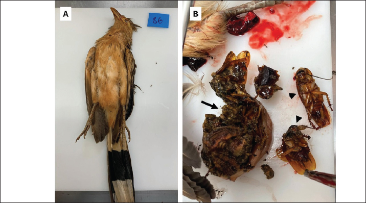
AbstractBackground: Sarcocystis species are protozoans that require intermediate and definitive hosts to complete their life cycle. Several species of Sarcocystis can infect birds and cause severe lesions. This study aimed to report the anatomopathological and molecular findings compatible with Sarcocystis spp. in guira cuckoos (Guira guira). Case Description: Two guira cuckoos were found dead and submitted for necropsy. Tissues were collected, fixed in 10% buffered formalin, and processed according to routine procedures. The sections were stained with hematoxylin and eosin. Nested polymerase chain reaction (PCR) targeting the 18s ribosomal ribonucleico acid region was performed to detect Sarcocystis spp., followed by digestion with restriction enzymes. Macroscopically, the lungs were edematous and dark red. One animal had partially digested cockroaches in the gizzard. Microscopically, diffuse pulmonary hemorrhage was associated with schizonts compatible with Sarcocystis. Schizonts associated with lymphocytic infiltrates were detected in the liver, skeletal muscle, and muscle tissues of the proventriculus. Encephalitis and gliosis were observed in the brain of one animal. Deoxyribonucleic acid amplification detected products compatible with Sarcocysts spp. in the tongue, heart, liver, pectoral muscle, brain, and esophagus. Conclusion: Together, the histopathological and molecular findings confirm systemic sarcocystosis in guira cuckoos and support their involvement in the local transmission cycle, with the presence of cockroaches suggesting a potential epidemiological link in parasite dissemination. Keywords: Histopathology, PCR-RFLP, Sarcocystosis, Wild bird. IntroductionSarcocystis spp. are protozoan parasites with heteroxenous life cycles that affect several animal species including humans (Fayer, 2004; Dubey et al., 2016; Rosenthal, 2021). Currently, approximately 200 species of Sarcocystis have been identified, 25% of which have a completely described life cycle (Rosenthal, 2021). Intermediate hosts, usually prey, become infected by ingesting sporocysts in the environment, and cysts develop mainly in the muscles and nervous tissues. Definitive hosts (predators) become infected by ingesting tissues containing sarcocysts from their prey, and sexual multiplication occurs in the small intestine through the formation of oocysts (Dubey et al., 2016). Birds play a fundamental role in the dissemination and maintenance of Sarcocystis, as they can participate as definitive and/or intermediate hosts depending on the animal species, and thus constitute an important link in the epidemiological chain of the disease (Llano et al., 2022; Rogers et al., 2022). Different species of Sarcocystis can infect birds; however, studies have shown that Sarcocystis neurona, Sarcocystis calchasi, and Sarcocystis falcatula are the main species, with S. falcatula being one of the most prevalent species in birds from different avian orders (Gondim et al., 2019; Llano et al., 2022; Rogers et al., 2022). Pulmonary sarcocystosis has been widely reported in birds; however, recent studies have described encephalitis in many avian species associated with S. calchasi and S. falcatula (Maier et al., 2015; Ushio et al., 2015; Bamac et al., 2020; Wilson et al., 2020). Other relevant changes in Sarcocystis infection in birds include meningoencephalitis due to the infiltration of macrophages and lymphocytes, perivascular cuffing, gliosis, and schizonts (Olson et al., 2007; Da Silva et al., 2009; Wünschmann et al., 2010). A noteworthy challenge is the difficulty in differentiating Sarcocystis species, mainly due to the genetic similarity between S. neurona and S. falcatula. Furthermore, there is a gap in understanding the correlation between isolated species of opossums and infection of birds with particular species of Sarcocystis. The present study aimed to report a case of sarcocystosis that caused encephalitis in two free-living guira cuckoos (Guira guira) in Brazil. Case DetailsTwo free-living guira cuckoos (G. guira; order Cuculiformes; Gmelin, 1788) were found dead near the campus of the Universidade Estadual de Londrina and were submitted for standard postmortem analysis. Multiple tissue samples were collected for histological and molecular analyses. Tissue samples were fixed in a 10% neutral buffered formalin solution for 24 hours, then transferred to a 70% ethyl alcohol solution, and embedded in paraffin. Formalin-fixed paraffin-embedded tissues were sectioned at 5 μm thickness and stained with hematoxylin–eosin (HE) for histopathologic analysis. The procedures with the animals were approved by the Animal Use Ethics Committee of the Universidade Estadual de Londrina under number 038.2020 and by the Biodiversity Authorization and Information System (SISBIO) under number 75997–1. Deoxyribonucleic acid (DNA) extraction was performed using the phenol–chloroform–isoamyl alcohol protocol as previously described (Sambrook et al., 1989). The DNA samples were stored at −20°C until molecular analysis. Amplification of the 18S ribosomal RNA gene [18S ribosomal ribonucleic acid (rRNA)] was performed for the molecular detection of Sarcocystis DNA and other apicomplexans (Da Silva et al., 2009). Polymerase chain reaction (PCR) reactions for amplification of the 18S rRNA gene were performed in a final volume of 25 µl, containing 12.5 µl of 2.5× Master Mix, 0.15 µl of each primer (48F and 359R), 0.25 µl of Taq DNA polymerase, 2 µl of template DNA, and 9.95 µl of ultrapure water. Amplification conditions consisted of an initial denaturation at 95°C for 4 minutes, followed by 30 cycles of 94°C for 30 seconds, 55°C for 1 minute, and 72°C for 2 minutes. Nested PCR was performed using 25 mM outer primers (Tg18s48F and Tg18s359R) and 50 mM inner primers (Tg18s58F and Tg18s348). The expected amplicon size was 290 base pairs (bp) for S. neurona, Neospora caninum, Hammondia hammondi, and Toxoplasma gondii and 310 bp for other Sarcocystis species. For the nested PCR reaction, the final volume was maintained at 25 µl, using 12.5 µl of 2.5× Master Mix, 0.15 µl of each primer (58F and 348R), 0.25 µl of Taq DNA polymerase, 2 µl of the product from the previous PCR as template, and 8.95 µl of ultrapure water. Thermocycling conditions were initial denaturation at 95°C for 4 minutes, followed by 35 cycles of 94°C for 30 seconds, 55°C for 1 minute, and 72°C for 90 seconds. Enzymatic digestion using the enzymes DdeI, HPY188III, and MspI was performed to analyze restriction fragment length polymorphisms (RFLP) and differentiate T. gondii, N. caninum, and Sarcocystis species. RFLP analysis was performed in a final volume of 20 µl, containing 0.2 µl of each restriction enzyme (Hpy188III, DdeI, and MspI), 0.2 µl of BSA (100×), 2 µl of 10× NEB2 buffer, 3 µl of the amplified PCR product, and 14.2 µl of ultrapure water. Digestion was carried out at 37°C for 60 minutes, and the resulting fragments were visualized on a 3% agarose gel. The amplified products were visualized using electrophoresis on a 1.5% agarose gel stained with SYBR Safe DNA Gel Stain (Thermo Fisher Scientific, Waltham, MA, USA). Gross examination revealed one male and one female guira cuckoo, both in good nutritional condition (Fig. 1A) and moderate carcass preservation. Internal evaluation of the female animal showed preserved visceral topography and a discrete accumulation of blood and clots in the coelomic cavity. In both animals, the lungs were non-collapsing and edematous, and significant amounts of blood oozed from the cut surfaces. In the female, several partially digested cockroaches were found in the gizzard (Fig. 1B).
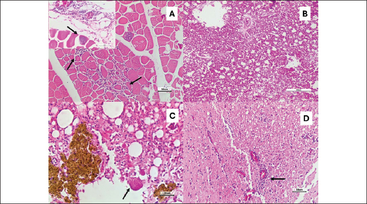
Fig. 1. Postmortem analysis of free-living guira cuckoo (Guira guira). A: Species identification. Guira cuckoo found dead. There are no external abnormalities noted, and the carcass is moderately well preserved. B: Gizzard. The sectioned gizzard (arrow) contained partially digested arthropods (cockroaches, arrowheads). Histological evaluation revealed marked diffuse hemorrhage in the lung in both animals and discrete multifocal lymphocytic myositis in the pectoral skeletal muscle. Furthermore, intralesional schizonts compatible with Sarcocystis spp. were observed in both tissues (Fig. 2). Schizonts associated with moderate multifocal lymphocytic infiltration were detected in the liver and muscle tissue of the proventriculus. In the brain, multifocal discrete lymphocytic encephalitis and multifocal moderate gliosis were observed in one animal (Fig 2).
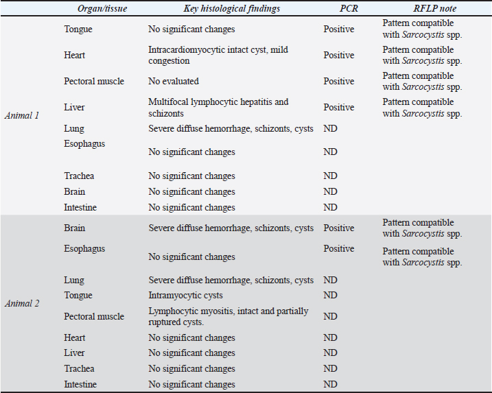
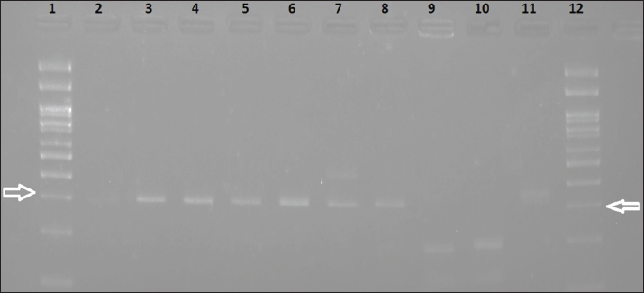
Fig. 2. Systemic sarcocystosis in a guira cuckoo (Guira guira). A: Pectoral muscle. Lymphocytic myositis (arrows) of skeletal muscle accompanied by an intramyocytic cyst containing numerous bradyzoites with morphology consistent with Sarcocystis spp. (Insert). HE, scale bar 50 µm. B: Multifocal and severe pulmonary hemorrhage. HE, scale bar 100 µm. C. Pulmonary hemosiderosis accompanied by extracellular cysts (arrows). HE, scale bar 20 µm. D. In the cerebrum, there is an increased number of glial cells (gliosis) with a discrete inflammatory infiltrate composed of lymphocytes and plasma cells expanding the perivascular region (arrow). HE, scale bar 50 µm. Amplified products with an expected fragment size between 290 and 310 bp, compatible with Sarcocystis spp., were detected in the tongue, heart, liver, pectoral skeletal muscle, brain, and esophagus. A total of 18 tissue samples were analyzed, of which six tested positive (Table 1). Gel images and RFLP band patterns are shown in Figure 3. The main histological changes and the results of the molecular analysis are presented in Table 1. Table 1. Results of the main histopathological findings and molecular analysis of two guira cuckoos (Guira guira).
DiscussionReports of detecting Sarcocystis spp. in wild birds are limited because of factors such as difficulty in obtaining samples and a lack of detailed clinical history. The objective of this study was to integrate different diagnostic methods using two free-ranging animals in good condition. Sarcocystis infection occurs in birds when they ingest sporocysts in contaminated water or food excreted by definitive hosts (Olson et al., 2007). In the intermediate host, the disease is associated with tissue destruction caused by the rapid asexual reproduction of the protozoan, also known as schizogony, which occurs after an oral infection (Wünschmann et al., 2010). In birds, one of the most characteristic clinical manifestations of S. falcatula infection is severe interstitial pneumonia, resulting from the rupture of infected pulmonary endothelial cells (Suedmeyer et al., 2001). In the present study, the main alterations observed in the affected animals were recorded in the lungs and nervous system. This observation agrees with previous studies that documented the pulmonary and neurological manifestations associated with S. falcatula infection (Suedmeyer et al., 2001; Ushio et al., 2015). Additionally, we observed inflammatory changes ranging from mild to moderate intensity in multiple organs, supporting the occurrence of a systemic infection. A recent study by Llano et al. (2022) identified a new species of Sarcocystis in G. guira (Cuculiformes: Cuculidae), demonstrating a genetic relationship of 92.65% with S. falcatula and 93.55% with Sarcocystis lindsayi (AF387164), reinforcing the importance of this bird in maintaining the parasite in the environment (Olias et al., 2014). Based on the research, our work is one of the few reports on the detection of Sarcocystis spp. in guira cuckoo, obtained through histological findings in various tissues, conventional PCR, and restriction enzymes, highlighting the relevance of accurate diagnosis by multiple methods. Another noteworthy point was the presence of cockroaches in the stomach contents of the birds analyzed, a factor that may be indicative of paratenic hosts for Sarcocystis muris. Although infection by S. muris is rare in birds, it has been experimentally documented by serological diagnosis and microscopic visualization (Tillmann et al., 1999). In a previous study, Clubb and Frenkel (1992) investigated the death of psittacines in an aviary. They concluded that cockroaches play a role in the transmission of S. falcatula from opossums to birds and are the etiological agents responsible for pulmonary sarcocystosis in birds. As insects are part of the natural diet of many birds, the presence of cockroaches should be considered a relevant factor in the epidemiology of the disease, as they may facilitate the transmission of the protozoan to new populations of birds. In the present study, the analyzed species is a carnivorous bird capable of acting both as prey and as a predator, which increases its potential participation in the biological cycle as a definitive and/or intermediate host for different Sarcocystis species. In this context, such characteristics make this species a relevant model for future investigations aimed at elucidating epidemiological aspects and host–parasite interactions involving Sarcocystis in natural settings. Furthermore, the genetic similarity between S. falcatula and S. neurona was demonstrated by analyzing 18S ribosomal RNA gene sequences, which improved our understanding of the evolution and transmission of these parasites (Tanhauser et al., 1999; Valadas et al., 2016). Although they share genetic similarities, the two species can be differentiated based on their biological characteristics, which is crucial for the diagnosis and control of infections (Box et al., 1984; Dame et al., 1995). Finally, the transmission dynamics of sarcocystosis in birds are influenced by the diversity of intermediate and definitive hosts, which make the biological cycle of Sarcocystis complex and illustrate the wide range of hosts that can participate in the biological cycle of these parasites, complicating the understanding and control of this parasitosis (Fenger et al., 1994). This study has some limitations that should be considered. The high diversity and close genetic relatedness among Sarcocystis spp. hinder precise species differentiation. In addition, most available data rely on indirect immunofluorescence for antibody detection, and the lack of commercially available antibodies limits the use of tissue-based techniques such as immunohistochemistry and in situ hybridization. Finally, the small sample size (n=2) restricts the broader generalization of these findings. The present study highlights the complexity of sarcocystosis in birds and underscores the importance of complementary diagnostic approaches, as well as a better understanding of the hosts and species involved. An increased number of diagnosed cases and diversification of research methods are essential for advancing our knowledge about the epidemiology of Sarcocystis in free-living animals. Furthermore, our findings open perspectives for future studies based on the combined analysis of avian stomach contents and molecular detection approaches. Such strategies may contribute to a better understanding of transmission dynamics, strengthen causal inferences, and clarify the role of different hosts in the parasite’s life cycle. ConclusionUsing complementary methods (histopathology, nested PCR, and RFLP), we confirmed systemic sarcocystosis in two free-living guira cuckoos that were found dead. The presence of cockroaches in the gizzard of one of the birds, along with multi-organ molecular positivity, reinforces the epidemiological relevance of this species in local Sarcocystis cycles. Future investigations incorporating higher-resolution genetic markers (e.g., ITS1, cox1) and expanded case accrual will be valuable to resolve species-level identity and clarify transmission pathways. Conflict of interestNot necessary for this manuscript. FundingDouglas A. Silva received a Brazilian Federal Agency for Support and Evaluation of Graduate Education (CAPES) fellowship. Author´s contributionD. A. Silva collected the data and drafted the manuscript; M. I. P. Sogari conducted the analysis; A. A. S. Baptista performed formal analysis; I. T. Navarro performed formal analysis; J. L. Garcia performed formal analysis; L. D. Barros interpreted the results; A. P. F. R. L. Bracarense conceived the study and revised the manuscript. All authors critically revised the manuscript and approved the final version. Data availabilityAll data supporting the findings of this study are available within the manuscript. ReferencesBamac, O.E., Rogers, K.H., Arranz-Solís, D., Saeij, J.P.J., Lewis, S., Duerr, R., Skoglund, J., Peronne, L. and Mete, A. 2020. Protozoal encephalitis associated with Sarcocystis calchasi and S. falcatula during an epizootic involving Brandt’s cormorants (Phalacrocorax penicillatus) in coastal Southern California, USA. Int. J. Parasitol. Parasites. Wildl. 12, 185–191; doi:10.1016/j.ijppaw.2020.06.005 Box, E.D., Meier, J.L. and Smith, J.H. 1984. Description of Sarcocystis falcatula Stiles, 1893, a parasite of birds and opossums. J. Protozool. 31(4), 521–524; doi:10.1111/j.1550-7408.1984.tb05495.x Clubb, S.L. and Frenkel, J.K. 1992. Sarcocystis falcatula of opossums: transmission by cockroaches with fatal pulmonary disease in psittacine birds. J. Parasitol. 78(1), 116–124; doi:10.2307/3283697 Da Silva, R.C., Su, C. and Langoni, H. 2009. First identification of Sarcocystis tenella (Railliet, 1886) Moulé, 1886 (Protozoa: apicomplexa) by PCR in naturally infected sheep from Brazil. Vet. Parasitol. 165(3-4), 332–336; doi:10.1016/j.vetpar.2009.07.016 Dame, J.B., MacKay, R.J., Yowell, C.A., Cutler, T.J., Marsh, A. and Greiner, E.C. 1995. Sarcocystis falcatula from passerine and psittacine birds: synonymy with Sarcocystis neurona, agent of equine protozoal myeloencephalitis. J. Parasitol. 81(6), 930–935; doi:10.2307/3284044 Dubey, J.P., Calero-Bernal, R., Rosenthal, B.M., Speer, C.A. and Fayer, R. 2016. Sarcocystosis of animals and humans, 2nd ed. Boca Raton, FL: cRC Press, p: 501. Fayer, R. 2004. Sarcocystis spp. in human infections. Clin. Microbiol. 17(4), 894–902; doi:10.1128/cmr.17.4.894-902.2004 Fenger, C.K., Granstrom, D.E., Langemeier, J.L., Gajadhar, A., Cothran, G., Tramontin, R.R., Stamper, S. and Dubey, J.P. 1994. Phylogenetic relationship of Sarcocystis neurona to other members of the family sarcocystidae based on small subunit ribosomal RNA gene sequence. J. Parasitol. 86(6), 966–975; doi:10.2307/3283447 Gondim, L.F.P., Soares, R.M., Tavares, A.S., Borges-Silva, W., De Jesus, R.F., Llano, H.A.B. and Gondim, L.Q. 2019. Sarcocystis falcatula-like derived from opossum in Northeastern Brazil: in vitro propagation in avian cells, molecular characterization and bioassay in birds. Int. J. Parasitol. Parasites Wildl. 10, 132–137; doi:10.1016/j.ijppaw.2019.08.008 Llano, H.A.B., Zavatieri Polato, H., Borges Keid, L., Ferreira de Souza Oliveira TM., Zwarg, T., De Oliveira, A.S., Sanches, T.C., Joppert, A.M., Gondim, L.F.P. and Martins Soares, R. 2022. Molecular screening for Sarcocystidae in muscles of wild birds from Brazil suggests a plethora of intermediate hosts for Sarcocystis falcatula. Int. J. Parasitol. Parasites. Wildl. 17, 230–238; doi:10.1016/j.ijppaw.2022.03.002 Maier, K., Olias, P., Enderlein, D., Klopfleisch, R., Mayr, S.L., Gruber, A.D. and Lierz, M. 2015. Parasite distribution and early-stage encephalitis in Sarcocystis calchasi infections in domestic pigeons (Columba livia f. domestica). Avian. Pathol. 44(1), 5–12; doi:10.1080/03079457.2014.978263 Olias, P., Maier, K., Wuenschmann, A., Reed, L., Armién, A.G., Shaw, D.P., Gruber, A.D. and Lierz, M. 2014. Sarcocystis calchasi has an expanded host range and induces neurological disease in cockatiels (Nymphicus hollandicus) and North American rock pigeons (Columbia livia f. dom.). Vet. Parasitol. 200(1-2), 59–65; doi:10.1016/j.vetpar.2013.11.012 Olson, E.J., Wünschmann, A. and Dubey, J.P. 2007. Sarcocystis sp.-associated meningoencephalitis in a bald eagle (Haliaeetus leucocephalus). J. Vet. Diagnostic. Investig. 19(5), 564–568; doi:10.1177/104063870701900519 Rogers, K.H., Arranz-Solís, D., Saeij, J.P.J., Lewis, S. and Mete, A. 2022. Sarcocystis calchasi and other Sarcocystidae detected in predatory birds in California, USA. Int. J. Parasitol. Parasites. Wildl. 17, 91–99; doi:10.1016/j.ijppaw.2021.12.008 Rosenthal, B.M. 2021. Zoonotic sarcocystis. Res. Vet. Sci. 136, 151–157; doi:10.1016/j.rvsc.2021.02.008 Sambrook, J., Fritsch, E.F. and Maniatis, T. 1989. Molecular cloning a laboratory manual. New York, NY: Cold Spring Harb Lab Press. Suedmeyer, W.K., Bermudez, A.J., Barr, B.C. and Marsh, A.E. 2001. Acute pulmonary Sarcocystis falcatula-like infection in three Victoria crowned pigeons (Goura victoria) housed indoors. J. Zoo. Wild. Med. 1042(2), 252–256. Tanhauser, S.M., Yowell, C.A., Cutler, T.J., Greiner, E.C., MacKay, R.J. and Dame, J.B. 1999. Multiple DNA markers differentiate Sarcocystis neurona and Sarcocystis falcatula. J. Parasitol. 85(2), 221–228; doi:10.2307/3285623 Tillmann, T., Kamino, K. and Mohr, U. 1999. Sarcocystis muris—a rare case in laboratory mice. Lab. Anim. 33(4), 390–392; doi:10.1258/002367799780487869 Ushio, N., Watanabe, K.I., Chambers, J.K., Shibato, T., Nakayama, H. and Uchida, K. 2015. Sarcocystis calchasi encephalitis in a rock pigeon. J. Vet. Med. Sci. 77(11), 1523–1526. Valadas, S.Y.O.B., Da Silva, J.I.G., Lopes, E.G., Keid, L.B., Zwarg, T., De Oliveira, A.S., Sanches, T.C., Joppert, A.M., Pena, H.F.J., Oliveira, T.M.F.S., Ferreira, H.L. and Soares, R.M. 2016. Diversity of Sarcocystis spp shed by opossums in Brazil inferred with phylogenetic analysis of DNA coding ITS1, cytochrome B, and surface antigens. Exp. Parasitol. 164, 71–78; doi:10.1016/j.exppara.2016.02.008 Wilson, T.M., Sousa, S.K.H., Paludo, G.R., De Melo, C.B., Llano, H.A.B., Soares, R.M. and Castro, M.B. 2020. An undescribed species of Sarcocystis associated with necrotizing meningoencephalitis in naturally infected backyard chickens in the Midwest of Brazil. Parasitol. Int. 76, 102098; doi:10.1016/j.parint.2020.102098 Wünschmann, A., Rejmanek, D., Conrad, P.A., Hall, N., Cruz-Martinez, L., Vaughn, S.B. and Barr, B.C. 2010. Natural fatal Sarcocystis falcatula infections in free-ranging eagles in North America. J. Vet. Diagnostic. Investig. 22(2), 282–289; doi:10.1177/104063871002200222
Fig. 3. Agarose gel of guira cuckoo samples after RFLP for differentiation among Toxoplasma gondii, Neospora caninum, Hammondia, and Sarcocystis spp. Lanes 1 and 12 show the 100 bp molecular weight marker. Arrows indicate the expected fragment size between 290 and 310 bp, compatible with Sarcocystis spp. DNA extracted from tongue, heart, liver, and muscle of Animal 1 are shown in lanes 3, 4, 5, and 6, respectively. Lanes 7 and 8 correspond to brain and esophagus of Animal 2. Positive controls for Toxoplasma gondii, Neospora caninum, and Sarcocystis spp. are shown in lanes 9, 10, and 11, respectively. The negative control is shown in lane 2. | ||
| How to Cite this Article |
| Pubmed Style Silva DA, Sogari MIP, Baptista AAS, Navarro IT, Garcia JL, Barros LDD, . Histopathological and molecular evidence of systemic Sarcocystis spp. infection in a free-living Guira Cuckoo (Guira guira). Open Vet. J.. 2026; 16(7): 4992-4998. doi:10.5455/OVJ.2026.v16.i7.78 Web Style Silva DA, Sogari MIP, Baptista AAS, Navarro IT, Garcia JL, Barros LDD, . Histopathological and molecular evidence of systemic Sarcocystis spp. infection in a free-living Guira Cuckoo (Guira guira). https://www.openveterinaryjournal.com/?mno=309029 [Access: July 27, 2026]. doi:10.5455/OVJ.2026.v16.i7.78 AMA (American Medical Association) Style Silva DA, Sogari MIP, Baptista AAS, Navarro IT, Garcia JL, Barros LDD, . Histopathological and molecular evidence of systemic Sarcocystis spp. infection in a free-living Guira Cuckoo (Guira guira). Open Vet. J.. 2026; 16(7): 4992-4998. doi:10.5455/OVJ.2026.v16.i7.78 Vancouver/ICMJE Style Silva DA, Sogari MIP, Baptista AAS, Navarro IT, Garcia JL, Barros LDD, . Histopathological and molecular evidence of systemic Sarcocystis spp. infection in a free-living Guira Cuckoo (Guira guira). Open Vet. J.. (2026), [cited July 27, 2026]; 16(7): 4992-4998. doi:10.5455/OVJ.2026.v16.i7.78 Harvard Style Silva, D. A., Sogari, . M. I. P., Baptista, . A. A. S., Navarro, . I. T., Garcia, . J. L., Barros, . L. D. D. & (2026) Histopathological and molecular evidence of systemic Sarcocystis spp. infection in a free-living Guira Cuckoo (Guira guira). Open Vet. J., 16 (7), 4992-4998. doi:10.5455/OVJ.2026.v16.i7.78 Turabian Style Silva, Douglas Aparecido, Maria Izabel Pedra Sogari, Ana Angelita Sampaio Baptista, Italmar Teodorico Navarro, João Luis Garcia, Luiz Daniel De Barros, and Ana Paula Frederico Rodrigues Loureiro Bracarense. 2026. Histopathological and molecular evidence of systemic Sarcocystis spp. infection in a free-living Guira Cuckoo (Guira guira). Open Veterinary Journal, 16 (7), 4992-4998. doi:10.5455/OVJ.2026.v16.i7.78 Chicago Style Silva, Douglas Aparecido, Maria Izabel Pedra Sogari, Ana Angelita Sampaio Baptista, Italmar Teodorico Navarro, João Luis Garcia, Luiz Daniel De Barros, and Ana Paula Frederico Rodrigues Loureiro Bracarense. "Histopathological and molecular evidence of systemic Sarcocystis spp. infection in a free-living Guira Cuckoo (Guira guira)." Open Veterinary Journal 16 (2026), 4992-4998. doi:10.5455/OVJ.2026.v16.i7.78 MLA (The Modern Language Association) Style Silva, Douglas Aparecido, Maria Izabel Pedra Sogari, Ana Angelita Sampaio Baptista, Italmar Teodorico Navarro, João Luis Garcia, Luiz Daniel De Barros, and Ana Paula Frederico Rodrigues Loureiro Bracarense. "Histopathological and molecular evidence of systemic Sarcocystis spp. infection in a free-living Guira Cuckoo (Guira guira)." Open Veterinary Journal 16.7 (2026), 4992-4998. Print. doi:10.5455/OVJ.2026.v16.i7.78 APA (American Psychological Association) Style Silva, D. A., Sogari, . M. I. P., Baptista, . A. A. S., Navarro, . I. T., Garcia, . J. L., Barros, . L. D. D. & (2026) Histopathological and molecular evidence of systemic Sarcocystis spp. infection in a free-living Guira Cuckoo (Guira guira). Open Veterinary Journal, 16 (7), 4992-4998. doi:10.5455/OVJ.2026.v16.i7.78 |


