Open Veterinary Journal, (2026), Vol. 16(7): 4466-4478
Research Article
10.5455/OVJ.2026.v16.i7.31
Role of Ajwa date (Phoenix dactylifera L.) extract against doxorubicin-induced genotoxicity and nephrotoxicity in female mice bearing Ehrlich Ascites Carcinoma
Sabha E. Elballat1*, Ahmed M. Aldawek2, Fawzeya A. Zayed1, Iman E. El-Araby3 and
Sara Ahmed El-Said1
1Zoology Department, Faculty of Science, Zagazig University, Zagazig, Egypt
2Department of Pathology and Clinical Pathology, Faculty of Veterinary Medicine, University of Tripoli, Tripoli, Libya
3Animal Wealth Development, Faculty of Veterinary Medicine, Zagazig University, Zagazig, Egypt
*Corresponding Author: Sabha E. Elballat. Zoology Department, Faculty of Science, Zagazig University, Zagazig, Egypt. Email: Sabhael [at] yahoo.com
Submitted: 06/02/2026 Revised: 30/05/2026 Accepted: 13/06/2026 Published: 11/07/2026
© 2025 Open Veterinary Journal
This is an Open Access article distributed under the terms of the Creative Commons Attribution-Non Commercial-No Derivatives License (http://creativecommons.org/licenses/by-nc-nd/4.0/), which permits non-commercial re-use, distribution, and reproduction in any medium, provided the original work is properly cited, and is not altered, transformed, or built upon in any way.
Abstract
Background: Doxorubicin (DOX), an anthracycline antibiotic widely used in cancer chemotherapy, is associated with toxic dose-dependent adverse effects affecting normal tissues. Ehrlich ascites carcinoma (EAC) is a transplantable murine mammary adenocarcinoma commonly used as an experimental tumor model. Ajwa date (Phoenix dactylifera L.) extract (ADE) is rich in bioactive phytochemicals with potent antioxidant and anticancer properties.
Aim: The present study aimed to evaluate the genotoxic (chromosomal aberrations) and nephrotoxic effects induced by DOX and to investigate the potential protective and ameliorative role of ADE in EAC-bearing adult female mice.
Methods: A total of 135 adult female mice were divided into two main groups: a negative control group (n=15) and an EAC-bearing group (n=120). EAC-inoculated mice were further subdivided into eight experimental groups, including EAC-positive control, DOX-treated, ADE-treated (10, 20, and 40 mg/gm), and combined DOX + ADE-treated groups. DOX (0.00026 mg/gm body weight) was administered intraperitoneally once per week for 30 days, while ADE was administered orally once daily for 30 consecutive days. At the end of the experiment, chromosomal aberrations in bone marrow cells and histopathological alterations in the kidney and tumor tissues were assessed.
Results: DOX significantly induced chromosomal aberrations and pathological alterations in EAC-bearing mice. Oral ADE administration markedly reduced DOX-induced genotoxicity and nephrotoxicity. Among the tested doses, ADE at 20 mg/gm showed the most pronounced protective effect when combined with DOX treatment.
Conclusion: Ajwa date extract is a safe and effective natural supplement that mitigates DOX-induced cytotoxicity while enhancing its therapeutic efficacy. The combined treatment of DOX with ADE at a dose of 20 mg/gm provided the best protective and therapeutic outcomes.
Keywords: Ajwa date, Chromosomal aberrations, Doxorubicin, Ehrlich ascites carcinoma, Mus musculus.
Introduction
Doxorubicin (DOX) is a widely used chemotherapeutic agent with potent antitumor activity against various solid and hematological malignancies. Its anticancer efficacy is mediated through multiple mechanisms, including DNA intercalation, inhibition of topoisomerase II, and generation of reactive oxygen species (ROS). However, DOX treatment is frequently limited by severe dose-dependent toxic effects on normal tissues, including genotoxicity, nephrotoxicity, and cardiotoxicity (Micallef et al., 2020). Ehrlich ascites carcinoma (EAC) is a spontaneous murine mammary adenocarcinoma characterized by rapid proliferation, high transplantability, and aggressive metastatic behavior. It is commonly used as an experimental tumor model for evaluating anticancer agents and studying tumor biology (Frajacomo et al., 2016). EAC cells proliferate within the peritoneal cavity when injected intraperitoneally, forming ascitic tumors that significantly compromise host survival (Mishra et al., 2018). Ajwa dates (Phoenix dactylifera L.) are a premium variety of dates cultivated mainly in Al-Madinah Al-Munawwarah, Saudi Arabia. They are rich in dietary fiber, vitamins, essential minerals, flavonoids, and polyphenolic compounds, which exhibit strong antioxidant, anti-inflammatory, and anticancer activities (Farhana and Roushney, 2021; Shahbaz et al., 2022). Several studies have reported the protective effects of Ajwa date extract against chemotherapy-induced organ toxicity and oxidative stress.
Therefore, this study was designed to investigate the genotoxic and pathological effects of DOX in EAC-bearing female mice and to evaluate the potential protective and ameliorative role of Ajwa date extract administered at different doses.
Materials and Methods
Chemicals
DOX was purchased from Khandelwal Laboratories Pvt. Ltd., Factory Thane, India (lyophilized DOX 50 mg for intravenous injection). DOX hydrochloride (5,12-Naphthacenedione,10-[(3-amino-2,3,6-trideoxy-Llyxohexopyranosyl) oxy]-7,8,9,10-tetrahydro-6,8,11-trihydroxy-8-(hydroxylacetyl)-1-methoxyhydrochloride (8S-cis).
EAC preparation
The Institutional Animal Ethics Committee approved the experimental animal protocol. EAC cells were collected from donor mice (Egyptian National Cancer Institute, Cairo University) and carefully suspended in sterile isotonic saline; 2.5 million cells were transplanted (intraperitoneally) into healthy mice. Mice were injected with EAC cells by successive transplantation of 1 × 10⁶ cells/mouse in the peritoneal cavity (Kabel, 2014).
The Trypan blue viability test
Cell viability was assessed before inoculation using the Trypan Blue exclusion assay (it was explained in our previous paper (Sara et at., 2025). Trypan blue is a stain usually used for detecting living cells, where cells without interactions with the dye are viable (Freshney, 1987).
Date fruit collection and extract preparation
Semi-dry Ajwa date fruits were purchased from the local market in Al-Madinah Al-Munawwara City, Saudi Arabia. Ajwa date extract (ADE) was prepared as described elsewhere with some modifications (Khan et al., 2021). In brief, the seeds were removed manually and discarded. Then, the flesh was washed with double-distilled water, dried, and ground into fine particles. The dried powder was homogenized in 95% ethyl alcohol (Sigma-Aldrich) at a ratio of 1:3 (W/V), mixed vigorously, and then stored at room temperature for 1 week. The mixture was filtered through Wattman filter paper grade 520 and then centrifuged at 4000 ×g for 15 minutes. A rotary evaporator (Buchi Rotavapor R-205, Switzerland) was used to evaporate the supernatant at 30°C. In a water bath (Lab1st-GX).
Phytoconstituents of the Ajwa dates
phytoconstituents of Ajwa dates include glycerol, D‐Pinitol, 4TMS derivative, pentakis (trimethylsilyl) ether (isomer 1), tris (trimethylsilyl) ether, trimethylsilyloxime, 2‐pentenedioic acid, bis (trimethylsilyl) ester, L‐threitol, L‐(+)‐threose, D‐(+)‐Galactose, pentafluorobenzyloxime (isomer 1), D‐(+)‐Arabitol, D‐Sorbitol, 2‐[(trimethylsilyl)oxy]‐, D‐(−)‐tagatofuranose, pentakis (trimethylsilyl) ether, 6TMS derivative, D‐Mannitol, D‐glucopyranose L‐(+)‐Tartaric acid, palmitic acid, 3‐Heptadecen‐5‐yne, (Z), 9‐Octadecenoic acid, (E)‐TMS derivative, fumaric acid, di (2‐propylphenyl) ester, 5TMS derivative, α‐linoleic acid, 6TMS derivative, 4TMS derivative, TMS derivative, and stearic acid (Othman et al., 2023).
Experimental design and tissue sampling methods
A total of 135 adult healthy female mice (Mus musculus) weighing (20 ± 1 gm; about 10 weeks old were used throughout the study). Animals were obtained from the Animal House Research Center, Cairo, Egypt. All animals were housed in metal cages under hygienic conditions and maintained on a light/dark cycle (12/12 hours). They were given food and water ad libitum. The 135 mice were divided into 2 main groups. Group A: 15 mice were given an ordinary diet for 30 successive days and were kept as a negative control. Group B: 120 mice were injected intraperitoneally with a single dose of EAC (1 × 10⁶ cells/mouse) only, on the 1st day of the experiment. The injected animals were kept in cages for 7 days. By the 8th day, the symptoms of cancer appeared, such as skin rash, redness, and irritation, and tumors developed in several locations of the body. The EAC-injected mice were randomly divided again into 8 subgroups as follows. The 1st group was the EAC (+ve Control) group: (15 animals) from mice that were intraperitoneally injected with EAC. The 2nd group (DOX) was intraperitoneally injected with DOX (0.00026 mg/gm b.w.) once per week for 30 successive days. The mouse dose was calculated from the therapeutic human dose according to Paget and Barnes (1964) while the 3rd, 4th, and 5th groups (ADE-A, ADE-B, and ADE-C) were orally administered with 10, 20, and 40 mg/gm of Ajwa date extract once daily for 30 successive days, respectively. The 6th, 7th, and 8th groups DOX + ADE (A, B, and C) were orally administered with 10, 20, and 40 mg/gm of ADE once daily, followed by intraperitoneal injection with 0.00026 mg/gm b.w. of DOX once per week for 30 consecutive days.
Chromosomal aberration assay
Bone marrow was collected from the femur bones of each mouse (nine mice per group), and chromosomes from the white blood corpuscles were extracted according to the method described by Preston et al. (1987). The slides were stained with Giemsa stain (1.5%) for approximately 20 minutes. All slides were carefully scanned for metaphase spreads under (×10) and (×40) objectives of the light microscope. The very well-spread metaphases were examined under (×100) oil immersion, and chromosome photomicrographs were taken. A total of 50 metaphases per mouse were analyzed.
Histopathological examination
Histological preparations were performed according to the method of Bancroft and Gamble (2002) where six mice/group were sacrificed and dissected, their kidneys and tumor tissues were collected and directly submerged in 10% neutral formalin, processed, and stained with H&E for histopathological investigations.
Statistical analysis
All data were analyzed using the R Studio program (version 2026). Data are presented as mean ± SD. One-way analysis of variance (ANOVA) (Lars and Svante, 1989) followed by Tukey’s post-hoc test (Keselman and Rogan, 1977) revealed significant differences among the experimental groups (p < 0.05).
Ethical approval
The experiments and handling of the experimental animals were conducted in compliance with the guidelines set forth by the Research Ethics Committee at Zagazig University, Egypt (ZU-IACUC/1/F/111/2023).
Results
Morphological and behavioral observations
Toxicity of EACs
Female mice that were injected intraperitoneally with a single dose (1 × 10⁶ cells/mouse) of EAC were kept in cages and observed daily. On the eighth day after the injection, redness and tumors started to appear in different locations in and near the injection site (Fig. 1a). Two weeks after the injection, redness behind the ear and mouth, body ulcers, general body weakness, and increased tumor volume were observed in some mice (Fig. 1b). In the third week, the EAC (+ve control) group showed marked deterioration, whereas the other group showed noticeable improvement. In the fourth week, the total chemotherapy (DOX) group begins to decline and worsen with a general weakness and degradation.
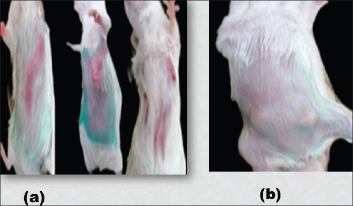
Fig. 1. (a) Variations in redness and tumors detected at the site of injection with Ehrlich carcinoma cells; (b) Mice with large tumors detected in different locations throughout the body.
Chromosomal aberrations in bone marrow cells
Aberrant metaphases in mice in the Ehrlich carcinoma cell (+ve control) group
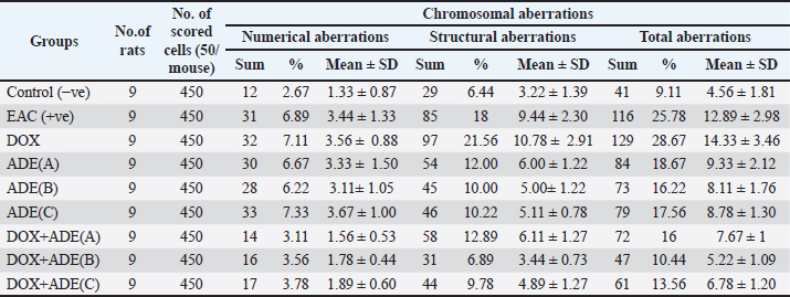
The percentage of total chromosomal aberrations (TCA), which included numerical aberrations (NA; changes in the chromosomal number) and structural aberrations (SA; changes in the chromosomal structure) from bone marrow cells of mice in the EAC (+ve control) group was increased and reached 25.78% compared with 9.11% in the −ve control group, which was extremely significant (p < 0.0001). The total NA included 3.56% aneuploid (AN; changes in the number of individual chromosomes 2n ± 1) and 3.33% polyploid (Po; presence of more than 2 genomes). It reached 6.89% compared with 2.67% in the control group. The total SA could be scored in 85 of 450 occasions compared with 29 among similar numbers of −ve controls. Telomeric deletion (T, loss of a part of a chromosome from its terminal end)) was the most abundant aberration among all scored aberrations, reaching 27 (6%) versus 7 (1.56) in the −ve control. ring (R), telomeric exchange (T.ex), gap (G), and dicentric (Di) were also recorded (Table 1 and Fig. 2).
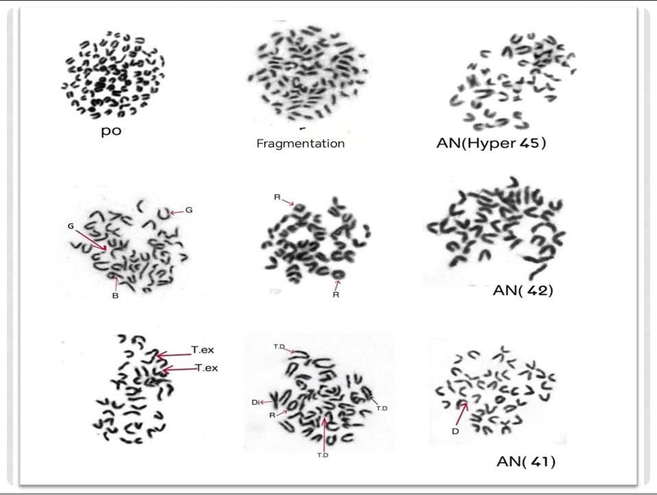
Fig. 2. Metaphases from bone marrow cells of adult female mice in the EAC (+ve control) group showing: NA in the form of aneuploid (AN) and polyploid (Po) in addition to SA represented by telomeric deletion (T.D); ring (R); telomeric exchange (T.ex); gap (G); and dicentric (Di). Total Magnification=1,000×.
Table 1. Comparison between the frequencies of chromosomal aberrations in bone marrow cells of adult female mice in different experimental groups.
Aberrant metaphases in mice in the DOX group
The TCA from mice in the DOX group was markedly increased. The percentage of TCA reached 28.67% compared with 9.11% in the control group, and these results were extremely significant at (p < 0.0001). NA, including aneuploidy (AN), was more affected and reached 19 occasions (4.22%) compared with 7 occasions (1.56%) in the −ve control group. In addition, polyploid (Po) was also recorded in 13 occasions (2.89%) versus 5 (1.11%) among the −ve control group. Total SA was also increased, and metaphase with SA could be spotted on 97 occasions compared with 29 in the control group. The most abundant SAs were telomeric break (T.B), telomeric exchange (T.ex), and centric fusions (C.F) (Table 1 and Fig. 3).
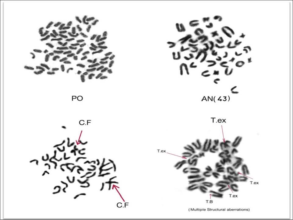
Fig. 3. Several metaphases from bone marrow cells of adult female mice in the DOX group, with NA in the form of AN and polyploid (Po), in addition to SA, which are represented by telomeric break (T.B); telomeric exchange(T.ex); and centric fusions (C.F). Total Magnification=1,000×.
Aberrant metaphases in mice in the ADE (A) group
As shown in Table 1, the TCA decreased and reached 18.67% compared with 25.78% in the +ve control, but was still higher than (9.11%) in the -ve control. The NA, polyploid (17 occasions), was more affected than aneuploid (13) compared with the control. Metaphases with SA could be spotted on 54 of 450 occasions versus 29 among similar numbers from the control group. The most abundant SAs were ring (R), telomeric deletion (T.D), telomeric exchange (T.ex), acentric fragment (Ac.f), and centric fusions (C.F) (Fig. 4).
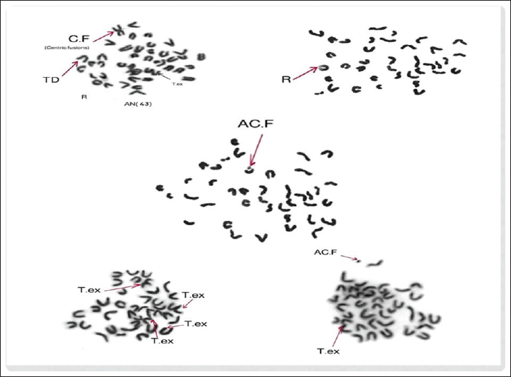
Fig. 4. Several metaphases from bone marrow cells of adult female mice in ADE (A), (B), and (C) showing the most abundant SA represented by ring (R); telomeric deletion (T.D); telomeric exchange (T.ex); acentric fragment (Ac.f); and centric fusions (C.F.). Total Magnification=1,000×.
Aberrant metaphases in mice in the ADE (B) group
TCA was significantly (p < 0.001) decreased to reach 16.22% compared with 25.78% in the +ve control and 18.67% in the ADE (A) group, but the value was still higher than 9.11% in the −ve control. As for the NA, the polyploid was also more affected and scored in 19 occasions compared to 17 in the ADE (A) group. Metaphases that included aneuploid were recorded in 9 occasions as compared with 13 occasions in group ADE (A).
The frequency of total SA also decreased to 10% compared with 12% in group ADE (A). The most abundant aberrations were ring (R), telomeric deletion (T.D), telomeric exchange (T.ex), acentric fragment (Ac.f), and centric fusions (C.F.) (Table 1 and Fig. 4).
Aberrant metaphases in mice in the ADE (C) group
The TCA in this group significantly (p < 0.01) reduced to 17.56% compared with 25.78% in the +ve control and 18.67% in the ADE (A) group, but the value was still higher than 9.11% in the −ve control and 16.22% in the ADE (B) group. The total NA was increased in 33 occasions versus 30 and 28 occasions in groups ADE (A) and ADE (B), respectively. Furthermore, polyploid was more affected and scored in 20 occasions versus 17 and 19 occasions among groups A and B, respectively. The metaphases that included aneuploid records on 13 occasions, as in group ADE (A). The frequency of total SA was near that of group ADE (B), reaching 10.22% compared with 10% and 12% in groups ADE (B) and ADE (A), respectively. Various forms of SA are also recorded, as ring (R); telomeric deletion (T.D); telomeric exchange (T.ex); acentric fragment (Ac.f); and centric fusions (C.F.) (Table 1 and Fig. 4).
Aberrant metaphases in mice in the DOX + ADE (A) group
The TCA frequency recorded an extremely significant (p < 0.0001) decrease and reached 16% compared with that of mice in the DOX group (28.67%). The total NA also decreased and recorded only 3.11% compared with 7.11% in the DOX group. The frequency of total SA also decreased to 12.89% compared with 21.56% in the DOX group. The recorded SAs were as follows: ring (R), break (B), gap (G), acentric fragment (Ac.f), and exchange (Ex.); they appeared with a frequency less than that recorded in the DOX group (Table 1 and Fig. 5).
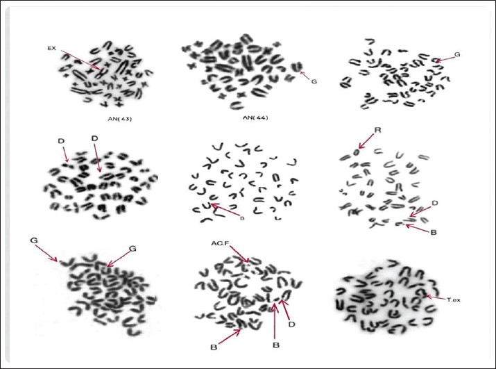
Fig. 5. Several metaphases from bone marrow cells of adult female mice in (DOX+ ADE (A), (B), and (C) groups showing: NA as AN in addition to the most abundant forms of SA like; ring (R); break (B); gap (G); acentric fragment (Ac.f); and exchange (Ex.) Total Magnification=1,000×.
Aberrant metaphases in mice in the DOX + ADE (B) group
This group showed an extremely significant (p < 0.0001) decrease in the frequency of TCA that reached 10.44% compared with 28.67% in the DOX group. The total NA also decreased; they were recorded in only 16 occasions compared with 32 among the DOX group. Moreover, the total SA decreased to 31 occasions compared to 97 occasions in the DOX group (Table 1 and Fig. 5).
Aberrant metaphases in mice in the DOX + ADE (C) group
The frequency of TCA was highly significant (p < 0.0001), decreased to 13.56% compared with 28.67% in the DOX group, but this value was still higher than that recorded in the DOX + ADE (B) group (10.44%). Moreover, the frequency of TCA in this group was higher than that of the −ve control group (9.11%). The total NA frequency decreased to 3.78% compared with 7.11% in the DOX group. The total SA also decreased to 9.78% compared with 21.56% in the DOX group and 12.89% in the DOX + ADE (A) group. The value was higher than that of DOX + ADE (B), which recorded 6.89 % (Table 1 and Fig. 5).
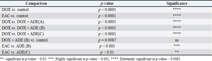
All data are presented as mean ± SD. One-way ANOVA revealed a highly significant difference among the experimental groups (F(8, 72)=22.94, p < 2 × 10⁻¹⁶/(p < 0.0001). DOX treatment significantly increased TCAs (0.287 ± 0.069 per cell) compared to (0.091 ± 0.038 per cell) in the control group (p < 0.0001), confirming its genotoxic effect. Similarly, EAC-bearing mice exhibited a significant increase in chromosomal damage (0.258 ± 0.060 per cell) relative to controls (p < 0.0001). Co-treatment with ADE significantly reduced chromosomal aberrations (0.153 ± 0.020, 0.104 ± 0.022 and 0.136 ± 0.024 in DOX + ADE A, B, and C mice, respectively) compared with DOX-treated mice (p < 0.0001 for all comparisons with the DOX group). Notably, ADE (B) restored chromosomal aberration levels to values comparable to the control group, with no significant difference observed (p=0.9987), indicating a strong protective effect (Tables 2, 3, and Fig. 6).
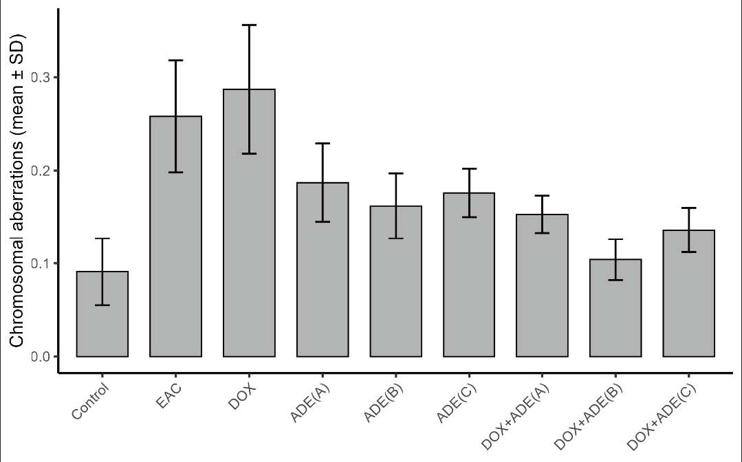
Fig. 6. Effect of doxorubicin and/or ADE administration on chromosomal aberration frequency (mean ± SD) in all experimental groups. Data are presented as mean ± SD. One-way analysis of variance followed by Tukey’s post hoc test was used to assess statistical significance. DOX significantly increased chromosomal aberrations compared with the control group. EAC also showed elevated levels relative to the control. Co-treatment with ADE reduced DOX-induced damage in a dose-dependent manner. ADE (B) restored values comparable to control with no significant difference. Statistical significance is indicated as *p < 0.05, **p < 0.01, ***p < 0.001, and ns=not significant.
Table 2. Comparison between the frequencies of total chromosomal aberrations per mouse cell in all experimental groups.
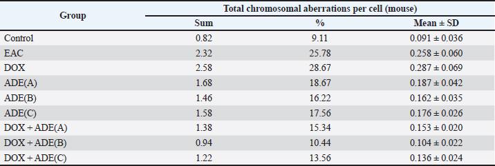
Table 3. Pairwise comparisons between the frequencies of total chromosomal aberrations per mouse cell in all experimental groups.
Histopathological findings
A: Ehrlich ascites carcinoma
B, C, and D: Ajwa date extract (A, B, and C)
E: Chemotherapy (DOX) A
F, G, and H: DOX + Ajwa date extract (A, B, and C)
At 30 days after treatment, there was a marked reduction of neoplastic invasive cells with the presence of islets of neoplastic apoptotic cells admixed with lymphocytes in between massive necrotic debris in the DOX + ADE (10 mg/gm) and DOX + ADE (40 mg/gm) groups (Fig. 7B, C, and E). The treatment group by DOX + ADE (20 mg/gm) and (40 mg/gm) combinations (Fig. 7D and F) showed similarities in their results and were better than the treatment group by DOX + ADE (10 mg/gm). The best curative results were seen in the group treated with DOX + ADE (20 mg/gm) combination (Fig. 7G) compared with the group treated with DOX + ADE (40 mg/gm) combination (Fig. 7H). In addition, the normal architecture of myofibers with mild interstitial exudate and remnant of apoptotic neoplastic cells was observed in the DOX + ADE (40 mg/gm) combination (Fig. 7H) 30 days after treatment.
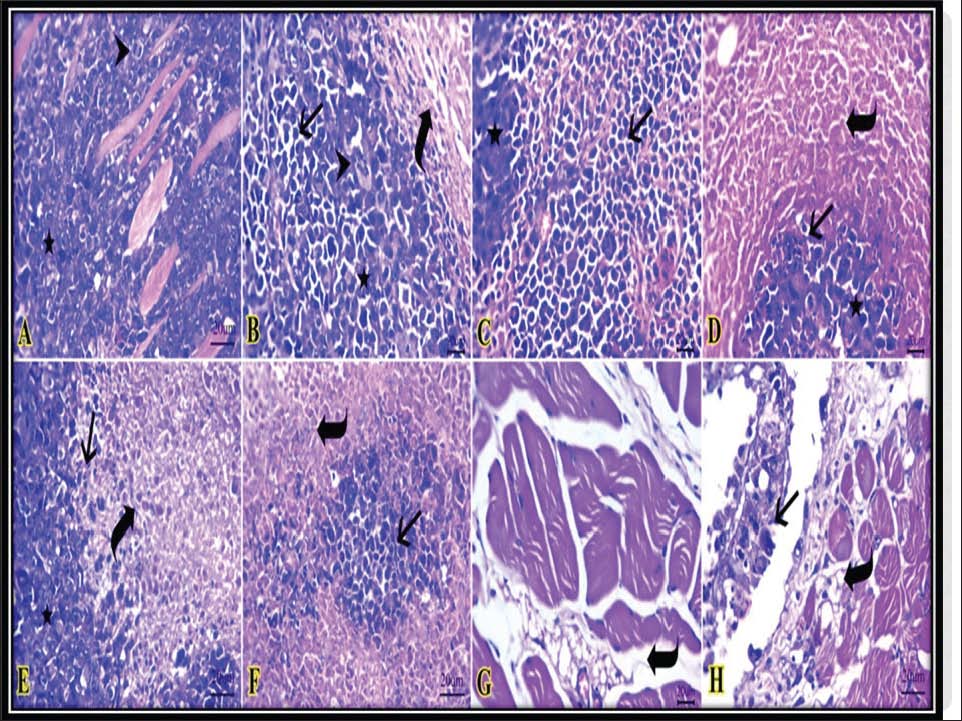
Fig. 7. Representative photomicrograph of H&E-stained sections of Ehrlich ascites carcinoma (EAC) at the skin and treated groups for 30 days showing: invaded neoplastic mass (star) toward the musculature with obvious mitotic activity (arrowhead) in the control +ve group (A). Reduction of neoplastic mass volume (star) with bizarre-shaped cells (arrowhead), islets of neoplastic apoptotic cells admixed with lymphocytes (arrow) in between massive necrotic debris (curved arrow) in the chemo group and extract group at 10, 40 mg/gm (B, C, and E). Islets of neoplastic apoptotic cells (star) are admixed with lymphocytes (arrow) in between massive necrotic debris (curved arrow) in the extract group at 20 and 10 mg/gm chemo + extract combination (D and F). Apparent normal architecture of myofibers with mild interstitial edema (curved arrow) in both the 20 and 40 mg/gm chemo +extract combination groups (G and H), beside the presence of a remnant of apoptotic neoplastic cells (arrow) in the 40 mg/gm chemo +extract combination group (3H). Scale bar 20 μm. Total magnification=400.
The kidney showed neoplastic-infiltrated cells attached to the perirenal serosal surface, which formed from pleomorphic cells with some apoptotic cells and necrosis in the EAC (+ve control) group (Fig. 8A). The DOX-treated group for 30 days showed the presence of necrosis in a few tubular epithelium and mild perivascular round cell infiltrations (Fig. 8B). However, a few apoptotic tubular epithelium with hypocellular glomerular tufts were detected in the group treated with ADE at 10 mg/gm for 30 days (Fig. 8C). Preserved glomerular and tubular structures were seen after 30 days of treatment in the ADE group at 20, 40mg/gm (Fig. 8D, E) and combination groups of DOX+ ADE at 10, 20 and 40 mg/gm (Fig. 8F–H), respectively.
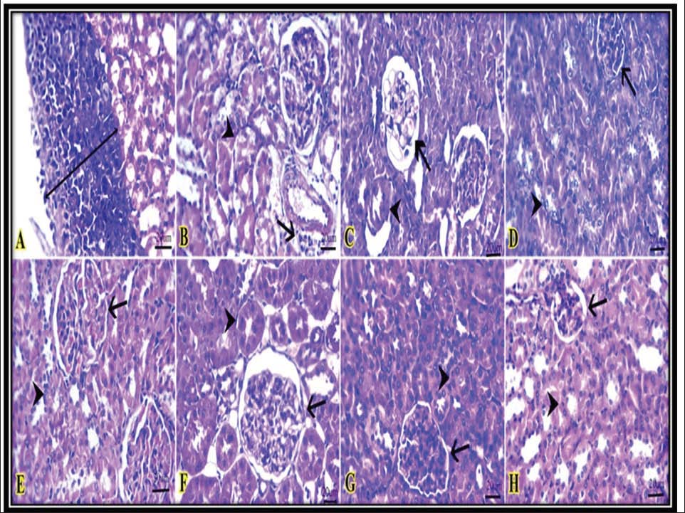
Fig. 8. Representative photomicrograph of H&E-stained sections of the kidneys of mice with Ehrlich ascites carcinoma (EAC) and those treated for 30 days, showing: neoplastic cells aggregate attached to the perirenal serosal surface (double-headed arrow) in the control + ve group (A). Necrosis in a less tubular epithelium (arrowheads) and mild perivascular round cell infiltration (arrow) in the chemotherapy group (B). Apoptotic few tubular epithelium (arrowhead) with hypocellular glomerular tufts (arrow) in the group treated with 10 mg/gm extract for 30 days (C). Glomerular (arrows) and tubular structures (arrowheads) were preserved after 30 days of treatment in the extract group at 20 and 40 mg/gm and in the combination group of chemo + extract at 10, 20, and 40 mg/gm (D–H), respectively. Scale bar 20 μm. Total magnification=400.
Discussion
A single dose of EAC cells (1 × 10⁶cell/mouse), the present study observed marked erythema and tumor development in and near the injection site. Additional pathological signs were evident after 2 weeks, including redness behind the ears and mouth, body ulcers, general weakness, increased tumor volume, and elevated mortality rates. By the third week, the condition of the EAC-positive control group had markedly deteriorated, whereas the ADE-treated groups showed noticeable improvement. The DOX-treated group exhibited a decline in health status during the fourth week, characterized by increased weakness and tissue degeneration. These morphological and behavioral alterations were most pronounced in the EAC-positive control group, likely because of the carcinogenic and metastatic nature of EAC cells, which promote tumor formation and increase mortality. These findings are consistent with those of Sneha et al. (2018) and Saleh et al. (2022) who described EAC as a highly malignant and rapidly proliferating tumor model capable of metastasizing to multiple organs, including the liver, lungs, spleen, kidney, diaphragm, adrenal glands, and blood.
By week 4, the DOX-treated group showed greater deterioration than the other groups, which may be attributed to the combined effects of EAC-induced tumor burden and DOX toxicity. This agrees with the findings of Qiu et al. (2021) who reported that anthracyclines such as DOX, although effective anticancer agents, exert severe systemic toxicity. EAC induces free neoplastic cells in the peritoneal ascitic fluid and causes significant metabolic and morphological disruptions, including reduced mitochondrial number, decreased RNA and DNA biosynthesis, and protein synthesis inhibition (Mishra et al., 2018; Alotaibi et al., 2020).
The cytogenetic analysis of bone marrow cells revealed that the EAC-positive control group exhibited a highly significant increase in TCA (p < 0.0001) compared with the negative control. NA were elevated, and polyploidy was more affected than aneuploidy. The number of SA was also markedly increased, with TD being the most frequent abnormality, followed by ring chromosomes. These results confirm the strong genotoxic effect of EAC, in agreement with Fenech (2000) who demonstrated that EAC significantly increases the frequency of chromosomal aberrations. The observed chromatin loss may result from chromosomal structure damage, spindle apparatus dysfunction, or centromere–kinetochore complex disruption. In addition, EAC-induced oxidative stress can lead to DNA damage and genetic instability (Moeller et al., 2004; Corn and Wafik, 2007).
Female mice bearing EAC and treated with a therapeutic dose of DOX (0.00026 mg/gm b.w. intraperitoneally, once per week for 30 days) exhibited a highly significant increase in TCA (p < 0.0001) compared with the control group. This marked increase in chromosomal damage is attributable to the cytotoxic mechanisms of DOX, including DNA intercalation, DNA and RNA synthesis inhibition, topoisomerase II inhibition, and ROS generation. These findings are supported by Quiles (2002); Lee et al. (2002); Wallace et al. (2020); Johnson-Arbor and James (2022) and Giacomini et al. (2023) who reported that DOX induces DNA breakage and chromosomal instability in both cancerous and normal cells.
The percentage of numerical aberrations was markedly increased in the DOX-treated group with aneuploidy being the predominant type. This may be related to mitochondrial dysfunction and spindle dysregulation, leading to improper chromosome segregation, as noted by Schirone et al. (2022). Structural aberrations were also increased, particularly telomeric break (T.B), telomeric exchange (T.ex), and centric fusions (C.F). This aligns with the findings of Shi et al. (2023) who demonstrated that DOX induces structural chromosomal damage primarily through oxidative stress and free radical formation. The exposure of cells to ROS causes genetic aberrations, which may be numerical, such as chromosomal gain/loss, or structural, such as deletion (Pang, 1995; Osataphan et al., 2020). In addition, Bingöl et al. (2014) reported that DOX induced a high frequency of SA in all treated animals (translocations, chromatid breaks, gaps, exchanges, isochromatid breaks, acentric fragments, and triradial figures) compared with control animals, with chromatid breaks being the most abundant SA.
Mice treated with ADE at doses of 10, 20, and 40 mg/gm/day for 30 days (groups A, B, and C) showed a significant reduction in total chromosomal aberrations compared with the EAC-positive control and DOX-treated groups. Although chromosomal aberration levels remained higher than those of the negative control, ADE markedly improved both numerical and structural aberrations, indicating its protective role. This protective effect is attributed to the antioxidant and anticancer properties of ADE, which is rich in flavonoids, polyphenols, and glycosides (Farhana and Roushney, 2021; Shahbaz et al., 2022; Shi et al., 2023). Among the tested doses, ADE at 20 mg/gm/day demonstrated the most effective protective and ameliorative effect, consistent with the findings of Ahmed et al. (2022) who reported that ADE contains high levels of lutein, beta-carotene, quercetin, rutin, and anthocyanins that help regulate intracellular ROS and reduce DOX cytotoxicity. Furthermore, mice treated with combined DOX + ADE exhibited clear improvement in chromosomal integrity, with both NA and SA showing nonsignificant differences from the negative control at higher ADE doses. This suggests that ADE has a synergistic protective effect against DOX-induced genotoxicity. Najm et al. (2021) and Yasin et al. (2015) similarly reported that Ajwa date extract acts as a potent natural antioxidant and anticancer agent that enhances cellular defense mechanisms while protecting normal tissues during chemotherapy.
Histopathological findings supported the cytogenetic results. ADE-treated mice exhibited improved renal architecture with reduced mesangial hypertrophy, decreased karyomegaly, and preserved glomerular and tubular structures, consistent with the findings of Fatani et al. (2022) and Abdelghffar et al. (2022). DOX-treated mice exhibited reduced neoplastic invasive cells but also increased lymphocytic infiltration and apoptosis. The combined DOX + ADE group exhibited the greatest tumor reduction and highest apoptotic activity, with the surrounding normal tissue and kidney structure preserved.
Conclusion
This study demonstrates that Ajwa date extract is a safe and effective natural supplement that mitigates DOX-induced genotoxicity and nephrotoxicity in EAC-bearing mice. The most pronounced protective and therapeutic effects were achieved with ADE at 20 mg/gm/day along with DOX (0.00026 mg/gm b.w.) treatment for 30 days.
Acknowledgments
The authors acknowledge the Faculty of Science and the Faculty of Veterinary Medicine, Zagazig University, for providing the facilities and resources required to conduct this research.
Conflict of interest
The authors declare no competing financial interests or personal relationships that could have appeared to influence the work reported in this paper.
Funding
This research did not receive any specific grant from funding agencies in the public, commercial, or not-for-profit sectors.
Authors’ contributions
All authors (Sabha E. Elballat, Fawzeya A. Zayed, Iman E. El-Araby, Ahmed M. Aldawek, and Sara Ahmed El-Said) have worked hard to generate this paper. All authors coordinated in applying the experiment, in data analysis, writing, and revision of the manuscript.
Data availability
The datasets generated and/or analyzed during the current study are available from the corresponding author on reasonable request.
References
Abdelghffar, E.A., Obaid, W.A., Mohammedsaleh, Z.M., Ouchari, W., Eldahshan, O.A. and Sobeh, M. 2022. Ajwa dates (Phoenix dactylifera LL.) attenuate cisplatin-induced nephrotoxicity in rats via augmenting Nrf2, modulating NADPH oxidase-4 and mitigating inflammatory/apoptotic mediators. Biomed. Pharmacotherapy. 156, 113836.
Ahmed, R., Abdulrahman, A. and Abdullah, F.A. 2022. Nutritional and medicinal role of Ajwa date: an overview. J. Funct. Foods. 85, 104694.
Alotaibi, H., Alsaleh, R. and Fathy, M. 2020. Metabolic alterations in ehrlich ascites carcinoma: a morphological study. Saudi. J. Biol. Sci. 27(10), 2645–2651.
Bancroft, J.D. and Gamble, M. 2002. Theory and practice of histological techniques 5th ed. Edinburgh, UK: Churchill Livingstone.
Bingöl, G., Gülkaç, M.D., Dillioğlugil, M.O., Polat, F. and Kanli, A.O. 2014. Effect of resveratrol on chromosomal aberrations induced by doxorubicin in rat bone marrow cells. Mutation Research/Genetic Toxicol. Environ. Mutagenesis 766, 1–4.
Corn, P.G. and Wafik, S.E. 2007. Microarray analysis of p53 target gene expression patterns in the HCT116 colorectal cancer cell line. Oncogene 26, 5347–5357.
Farhana, S.L. and Roushney, M.F. 2021.Bangladeshi wild date palm fruits (Phoenix sylvestris): promising source of anti-cancer agents for hepatocellular carcinoma treatment. Int. J. Appl. Sci. Biotechnol. 9(1), 32–37.
Fatani, A.M., Baothman, O.A.S., Shash, L.S., Abuaraki, H.A., Zeyadi, M.A., Hosawi, S.B., Altayb, H.N. and Abo-Golayel, M.K. 2022. Hepatoprotective effect of date palm fruit extract against doxorubicin intoxication in Wistar rats: in vivo and in silico studies. Asian. Pacific. J. Trop. Biomed. 12(8), 357–366.
Fenech, M. 2000. The in vitro micronucleus technique. Mutation. Research/Fundamental. Mol. Mech. Mutagenesis. 455(1–2), 81–95.
Frajacomo, S.A., Pinto, M.C. and Barbosa, F. 2016. Ehrlich ascites carcinoma: historical insights and contemporary applications. Exp. Oncol. 38(2), 89–95.
Freshney, R.I. 1987. Culture of animal cells: a manual of basic technique 2nd ed [Internet]. New York, NY: Alan R. Liss, Inc. p: 117.
Giacomini, E., Elisa, B., Enke, B., Simona, V., Laura, P. and Andrea, C. 2023. Anthracyclines: mechanisms of action, redox signaling and ongoing strategies to improve efficacy and reduce toxicity. Int. J. Mol. Sci. 24(2), 1582.
Johnson-Arbor, K. and James, D. 2022. Doxorubicin [Internet]. StatPearls. Treasure Island, FL: StatPearls Publishing. Available via https://www.ncbi.nlm.nih.gov/books/NBK482221/ (Accessed 2026 Jun 25).
Kabel, A.M. 2014. Effect of combination between methotrexate and histone deacetylase inhibitors on transplantable tumor model. Am. J. Med. Stud. 2(1), 12–18.
Keselman, H.J. and Rogan, J.C. 1977. The Tukey multiple comparison test: 1953-1976. Psychol. Bull. 84(5), 1050–1056.
Khan, M.A., Siddiqui, S., Ahmad, I., Singh, R., Mishra, D.P., Srivastava, A.N. and Ahmad, R. 2021. Phytochemicals from Ajwa dates pulp extract induce apoptosis in human triple-negative breast cancer by inhibiting AKT/mTOR pathway and modulating Bcl-2 family proteins. Sci. Rep. 11(1), 10322.
Lars, S. and Svante, W. 1989. Analysis of variance (ANOVA). Chemometrics. Intell. Lab. Syst. 6(4), 259–272.
Lee, S., Baek, M., Kim, H.Y., Ha, J.H. and Jeoung, D.I. 2002. Mechanism of doxorubicin-induced cell death and expression profile analysis. Biotechnol. Lett. 24(14), 1147–1151.
Micallef, J., Robert, R.M., Camille, J.T. and Abigail, R.S. 2020. Molecular mechanisms of doxorubicin-induced DNA damage: an overview of interactions with DNA and topoisomerase II. Front. Mol. Biosciences 7, 559251.
Mishra, A., Sinha, A. and Sharma, R. 2018. Biological behavior of Ehrlich ascites carcinoma in Swiss albino mice. J. Cancer Res. Therapeutics 14(2), 347–353.
Moeller, B.J., Cao, Y., Li, C.Y. and Dewhirst, M.W. 2004. Radiation activates HIF-1 to regulate vascular radiosensitivity in tumors: role of reoxygenation, free radicals, and stress granules. Cancer. Cell. 5(5), 429–441.
Najm, W.M., Abdullah, A. and Firas, H.S. 2021. Ajwa Date (Phoenix dactylifera L.): ethnopharmacological significance and bioactive compounds for functional food development. Pharm. Biol. 59(1), 958–967.
Osataphan, N., Chawalit, P., Rapeeporn, C. and Siriporn, C.C. 2020. Doxorubicin and Iron: a Dangerous Liaison in Cardiotoxicity. Pharmacol. Res. 155, 104741.
Othman, A.S., Hisham, N., Mustafa, A., Salman, B. and Mohamed, K. 2023. Phytochemical analysis and nephroprotective potential of Ajwa date in doxorubicin‐induced nephrotoxicity rats: biochemical and molecular docking approaches. Food Sci. Nutr. 11(3), 1584–1598.
Paget, G.E. and Barnes, J.M. 1964. Toxicity tests. Evaluation of drug activities: Pharmacometrics 1, 13–65.
Preston, R.J., Dean, B.J., Galloway, S., Holden, H., Mcfee, A.F. and Shelby, M. 1987. Mammalian in vivo cytogenetic assays. Analysis of chromosome aberrations in bone marrow cells. Mutat. Res. 189, 157–165.
Qiu, Y., Jing, L., Xiangdong, W. and Junjie, X. 2021. Cardiotoxicity of anthracycline chemotherapeutics: molecular mechanisms, prevention strategies and future perspectives. Mol. Cancer. 20, 102.
Quiles, J. 2002. Antioxidant nutrients and adriamycin toxicity. Toxicology 180(1), 79–95.
Saleh, N., Allam, T., Abdelfattah, A. and El-Borai, N. 2022. Review on Ehrlich Ascites Carcinoma in mice and cancer treatment with special reference to the potential protective and therapeutic effects of hesperidin versus cisplatin. J. Curr. Vet. Res. 4(1), 47–57.
Sara, A., Sabha, A., Iman, E., Fawzeya, A. and Maher, S. 2025. In vivo, effects of combined treatment of female mice with Phoenix dactylifera extract and doxorubicin on reducing neighboring cell death during hepatic cancer therapy. J. Biosci. Appl. Res. 11(1), 88–100.
Schirone, L., D’Ambrosio, L., Forte, M., Genovese, R., Schiavon, S., Spinosa, G., Iacovone, G., Valenti, V., Frati, G. and Sciarretta, S. 2022. Mitochondria and doxorubicin-induced cardiomyopathy: a complex interplay. Cells 11(13), 2000.
Shahbaz, K., Asif, J.A., Liszen, T., Nurul, A.A. and Alam, M.K. 2022. Cytotoxic and antioxidant effects of Phoenix dactylifera L. (Ajwa date extract) on oral squamous cell carcinoma cell line. BioMed. Res. Int. 1(1), 5792830.
Shi, S., Chen, Y., Luo, Z., Nie, G. and Dai, Y. 2023. Role of oxidative stress and inflammation-related signaling pathways in doxorubicin-induced cardiomyopathy. Cell Commun. Signal. 21(1), 61.
Sneha, K., Meenakshi, G. and Rajan, R. 2018. Cachexia and metastasis in Ehrlich ascites carcinoma: morphological and molecular correlation. Indian. J. Exp. Biol. 56(12), 881–887.
Wallace, K.B., Sardão, V.A. and Oliveira, P.J. 2020. Mitochondrial determinants of doxorubicin-induced cardiomyopathy. Circulat. Res. 126(7), 926–941.
Yasin, F.M., Mohd, H.M. and Nafeeza, M.I. 2015. Health-promoting phytochemicals from Ajwa date fruit and potential benefits in cardiovascular diseases. J. Food Biochem. 39(6), 785–795.








