Open Veterinary Journal, (2026), Vol. 16(7): 4112-4131
Review Article
10.5455/OVJ.2026.v16.i7.3
Filariasis at the human–animal interface: Implications for public and veterinary health
Muhammad Nirwan1, Yunita Amraeni2*, Aswin Rafif Khairullah3, Shinta Shinta1, Raflizar Raflizar1, Sri Seprianto Maddusa4, Erwin Azizi Jayadipraja5, La Ode Tasrun5, Ari Tjahyadi Rafiuddin5,
Syahputra Wibowo6, Sri Suryatmiati Prihandani3 and Bima Putra Pratama7
1Research Center for Public Health and Nutrition, National Research and Innovation Agency (BRIN), Bogor, Indonesia
2Department of Public Health, Faculty of Health Sciences, Universitas Pembangunan Nasional “Veteran” Jakarta, South Jakarta, Indonesia
3Research Center for Veterinary Science, National Research and Innovation Agency (BRIN), Bogor, Indonesia
4Faculty of Public Health, Universitas Sam Ratulangi, Manado, Indonesia
5Universitas Mandala Waluya, Kendari, Indonesia
6Eijkman Research Center for Molecular Biology, National Research and Innovation Agency (BRIN), Bogor, Indonesia
7Research Center for Process Technology, National Research and Innovation Agency (BRIN), South Tangerang, Indonesia
*Corresponding Author: Yunita Amraeni. Department of Public Health, Faculty of Health Sciences, Universitas Pembangunan Nasional “Veteran” Jakarta, South Jakarta, Indonesia. Email: yunitaraeni [at] upnvj.ac.id
Submitted: 08/02/2026 Revised: 30/05/2026 Accepted: 10/06/2026 Published: 02/07/2026
© 2026 Open Veterinary Journal
This is an Open Access article distributed under the terms of the Creative Commons Attribution-Non Commercial-No Derivatives License (http://creativecommons.org/licenses/by-nc-nd/4.0/), which permits non-commercial re-use, distribution, and reproduction in any medium, provided the original work is properly cited, and is not altered, transformed, or built upon in any way.
ABSTRACT
Filariasis is a parasitic infectious disease transmitted by blood-sucking mosquito bites and remains a public health challenge in tropical and subtropical regions. This disease is caused by filarial worms from the nematode group, such as Wuchereria bancrofti, Brugia malayi, Brugia timori, and Dirofilaria spp., which can act as zoonotic reservoirs. In humans, filarial infection can cause clinical manifestations such as lymphedema, elephantiasis, and filarial hydrocele, whereas in animals, it can cause cardiopulmonary disorders or subcutaneous lesions, thus increasing the opportunity for interspecies transmission. Establishing a diagnosis of filariasis requires a comprehensive approach that includes clinical examination, microscopic detection of microfilariae, serological tests for antigens or antibodies, polymerase chain reaction-based molecular analysis, and imaging examinations such as ultrasound or radiology to identify adult worms. The risk of filariasis is influenced by the mosquito vector density, tropical environmental conditions that support the vector’s life cycle, the intensity of human–animal contact, the host’s immune status, and community behavior in preventing mosquito bites. Treatment involves the administration of antifilarial drugs, including diethylcarbamazine, ivermectin, and albendazole, along with the management of chronic complications through lymphatic system therapy and, if necessary, surgical intervention. Filariasis control will be more effective if it is implemented in an integrated manner through mass drug administration programs, vector control, treatment of animal reservoirs, increased public education, and ongoing surveillance. A One Health approach that connects human, animal, and environmental health aspects has proven to be essential in breaking the transmission chain, reducing the disease burden, and minimizing social and economic impacts. This review emphasizes the urgency of implementing interdisciplinary strategies to control filariasis to support sustainable public health in endemic areas.
Keywords: Disease, Filariasis, Mosquito vector, One Health, Zoonosis.
Introduction
Filariasis is a parasitic infectious disease classified as a neglected tropical disease and remains a significant public health problem in various tropical and subtropical regions (Chandy et al., 2011). It is caused by filarial worms from the nematode family Onchocercidae, which can infect both humans and animals, creating a cross-species health issue (Remya et al., 2025). In humans, lymphatic filariasis, caused by infection with Wuchereria bancrofti, Brugia malayi, and Brugia timori, attacks the lymphatic system and triggers lymphedema, elephantiasis, and filarial hydrocele (Babu and Nutman, 2012). The chronic and progressive course of the disease results in a decreased quality of life, reduced work productivity, and limited social participation, ultimately triggering social stigma and impacting the well-being of individuals and communities (Vasconez-Gonzalez et al., 2025).
Filariasis also has a significant impact on animal health, particularly caused by Dirofilaria immitis and Dirofilaria repens, which infect dogs, cats, and various other mammals (Mendoza-Roldan et al., 2021). Infection in animals can trigger cardiopulmonary disorders and the formation of subcutaneous lesions, making them a zoonotic reservoir, contributing to an increased risk of interspecies transmission (Simón et al., 2017). The existence of this animal reservoir adds complexity to filariasis control, necessitating the adoption of an integrated One Health approach that simultaneously encompasses human, animal, and environmental health (Igreja et al., 2025).
Filariasis is transmitted through the bite of blood-sucking female mosquitoes from various genera, such as Culex, Aedes, Anopheles, and Mansonia, which serve as intermediate hosts (Bhuvaneswari et al., 2023). Microfilariae circulating in the bloodstream are taken up by the mosquito during a blood meal and then develop into infective L3 larvae within the vector before being transmitted to humans or animals through subsequent bites (Rajamanickam and Babu, 2025). The ability of filarials to biologically adapt to their hosts and vectors allows them to survive in a variety of environmental conditions, including changes in temperature, humidity levels, and vector density (Rivera et al., 2025). Filariasis is difficult to eradicate, particularly in areas with high population density, inadequate environmental sanitation, and vector ecosystems that support the parasite life cycle (Galgamuwa et al., 2025).
Environmental, social, and biological factors play a crucial role in determining filariasis epidemiological patterns (Lestyoningrum et al., 2026). Tropical and subtropical climates, characterized by warm temperatures and high humidity, create optimal conditions for vector mosquito breeding, whereas high human population densities and the presence of animal reservoirs reinforce transmission cycle continuity (Ahmed et al., 2019). Host factors, such as immune status, nutritional status, and prior exposure history, influence infection rates and clinical manifestations (de Souza and Bockarie, 2025). Children and individuals with compromised immune systems are generally more susceptible to infection and at risk of developing more severe chronic complications (Singh et al, 2025a). The complexity of the interactions between humans, animals, and vectors underscores the importance of implementing an integrated approach to the prevention and control of filariasis (Ye et al., 2025).
The impact of filariasis extends beyond clinical aspects and has social and economic consequences (Wynd, 2007). Long-term complications, such as elephantiasis and lymphedema, can reduce work capacity, increase medical costs, and impact the well-being of families and the wider community (Mackenzie et al., 2024). Control efforts, which include mass drug administration (MDA) programs, vector control, and increased public education, require significant resource allocation (Wayangkau et al., 2025). Nevertheless, these investments have been proven to reduce prevalence rates, interrupt transmission chains, and improve the quality of life for communities in endemic areas (Nawan et al., 2025).
With the close involvement of humans, animals, and the environment, filariasis can be viewed as a disease representative of the One Health framework, requiring a multidisciplinary approach. A comprehensive understanding of the etiology, parasite life cycle, transmission mechanisms, risk factors, and health and economic impacts is the primary basis for developing effective control strategies. In line with this, this review aims to discuss aspects of filariasis epidemiology, pathogenesis, clinical presentation, diagnostic methods, therapeutic options, and prevention efforts, emphasizing its relevance to human and animal health, as well as its implications for public health policy and the implementation of control programs in endemic areas.
Etiology
Filariasis is a parasitic infectious disease caused by filarial worms of the nematode family Onchocercidae, which can infect both humans and animals (Srikacha and Pornpanom, 2025). The main causative agents of this disease in humans are W. bancrofti, B. malayi, and B. timori, which target the lymphatic system and cause manifestations such as lymphedema, hydrocele, and various other chronic complications (Rajamanickam and Babu, 2025). West bancrofti has the widest geographic distribution and is responsible for more than 90% of lymphatic filariasis cases globally (McNulty et al., 2013). On the contrary, B. malayi and B. timori have a more limited distribution, primarily endemic to Southeast Asia and Timor Island, with humans as the primary host in the transmission cycle (Cano et al., 2014).
Dirofilaria immitis and D. repens primarily cause animal filariasis by infecting dogs, cats, and various wild mammals (Grosbois et al., 2025). Dirofilaria immitis attacks the cardiopulmonary system and causes heartworm disease, whereas D. repens is generally associated with subcutaneous infections (Oliveira et al., 2021). Several other filarial species, such as Acanthocheilonema reconditum and Setaria spp., can be found as commensal parasites or cause mild infections in domestic animals (Vargas et al., 2023). Although their clinical impact is relatively limited, the presence of these species remains epidemiologically significant because they potentially act as reservoirs that influence cross-species transmission dynamics (Hafizu et al., 2025).
Filariasis is a vector-borne disease, specifically in female mosquitoes of the genera Culex, Aedes, Anopheles, and Mansonia (Famakinde, 2018). The type of vector depends on the filarial species and local ecological characteristics (Anjal et al., 2025). In its life cycle, the mosquito serves as an intermediate host where microfilariae develop into infective L3 larvae (Remya et al., 2025). Thus, transmission occurs through vector-mediated inoculation of infective larvae during mosquito blood feeding, representing the primary mode of infection and a key determinant in disease spread rather than a separate pathogenic mechanism discussed elsewhere (Gleave et al., 2016). The adaptability of filarial parasites to both their vector and host allows them to survive in a wide range of environmental conditions, including variations in temperature and humidity and differences in vector population density (Simón et al., 2017).
Ecological, biological, and social aspects play a crucial role in determining filariasis etiology (Galgamuwa et al., 2025). High human and animal population densities, intense human–animal interactions, and inadequate environmental sanitation can reinforce the transmission cycle (Storey, 1993). Variation between filarial species, differences in tissue tropism, and the ability of the parasite to modulate the host’s immune response contribute to the diversity of clinical manifestations, infection burden, and potential for zoonotic transmission (Rajamanickam and Babu, 2025).
Life cycle
Filariasis is a vector-borne disease that relies on the interaction between a vertebrate host and a mosquito as a vector, thus having a complex life cycle that influences epidemiological dynamics and zoonotic potential (Paily et al., 2009). The infection process begins when infective L3 larvae, which develop inside a female mosquito, are transferred to a vertebrate host through a bite (Erickson et al., 2009). After penetrating the skin, the larvae migrate through the subcutaneous tissue or vascular system to specific target organs, depending on the parasite type (Kilarski et al., 2019). Figure 1 illustrates the life cycle of filarial parasites at the human–animal interface, highlighting the role of mosquito vectors in transmitting infective larvae between animal reservoirs and human hosts.
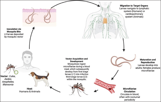
Fig. 1. Life cycle of filarial parasites at the human–animal interface.
In human filariasis, particularly caused by W. bancrofti and Brugia spp., infective L3 larvae migrate to the peripheral lymphatic vessels, lymph nodes, and mesenteric lymphatic plexus (McNulty et al., 2013). The larvae mature into adult male and female worms. The mature female worms then produce microfilariae that circulate in the peripheral blood and act as a source of infection for vector mosquitoes during blood feeding (Rajamanickam and Babu, 2025). This series of processes maintains the transmission cycle between humans and mosquitoes (Stephano et al., 2026).
In animal filariasis, such as D. immitis infection, infective L3 larvae migrate to the pleural cavity and right heart, where they develop into adult worms that release microfilariae into the bloodstream (Remya et al., 2025). The presence of these adult worms causes blood vessel obstruction, triggers endothelial inflammation, and leads to structural changes in cardiopulmonary tissue, leading to clinical manifestations, including right heart failure in dogs and cats (Hoch and Strickland, 2008). Circulating microfilariae in the bloodstream then serve as a source of infection for the mosquito vector during a blood meal, thus maintaining the filarial life cycle (Babu and Nutman, 2012).
Filariae possess biological adaptations that contribute to increased transmission success (Babayan et al., 2010). For example, W. bancrofti microfilariae exhibit nocturnal periodicity in human blood circulation in tropical regions, which aligns with the mosquito vector’s nocturnal activity patterns (Thanchomnang et al., 2013). Environmental factors, such as temperature, humidity, and vector population density, also influence larval development rate and degree of infectivity (Rekha et al., 2025). Furthermore, differences between filarial species determine target tissue location preferences, blood microfilaria levels, and the host’s potential for clinical complications (Pietrzak et al., 2024).
Epidemiology
Filariasis is a parasitic infectious disease with an uneven geographic distribution that is influenced by variations in filarial species, the presence and type of vector, environmental conditions, and socioeconomic factors (Cano et al., 2014). The main causative agents of filariasis in humans are W. bancrofti, B. malayi, and B. timori (Nutman, 2013). West bancrofti has the widest distribution range and is reported to be endemic in more than 80 countries in tropical and subtropical regions, including sub-Saharan Africa, South Asia, the Western Pacific region, and Latin America (McNulty et al., 2013). These endemic areas are generally characterized by tropical climates with high temperatures and humidity and adequate vector densities, particularly mosquitoes of the genera Culex, Anopheles, and Aedes, which support the parasite’s life cycle and ongoing transmission (Bhuvaneswari et al., 2023). In addition to environmental factors, socioeconomic conditions, such as inadequate sanitation, high population density, and limited access to mass treatment programs, also play a role in maintaining filariasis transmission at the community level (Lestyoningrum et al., 2026). Table 1 summarizes the geographic distribution patterns of filariasis in humans and animals, including the filarial species involved, primary hosts, endemic areas, vector types, and environmental and socio-economic factors influencing its spread.
Table 1. Geographic distribution and epidemiological determinants of human and animal filariasis.
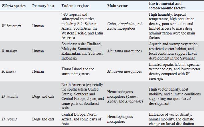
On the contrary, B. malayi has a more limited distribution and is primarily found in Southeast Asia, including Thailand, Malaysia, and Indonesia, particularly in Sumatra, Kalimantan, and Sulawesi (Aisyah et al., 2022). Meanwhile, B. timori is reportedly endemic to Timor Island and its surrounding areas (Supali et al., 2002). The specific ecological characteristics of their vectors strongly influence the distribution patterns of both species, particularly mosquitoes of the genus Mansonia, which breed in aquatic environments with vegetation and swampy areas (Alonso et al., 2022). This condition tends to localize the distribution of filarials to specific habitats that support the survival of the vector and the life cycle of the parasite (Taylor, 2002).
Animal filariasis, particularly caused by D. immitis and D. repens, exhibits varying geographic distribution patterns (Kaikuntod et al., 2021). Dirofilaria immitis, the causative agent of heartworm disease in dogs and cats, is primarily reported in North America—particularly the southeastern United States—as well as in southern and central Europe, Japan, and several countries in Southeast Asia (Noack et al., 2021). On the contrary, D. repens is more frequently associated with subcutaneous infections and is widely distributed in Central Europe, North Africa, and parts of Asia (Dimzas et al., 2024). The distribution of filarial infections in animals is strongly influenced by the density of blood-sucking vectors, host movement, and climate dynamics that determine larval survival and development (Remya et al., 2025). Furthermore, climate change, urbanization, deforestation, and increased human and animal mobility have contributed to the emergence of new endemic areas and the occurrence of sporadic cases in areas previously free of filariasis (Kwa, 2008).
Pathogenesis
The pathogenesis of filariasis is a consequence of a complex interaction between the filarial parasite, the vertebrate host, and the host’s immune response, which collectively determine the clinical presentation and severity of the disease (Babu and Nutman, 2012). The infection process begins when L3-stage filarial larvae, which are transmitted through the bite of a vector mosquito, penetrate the skin and migrate to specific target tissues (Rajamanickam and Babu, 2025). In human filariasis caused by W. bancrofti and Brugia spp., the larvae settle in the lymphatic system—including the lymph nodes, lymphatic vessels, and mesenteric lymphatic plexus—and then mature into adult male and female worms (Handa et al., 2024). The adult female worms then produce microfilariae that circulate in the peripheral blood, marking the infectious phase for the vector mosquito in subsequent transmission cycles (Fimbo et al., 2020).
The interaction between the parasite and the host immune response largely determines the clinical pathogenesis of filariasis (Chakraborty et al., 2013). Adult worms secrete various immunomodulatory molecules that inhibit T cell activation and reduce proinflammatory cytokine production, allowing the parasite to persist in lymphatic tissue for extended periods (Momčilović et al., 2025). Conversely, the death of adult worms or the degradation of microfilariae can trigger an acute inflammatory response that clinically manifests as episodic fever, lymphangitis, and local edema (Medeiros et al., 2022). Repeated exposure and chronic infection further contribute to lymphatic system fibrosis, which progresses to lymphedema and filarial hydrocele, the main complications of lymphatic filariasis (Pfarr et al., 2009).
In animal filariasis, such as D. immitis infection in dogs, infective L3-stage larvae migrate through the subcutaneous tissue to the pulmonary artery and right heart, where they develop into adult worms, causing vascular obstruction, endothelial inflammation, and structural changes in the cardiopulmonary system (Stancu et al., 2025). Chronic infection triggers the infiltration of eosinophils, macrophages, and other inflammatory cells, which contribute to tissue damage and granuloma formation (Ehrens et al., 2022). Adult worm accumulation in the pulmonary artery can subsequently lead to pulmonary hypertension, right heart failure, hepatomegaly, and pleural effusion (Cheepsattayakorn and Cheepsattayakorn, 2014).
The pathogenesis of filariasis is influenced by various factors, including infectious load or infective dose, host immune status and response, genetic factors, the intensity of human–animal interactions, and environmental conditions that determine vector density and larval survival (Damayanti et al., 2025). Furthermore, differences in host species contribute to variations in clinical presentation, with humans generally more susceptible to lymphatic system involvement, whereas domestic and wild animals are more likely to exhibit cardiopulmonary complications or subcutaneous lesions (Momčilović et al., 2025). Overall, this section focuses on mechanistic disease development, while epidemiological, transmission, and clinical details are intentionally separated into their respective sections to improve clarity and reduce redundancy.
Immune response
Filariasis infection triggers a complex and integrated immune response between the innate and adaptive immune systems, which plays a crucial role in determining the disease’s clinical course in both humans and animals (Sibi et al., 2021). Skin epithelial cells and macrophages immediately recognize the parasite through pattern recognition receptors after L3-stage filarial larvae enter the host through a mosquito bite, which then induces the release of pro-inflammatory cytokines, including IL-1, IL-6, and TNF-α (Laney et al., 2010). Although this initial response restricts larval movement, filariae are capable of secreting immunomodulatory molecules that inhibit T cell activation and increase the expression of anti-inflammatory mediators such as IL-10, allowing the parasite to persist in lymphatic or vascular tissues for extended periods (Babu and Nutman, 2012).
During the adaptive immune response phase, filarial infection shifts the host immune system toward a Th2-dominated profile, characterized by increased IL-4, IL-5, and IL-13 production, immunoglobulin E formation, and eosinophil activation (Ehrens et al., 2022). This Th2 response plays a role in controlling parasite burden, but simultaneously contributes to tissue fibrosis and chronic lymphedema through fibroblast activation and increased collagen deposition (Jiang et al., 2018). Conversely, the number of regulatory T cells tends to increase in chronic infection, suppressing excessive immune responses and reducing acute inflammation (Metenou and Nutman, 2013). This mechanism explains why many individuals with blood microfilariae remain asymptomatic despite prolonged infection (O’Regan et al., 2014).
A similar immune response pattern can be observed in animal filariasis, particularly that caused by D. immitis (Remya et al., 2025). Larvae and adult worms trigger eosinophil and macrophage infiltration in the pulmonary arteries and heart, which then induces a granulomatous reaction and potentially leads to structural changes in blood vessels and heart failure (Hidron et al., 2010). Eosinophils play a central role in degranulation and release of cytotoxic proteins, but filariae can develop defense mechanisms to avoid this response, including by expressing surface molecules that inhibit opsonization and phagocytosis (Ehrens et al., 2022).
The immune response to filariasis is also influenced by environmental conditions, the level of infection burden, and the intensity of interaction between humans and animals, which collectively explain the spectrum of clinical presentations in animals, ranging from asymptomatic infection to the appearance of severe manifestations, such as progressive lymphedema, hydrocele, or heart failure (Leggat et al., 2018).
Pathology
The pathological features of filariasis result from complex interactions between the filarial parasite, the host immune response, and target tissues, resulting in a broad spectrum of lesions ranging from asymptomatic conditions to chronic tissue damage (Nutman, 2013). In humans, lymphatic filariasis is caused by W. bancrofti and Brugia spp. Infection primarily involves the lymphatic system, including lymph nodes, peripheral lymphatic vessels, and the mesenteric lymphatic plexus (Medeiros et al., 2022). The presence of adult worms within the lymphatic vessels causes mechanical obstruction that leads to vessel dilation, lymphatic stasis, and tissue swelling (Chakraborty et al., 2013). Repeated lymph accumulation subsequently induces a chronic inflammatory response, fibrosis, and tissue structural changes, which clinically manifest as progressive lymphedema, elephantiasis, and filarial hydrocele (Mackenzie et al., 2024).
Histopathologically, human lesions are characterized by infiltration of various inflammatory cells, such as lymphocytes, eosinophils, and macrophages, accompanied by markedly increased collagen deposition in the walls of lymphatic vessels (Babu and Nutman, 2012). The death of microfilariae or the degradation of adult worms can trigger an acute immune response manifested as episodic fever, lymphangitis, and exacerbation of local inflammation (Medeiros et al., 2022). Furthermore, filariae can secrete immunomodulatory molecules that inhibit T cell activation and suppress the production of proinflammatory cytokines, allowing parasites to survive in host tissues for extended periods (Bhoj et al., 2022). Figure 2 shows the comparison of the clinical impact of filariasis in humans and animals, highlighting the similarities and differences in disease manifestations, pathological outcomes, and host responses.
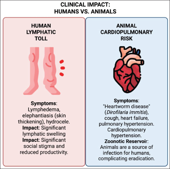
Fig. 2. Clinical impact of filariasis in humans and animals.
In animal filariasis caused by D. immitis, adult worms are located in the pulmonary artery and right heart, causing vascular obstruction, endothelial inflammation, and structural changes in the cardiopulmonary system (Mupanomunda et al., 1997). Macroscopic lesions include pulmonary artery dilation, increased pulmonary pressure, liver enlargement, and pleural effusion (Shukla et al., 2017). The pathology is characterized by inflammatory cell infiltration, eosinophilic granuloma formation, and fibrosis of the vascular wall (Fercoq et al., 2020). The accumulation of adult worms and microfilariae further increases the hemodynamic burden, contributing to progressive damage to the heart and lung tissue (Karunakaran et al., 2023).
The pathogenesis of filariasis is influenced by various factors, including infection level and duration, host immunological status, infecting filarial species, and interactions between humans and animals (Rajamanickam and Babu, 2025). Variations in host characteristics contribute to differences in clinical manifestations, with humans more often developing lymphedema and fibrosis of the lymphatic system, whereas infection is generally associated with cardiopulmonary disorders or the formation of subcutaneous lesions in animals (Bennuru and Nutman, 2009).
Clinical manifestations
The clinical presentation of filariasis is strongly influenced by the infecting filarial species, parasite distribution within the host, infection intensity, and host immune response characteristics (Pietrzak et al., 2024). Therefore, its manifestations range from asymptomatic to severe. In humans, infections by W. bancrofti and Brugia spp. primarily target the lymphatic system; therefore, clinical symptoms are generally related to impaired lymphatic function (Ritter et al., 2019). In the early phase of infection, most patients are asymptomatic even though microfilariae are circulating in the bloodstream, allowing these individuals to continue to act as reservoirs for the vector (Stolk et al., 2015).
Recurrent fever, lymphangitis, lymphadenitis, and inflammatory episodes in the extremities generally characterize the acute phase of filariasis (Babu and Nutman, 2012). The death of adult worms or microfilariae triggers the release of pro-inflammatory mediators, immune cell recruitment, and endothelial activation in the lymphatic vessels (Schroeder et al., 2012). Chronic infection leads to progressive lymphedema, lymphatic fibrosis, elephantiasis, and filarial hydrocele in men (Galgamuwa et al., 2025). The fibrosis process triggered by repeated immune responses to the presence of the parasite and ongoing tissue damage results in permanent lymphatic vessel dilation, subcutaneous tissue thickening, and skin hypertrophy (Shenoy, 2008).
In animal filariasis, such as infection caused by D. immitis, the parasite’s tissue tropism strongly influences the clinical presentation (Remya et al., 2025). Adult worms primarily localize in the right heart and pulmonary artery, triggering heartworm disease, which is characterized by persistent cough, decreased activity tolerance, shortness of breath, liver enlargement, ascites, and right heart failure in advanced cases (Palumbo and E, 2008). Dirofilaria repens subcutaneous infection generally results in the formation of subcutaneous nodules, local dermatitis, and a granulomatous inflammatory reaction (Wyatt et al., 2020). The pathogenesis of lesions involves the infiltration of eosinophils and other inflammatory cells, while the accumulation of adult worms and microfilariae contributes to gradual vascular remodeling (Ehrens et al., 2022).
Factors influencing the variation in clinical manifestations of filariasis include infection intensity, microfilariae frequency, host immune status, and coinfections with other pathogens (de Souza and Bockarie, 2025). Differences in host characteristics between humans and animals contribute to the clinical presentation, with humans more frequently experiencing chronic lymphatic disorders, whereas animals predominantly present with cardiopulmonary complications or subcutaneous lesion formation (Momčilović et al., 2025). Overall, the clinical spectrum reflects the downstream consequences of parasite localization and host immune response, whereas detailed mechanistic explanations are consolidated in the pathogenesis section to improve the flow of the manuscript and avoid redundancy.
Diagnosis
Filariasis diagnosis requires an integrated approach that combines clinical assessment with parasite identification techniques, serological examination, molecular methods, and imaging (Pietrzak et al., 2024). This is due to the diversity of clinical manifestations and the periodicity of microfilariae, which often complicates early infection detection (Singh et al, 2025b). In humans, filarial infections are often asymptomatic in the early stages; therefore, infected individuals continue to act as reservoirs for vector mosquitoes even if they do not show clinical signs (Sinha et al., 2023). Therefore, the choice of diagnostic strategy must be tailored to the filarial species, vector activity patterns, and purpose of the examination, whether for screening or epidemiological surveillance (Kelly-Hope et al., 2018). Table 2 summarizes various methods for diagnosing filariasis in humans and animals—including microscopic, serological, molecular, and imaging techniques—along with the target species, advantages, and limitations of each method.
Table 2. Diagnostic methods for filariasis in humans and animals.
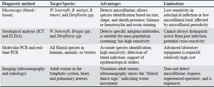
Microscopic examination of blood or tissue samples is a commonly used conventional technique for detecting microfilariae (Fink et al., 2011). Species such as W. bancrofti and Brugia spp. have specific periodicity patterns, which are generally nocturnal in tropical areas. Therefore, blood sampling times need to be adjusted to increase the test sensitivity (de Almeida and Freedman, 1999). Microfilariae can be observed using Giemsa or hematoxylin-eosin staining, which allows species identification based on morphological characteristics such as size, shape, and sheath presence (Mathison et al., 2019). Microfilariae of D. immitis or D. repens can be detected in both peripheral blood and subcutaneous tissue in animals, depending on the tropism of each species (Santhosh et al., 2025).
Serological approaches are used to identify filarial-specific antigens or antibodies, particularly in cases of subclinical infection or when blood microfilariae density is low (Pastor et al., 2021). Rapid immunochromatography (ICT) tests are widely used to detect W. bancrofti antigens, while enzyme-linked immunosorbent assay (ELISA) methods are used to detect antibodies against Brugia spp. and Dirofilaria spp. (Supali et al., 2002). For Brugia infections, the Brugia Rapid test has been used to detect filaria-specific IgG4 antibodies, including in individuals from areas highly endemic for Brugia timori (Supali et al., 2004). Serological methods have the advantage of high sensitivity and ease of application in large-scale population screening, although their limitation lies in the inability to distinguish between active infection and prior exposure (Avendaño and Patarroyo, 2020).
Molecular approaches, such as polymerase chain reaction (PCR) and real-time PCR, increase the sensitivity and specificity of filarial detection, even in cases with low microfilaria counts or latent infections (Pietrzak et al., 2024). These methods allow for the accurate identification of parasite species in humans, animals, and mosquito vectors, thus supporting epidemiological studies and assessing the effectiveness of control programs (Momčilović et al., 2019). Imaging techniques, including ultrasonography and radiology, are used to visualize adult worms in the lymphatic system, heart, or pulmonary arteries (Gurung et al., 2022). Ultrasonography demonstrates the “filarial dance sign,” a marker of adult worm movement, which helps confirm infection, especially when microfilariae are not microscopically detected (Arndts et al., 2012).
The selection of an appropriate diagnostic technique requires consideration of the filarial species, host type, infection burden, and purpose of the examination, whether for screening or surveillance (Riches et al., 2020). The combination of microscopic, serological, molecular, and imaging methods creates a comprehensive diagnostic approach, enabling early detection, accurate species identification, and control of filariasis transmission (Pietrzak et al., 2024).
Laboratory analyses are also essential for assessing disease severity and systemic involvement in canine heartworm disease (Noack et al., 2021). Routine hematological and biochemical evaluations, including complete blood count and serum biochemistry, may demonstrate eosinophilia, anemia, thrombocytopenia, elevated liver enzymes, azotemia, and serum protein profile alterations associated with cardiopulmonary and renal dysfunction (Babu and Nutman, 2012). Novel biomarkers have gained increasing importance in the diagnosis and prognosis of heartworm disease (Bennuru et al., 2018). Cardiac troponins (cTnI and cTnT) are sensitive indicators of myocardial injury, whereas N-terminal pro–B-type natriuretic peptide reflects cardiac stress and heart failure severity (Kanyal et al., 2025). Elevated D-dimer levels may indicate thromboembolic events and pulmonary vascular damage in advanced infections (Ishikawa et al., 2017). Therefore, the integration of conventional laboratory testing with emerging cardiac and coagulation biomarkers can improve the diagnostic accuracy, disease staging, prognostic assessment, and therapeutic monitoring of heartworm disease.
Differential diagnosis
The differential diagnosis of filariasis is important because its clinical symptoms can mimic other diseases in humans and animals (Pietrzak et al., 2024). In humans, conditions such as lymphedema, elephantiasis, and hydrocele can appear similar to non-parasitic disorders, such as congenital primary lymphedema, which is usually bilateral and present at birth without a history of vector exposure, or secondary lymphedema caused by bacterial infection, trauma, surgical procedures, or neoplasia (Shenoy, 2008). Furthermore, thickening of the subcutaneous tissue due to lipedema or lipohypertrophy can also produce a similar clinical picture, but microfilariae are not detected in the bloodstream (Medeiros et al., 2022).
Subcutaneous lesions caused by filarials, such as D. repens, can resemble parasitic granulomas caused by other species (e.g., Onchocerca or Loa loa), soft tissue neoplasia, or chronic bacterial or fungal infections (Bytyqi et al., 2024). Differentiation between these conditions can be achieved through histopathological evaluation, microfilariae identification, or parasite-specific DNA detection using molecular techniques (McNulty et al., 2013).
Dirofilaria immitis-induced cardiopulmonary manifestations in animals must be differentiated from congenital or degenerative heart disease, such as pulmonary stenosis or tricuspid valve degeneration, and non-parasitic pulmonary disorders, including bacterial pneumonia, fungal pneumonia, or cardiogenic pulmonary edema (Thompson, 2014). Infection with other worms, such as Angiostrongylus vasorum, can also cause clinical manifestations similar to heartworm disease because they attack the pulmonary arteries and heart (Colella et al., 2016). On the contrary, D. repens typically exhibits a different clinical spectrum, as it predominantly causes subcutaneous dirofilariasis and may present with aberrant or ectopic localizations, including subcutaneous nodules, ocular involvement, and occasionally atypical presentations such as herniated masses in soft tissues, which can lead to diagnostic confusion with neoplastic or inflammatory conditions (Momčilović et al., 2025).
Furthermore, the microscopic examination of microfilariae requires precision because the morphology of some species can overlap (Loyola et al., 2025). Morphological characteristics, such as size, shape, and sheath presence, should be combined with molecular techniques, such as PCR, to differentiate between human and animal filarial species and detect latent or subclinical infections (Thanomsub et al., 2000). Additional approaches, such as serology and imaging methods—including ultrasonography with the “filarial dance sign” phenomenon or cardiopulmonary radiological evaluation—also help differentiate filarial infections from non-parasitic lesions (Momčilović et al., 2019).
This multimodal approach allows for precise species identification, reduces the risk of misdiagnosis, and supports the selection of appropriate intervention strategies at the individual and population levels (Rajamanickam and Babu, 2025).
Transmission
Filariasis transmission is a complex biological process involving interactions between the filarial parasite, its vertebrate host, and its vector mosquito. It is influenced by ecological, social, and behavioral factors in both humans and animals (Remya et al., 2025). Filariasis is a vector-borne disease in which the infective L3 larvae are transmitted to the host through the bite of a female mosquito of the genus Culex, Aedes, Anopheles, or Mansonia, depending on the filarial species and its geographic distribution (Ridha et al., 2020). This mosquito-mediated transmission represents the primary mode of infection and is, therefore, not reexplained in detail in the pathogenesis and clinical manifestation sections to avoid redundancy. The mosquito acts as an intermediate host, where the microfilariae develop into L1 and then L3 larvae before being transmitted to the next host (Gordon et al., 2018).
In human filariasis, W. bancrofti exhibits a nocturnal periodicity of microfilariae, which adapts to the vector mosquito’s nocturnal activity (Sack, 2009). Brugia malayi and B. timori have similar patterns, although regional variations may occur depending on the vector mosquito’s behavior and habitat (Diekmann et al., 2025). Microfilariae circulating in human blood are taken up by mosquitoes during a blood meal and develop into L3-stage larvae in the mosquito’s gut and hexameric glands before being transmitted to a new host (Ghofur et al., 2024). Environmental factors, such as high humidity, favorable temperature, and mosquito population density, significantly influence the success of the filarial transmission cycle (Cano et al., 2014).
Animal filariasis, particularly that caused by D. immitis, is transmitted by hematophagous mosquitoes that ingest microfilariae from infected dogs or cats (Ivănescu et al., 2025). L1 larvae develop into infective L3 larvae within the mosquito, which can then be transmitted to other animals through bites (Riahi et al., 2021). Differences in tissue tropism explain the appearance of cardiopulmonary manifestations in animals, whereas the parasite’s survival is ensured by the cycle back to the vector mosquito (Morchón et al., 2012). Dirofilaria repens infection in both animals and humans follows a similar mechanism, but the larvae reside in the subcutaneous tissue (Genchi and Kramer, 2017).
The relationship between humans, animals, and vectors highlights the risk of cross-species transmission and zoonosis (Siriyasatien et al., 2023). Factors such as human mobility, domestic animal density, climate change, urbanization, and natural habitat degradation can increase contact with vectors and expand the distribution of filariasis endemic areas (Galgamuwa et al., 2025). In summary, detailed mechanistic descriptions of parasite development within the host are provided in the pathogenesis section to maintain clarity and avoid repetition across sections.
Risk factors
The complex interactions between the filarial parasite, its mosquito vector, its vertebrate host, and socioecological conditions, which influence the likelihood of infection, parasite load, and disease severity, are the risk factors for filariasis (Galgamuwa et al., 2025). Infection risk is higher in individuals or populations living in endemic areas, where vector mosquito densities are high and environmental sanitation is poor, increasing the likelihood of human or animal contact with infective L3 larvae (Kermelita et al., 2024).
Environmental factors play a crucial role in filariasis transmission (Lestyoningrum et al., 2026). Tropical and subtropical regions with warm temperatures, high humidity, and standing water provide ideal conditions for mosquito vectors (Lamy et al., 2023). The growth of Mansonia, Culex, Aedes, and Anopheles mosquito populations is supported by aquatic vegetation, swamps, and irrigation systems, thus reinforcing the filarial transmission cycle (Mulyaningsih et al., 2019). Furthermore, unplanned urbanization, high population density, and poor sanitation infrastructure increase human exposure to vectors and facilitate disease spread at the community level (Yadav et al., 2025).
Biological factors also influence the risk of infection at the individual level (Galgamuwa et al., 2025). Age, immune status, nutritional status, and history of previous infections determine the host’s ability to withstand or develop disease symptoms (Amewu et al., 2025). Children and individuals with compromised immune systems tend to have higher microfilariae loads and a higher risk of severe lymphatic complications (Mandanas, 2021). In domestic animals, high dog or cat population density, interaction with wild animals, and animal mobility increase the likelihood of Dirofilaria spp. transmission via mosquito vectors (Pękacz et al., 2025).
Human behavior also influences the risk of infection. Outdoor activities at night, minimal use of mosquito nets or repellents, and low compliance with MDA programs increase the chances of infection (Nawan et al., 2025). Furthermore, close interactions between humans and domestic animals or wild reservoirs can facilitate cross-species transmission, making filariasis a One Health issue that requires an integrated approach (Webster et al., 2016).
The genetics of the filarial species and the ability of the parasite to modulate the host’s response influence the success of infection (Bhoj et al., 2022). Some species produce immunomodulatory molecules that suppress the host’s immune system, allowing microfilariae to persist in the bloodstream longer and increasing the chance of transmission (Sureshan et al., 2025). Variations in mosquito vector species also determine transmission efficiency, microfilariae frequency patterns, and disease-endemic area distribution (Lupenza et al., 2021).
Public health importance
Filariasis is a parasitic disease that has a significant impact on public health, particularly in tropical and subtropical regions where it remains endemic (Azzahra et al., 2024). Human filarial infections, particularly those caused by W. bancrofti, B. malayi, and B. timori, can cause chronic morbidity in the form of lymphedema, elephantiasis, and filarial hydrocele, which impact the quality of life, productivity, and social participation of individuals (Vasconez-Gonzalez et al., 2025). The impact of filariasis is not only medical but also causes a psychosocial burden and stigma, thus affecting the community’s overall well-being (Barrett et al., 2024).
From an epidemiological perspective, filariasis exhibits an endemic pattern with continuous transmission, supported by the presence of vector mosquitoes (Culex, Aedes, Anopheles, and Mansonia) and human or animal reservoirs (Anjal et al., 2025). Asymptomatic individuals remain a source of infection for mosquitoes, so population-based disease control efforts, including MDA programs, active surveillance, and vector control, are necessary (Sinha et al., 2023).
Filariasis also impacts animal health, particularly those caused by D. immitis and D. repens, which can cause cardiopulmonary disease or subcutaneous lesions (Virruso et al., 2025). Domestic animals serve as potential reservoirs, increasing the risk of zoonotic and cross-species transmission (Naing et al., 2024). Therefore, public health interventions must implement a One Health approach that integrates human, animal, and environmental aspects (Igreja et al., 2025).
In addition to clinical impacts, filariasis has significant economic and social consequences (Vasconez-Gonzalez et al., 2025). In endemic countries, long-term care costs, reduced productivity, and the need for community-level interventions place a burden on health systems (Molyneux, 2003). Social stigma and mobility limitations resulting from chronic complications reduce individuals’ economic contributions and disrupt social interactions (Medeiros et al., 2022). Therefore, public health strategies should emphasize transmission prevention, early detection, appropriate treatment, community education, and vector control (Remya et al., 2025).
Treatment
Filariasis treatment aims to eradicate the parasite, prevent complications, and reduce transmission, with strategies tailored to the filarial species, disease severity, and host condition (Addiss and Brady, 2007). In humans, the main pharmacological therapies include the use of antifilarial drugs, such as diethylcarbamazine (DEC), ivermectin, and albendazole, either alone or in combination (Macfarlane et al., 2019). Combination therapy is more effective in reducing the number of microfilariae and inhibiting the development of adult worms, thereby reducing the risk of onward transmission (Mayfield et al., 2025). Table 3 summarizes filariasis treatment strategies in humans and animals, including pharmacological therapy, drug combinations, complication management, surgical intervention, and population-based approaches.
Table 3. Treatment strategies for human and animal filariasis.
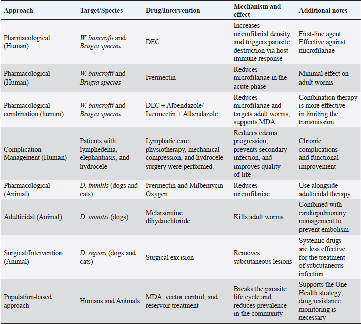
DEC is a first-line drug for W. bancrofti and Brugia spp. It works by increasing the concentration of microfilariae in the bloodstream and triggering parasite destruction through the activation of the host immune response (McGarry et al., 2005). Ivermectin is effective in reducing the number of microfilariae in the acute phase but has a limited effect on adult worms (Martin et al., 2021). Albendazole is usually added to combination therapy to target adult worms, enhance the microfilaricidal effect, and support the implementation of MDA programs in endemic areas (Macfarlane et al., 2019). Figure 3 illustrates integrated filariasis control strategies based on the One Health approach, emphasizing the interconnected roles of humans, animal health, and environmental management.
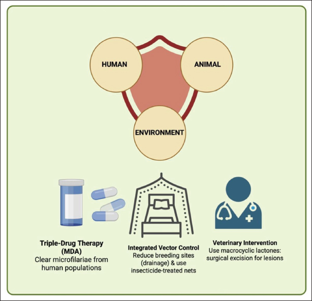
Fig. 3. One Health-based integrated control strategies for filariasis.
In addition to pharmacological therapy, the management of chronic complications, such as lymphedema, elephantiasis, and hydrocele, requires supportive interventions and surgery (Rajamanickam and Babu, 2025). Lymphedema treatment includes skin hygiene, physiotherapy exercises, and the use of mechanical compression to reduce secondary infection, inflammation, and edema progression (León-Díaz and Medina-Otero, 2025). Surgical treatment for hydrocele in men with filarial hydrocele can improve quality of life and prevent further complications (Betts et al., 2020).
Treatment for animal filariasis, particularly that caused by D. immitis, includes therapy for both microfilariae and adult worms (Diakou and Prichard, 2021). Melarsomine dihydrochloride is used to eradicate adult worms in dogs, while ivermectin or milbemycin oxime effectively reduces the number of microfilariae (Savadelis et al., 2022) to reduce the risk of pulmonary embolism (PE) and heart failure (HF) (Prichard, 2021). For D. repens-induced subcutaneous infections, surgical excision is usually necessary because systemic medications are less effective (Ahmed et al., 2022).
Treatment strategies for filtration should also include population and transmission control efforts, particularly within a One Health approach (Abokwara and Madubueze, 2025). Mass drug administration programs for humans, vector control, and treatment of animal reservoirs can interrupt the parasite’s life cycle and reduce community prevalence (Gyapong et al., 2018). The continuous monitoring of potential drug resistance and therapeutic effectiveness is crucial to ensure the long-term success of filariasis interventions (Plichart and Lemoine, 2013). Computational approaches, including in silico screening and molecular docking, can support the preliminary evaluation of bioactive compounds against inflammation-related targets, although further in vivo validation remains essential before therapeutic application (Budiastuti et al., 2025).
Control
Filariasis control emphasizes a holistic approach involving humans, animals, and vectors in accordance with the One Health principle, with the goal of breaking the transmission cycle and reducing disease prevalence (Danasekaran, 2024). Control strategies include pharmacological interventions, vector management, public education, and population monitoring tailored to the vector ecology, filarial species, and socioeconomic conditions in endemic areas (Galgamuwa et al., 2025).
Pharmacological interventions in humans are carried out through MDA programs that cover the entire population in endemic areas (Freitas et al., 2024). Drugs, such as DEC, ivermectin, and albendazole, are used to reduce the number of microfilariae in the blood, reduce the human reservoir, and prevent further transmission (Adinarayanan et al., 2007). This approach can also be combined with individual treatment in clinical cases to minimize the risk of developing chronic complications (Supali et al., 2002). The success of MDA programs depends heavily on population coverage, adherence, and drug resistance monitoring (Krentel et al., 2013).
Vector control is crucial because mosquitoes are crucial intermediate hosts in the filarial life cycle (Famakinde, 2018). Strategies include reducing mosquito breeding habitats by draining stagnant water, managing irrigation, and improving environmental sanitation; using residual insecticides, larvicides, and bed nets; and biological approaches, such as introducing mosquito larvae predators (Bera, 2025). These efforts aim to reduce mosquito population density and risk of filarial transmission to humans and reservoir animals (Erickson et al., 2013). A multifaceted approach has also been emphasized in other veterinary infectious diseases, such as classical swine fever, where laboratory diagnosis, surveillance, vaccination, and rapid outbreak response are required to reduce disease spread and economic losses (Khairullah et al., 2024).
The control of filariasis in animals primarily focuses on D. immitis and D. repens (Remya et al., 2025). Routine preventive therapy of domestic animals using macrocyclic lactones helps reduce circulating microfilariae, reduces the risk of transmission to mosquitoes, and minimizes zoonotic potential (Traversa et al., 2024). Monitoring animal reservoir populations is also crucial to prevent the formation of transmission hotspots (Mehta and Maharjan, 2026).
Public education and community participation are important nonpharmacological strategies to support successful filariasis control (Taylor et al., 2022). Raising awareness of mosquito bite protection, proper lymphatic drainage, and MDA program adherence can enhance the effectiveness of interventions (Wayangkau et al., 2025). Furthermore, outreach helps reduce social stigma against individuals with lymphedema or elephantiasis, thus encouraging greater engagement in public health programs (Maritim et al., 2021).
Surveillance and monitoring are crucial components of a long-term filariasis control strategy (Kelly-Hope et al., 2018). The early identification of new cases, monitoring compliance with the MDA program, evaluating mosquito population density, and detecting reservoir animals allow for timely adjustments to interventions (Srividya et al., 2019). This approach supports efforts to eliminate filariasis as a public health problem, reduce morbidity, and improve the quality of life of communities in endemic areas (Chiphwanya et al., 2024).
Filariasis control programs can effectively reduce disease prevalence, break transmission cycles, and prevent new endemics by combining pharmacological interventions, vector control, reservoir animal management, public education, and surveillance activities (de Souza and Bockarie, 2025). The One Health framework also highlights the importance of biodiversity and ecosystem preservation, as local biological resources such as Holothuria edulis have been reported to contribute to marine ecosystem balance while containing bioactive metabolites with antioxidant and anticancer potential (Misgiati et al., 2024). Natural bioactive compounds continue to receive attention for their pharmacological potential, including antioxidant, anti-inflammatory, antimicrobial, and metabolic regulatory activities, as reported in plant-derived compounds such as capsaicin and Indonesian bay leaf bioactives (Pratama et al., 2021; Pratama et al., 2022; Sukmanadi et al., 2025).
Conclusion
Filariasis is a parasitic disease that remains a public health problem in tropical and subtropical regions due to the complex interactions between the parasite, humans, animals, and mosquito vectors. In humans, infection can cause lymphedema, elephantiasis, and filarial hydrocele, thereby negatively affecting quality of life and productivity. Infection by Dirofilaria spp. serves as a reservoir in domestic animals, increasing the risk of cross-species or zoonotic transmission.
Early detection using clinical, microscopic, serological, molecular, and imaging methods is crucial for reducing morbidity and breaking the filariasis transmission cycle. Treatment and control approaches, including pharmacological therapy, complication management, vector control, treatment of reservoir animals, and public education, have proven effective in suppressing disease prevalence while mitigating its social and economic impacts.
The One Health approach, which integrates human, animal, and environmental aspects, is a crucial element in planning long-term interventions. A comprehensive understanding of the disease’s epidemiology, risk factors, transmission mechanisms, and impact allows for the design of appropriate, effective, and sustainable control strategies. Overall, these integrated interventions can reduce morbidity burden, prevent chronic complications, and improve public health.
Acknowledgments
The author expresses his gratitude for the support and contributions that helped in the preparation of this review article. The author independently carried out all writing, literature analysis, and manuscript preparation without external funding.
Conflict of interest
The authors declare no conflict of interest.
Funding
The author independently funded all writing and manuscript preparation activities.
Author’s contributions
M.N., Y.A., and A.R.K. drafted the manuscript. S.S., R.R., and S.S.M. revised and edited the manuscript. E.A.J., L.O.T., and A.T.R. prepared and critically checked this manuscript. S.W., S.S.P., and B.P.P. edited the references. All authors are major contributors to this work and have approved the final version of the manuscript.
Data availability
All references are open access, so data can be obtained from the internet.
References
Abokwara, A. and Madubueze, C.E. 2025. Optimizing the control of lymphatic filariasis: a mathematical modelling and cost-effectiveness analysis. Discov. Public. Health. 22(1), 703.
Addiss, D.G. and Brady, M.A. 2007. Morbidity management in the Global Programme to Eliminate Lymphatic Filariasis: a review of the scientific literature. Filaria J. 6(1), 2.
Adinarayanan, S., Critchley, J.A., Das, P.K. and Gelband, H. 2007. Diethylcarbamazine (DEC)-medicated salt for community-based control of lymphatic filariasis. Cochrane. Database. Syst. Rev. 1(1), CD003758.
Ahmed, N., Tonelli, L., Labagnara, G., Doglioni, C. and Pedica, F. 2022. Subcutaneous dirofilariosis in Italy: a diagnostic hypothesis to remind when the anamnesis is misleading. Pathologica 114(2), 174–177.
Ahmed, T., Hyder, M.Z., Liaqat, I. and Scholz, M. 2019. Climatic conditions: conventional and nanotechnology-based methods for the control of mosquito vectors causing human health issues. Int. J. Environ. Res. Public Health 16(17), 3165.
Aisyah, D.N., Kozlakidis, Z., Diva, H., Trimizi, S.N., Sianipar, L.R., Wijayanti, E., Avicena, A.M. and Adisasmito, W. 2022. The spatial-temporal distribution of chronic lymphatic filariasis in Indonesia: a 18-year registry-based analysis. Microbiol. Res. 13(4), 681–690.
Alonso, D.P., Alvarez, M.V.N., Amorim, J.A., De Sá, I.L.R., De Carvalho, D.P., Ribeiro, K.A.N., Ribolla, P.E.M. and Sallum, M.A.M. 2022. Mansonia spp. population genetics based on mitochondrion whole-genome sequencing alongside the Madeira River near Porto Velho, Rondonia, Brazil. Infect. Genet. Evol. 103(1), 105341.
Amewu, E.K.A., Amaglo, B., Osei-Poku, P., Abakah, A., Koney Shardow, A.L., Wright, L. and Kwarteng, A. 2025. Nutritional predictors of lymphatic filariasis progression: insights from a machine learning approach. PLoS One 20(3), 320640.
Anjal, K., Rawal, V., Pandey, S., Harshavarthini, M., Verma, A.K., Mohan, B., Gurha, S., Sondhiya, G., Ansari, A., Kombiah, S., Shrivastava, S., Barde, P.V. and Singh, P. 2025. Vector incrimination studies of lymphatic filariasis in rural areas of endemic Datia district of Madhya Pradesh, India. Trans. R. Soc. Trop. Med. Hyg. 119(9), 1059–1065.
Arndts, K., Deininger, S., Specht, S., Klarmann, U., Mand, S., Adjobimey, T., Debrah, A.Y., Batsa, L., Kwarteng, A., Epp, C., Taylor, M., Adjei, O., Layland, L.E. and Hoerauf, A. 2012. Elevated adaptive immune responses are associated with latent infections of Wuchereria bancrofti. PLos Negl. Trop. Dis. 6(4), e1611.
Avendaño, C. and Patarroyo, M.A. 2020. Loop-mediated isothermal amplification as point-of-care diagnosis for neglected parasitic infections. Int. J. Mol. Sci. 21(1), 7981.
Azzahra, M.K., Shafwah, D.A., Sondakh, C.S. and Adriyani, R. 2024. A review of bed nets usage and sewerage conditions as risk factors for lymphatic filariasis in developing countries. J. Environ. Health 16(1), 89–100.
Babayan, S.A., Read, A.F., Lawrence, R.A., Bain, O. and Allen, J.E. 2010. Filarial parasites develop faster and reproduce earlier in response to host immune effectors that determine filarial life expectancy. PLoS Biol. 8(10), e1000525.
Babu, S. and Nutman, T.B. 2012. Immunopathogenesis of lymphatic filarial disease. Semin. Immunopathol. 34(6), 847–861.
Barrett, C., Chiphwanya, J., Matipula, D.E., Douglass, J., Kelly-Hope, L.A. and Dean, L. 2024. Addressing the syndemic relationship between lymphatic filariasis and mental distress in Malawi: the potential of enhanced self-care. Trop. Med. Infect. Dis. 9(8), 172.
Bennuru, S. and Nutman, T.B. 2009. Lymphatics in human lymphatic filariasis: in vitro models of parasite-induced lymphatic remodeling. Lymphat. Res. Biol. 7(4), 215–219.
Bennuru, S., O’Connell, E.M., Drame, P.M. and Nutman, T.B. 2018. Mining filarial genomes for diagnostic and therapeutic targets. Trends Parasitol. 34(1), 80–90.
Bera, B. 2025. Mosquito ecology and disease transmission: implications for global health and vector control. Uttar. Pradesh. J. Zool. 46(8), 64–72.
Betts, H., Martindale, S., Chiphwanya, J., Mkwanda, S.Z., Matipula, D.E., Ndhlovu, P., Mackenzie, C., Taylor, M.J. and Kelly-Hope, L.A. 2020. Significant improvement in quality of life following surgery for hydrocoele caused by lymphatic filariasis in Malawi: a prospective cohort study. PLos Negl. Trop. Dis. 14(5), 8314.
Bhoj, P., Togre, N., Khatri, V. and Goswami, K. 2022. Harnessing immune evasion strategy of lymphatic filariae: a therapeutic approach against inflammatory and infective pathology. Vaccines (Basel) 10(8), 1235.
Bhuvaneswari, A., Shriram, A.N., Raju, K.H.K. and Kumar, A. 2023. Mosquitoes, lymphatic filariasis, and public health: a systematic review of Anopheles and Aedes surveillance strategies. Pathogens 12(12), 1406.
Budiastuti, Helmi, V.N., Effendi, M.H., Plumeriastuti, H., Khairullah, A.R., Ugbo, E.N., Tyasningsih, W., Awwanah, M., Pratama, B.P., Prasetyo, A., Moses, I.B. and Ahmad, R.Z. 2025. In silico analysis of metabolite compounds from the essential oil of Cinnamomum burmannii bark with COX-1 and COX-2 as target molecules. Trends Sci. 22(9), 10292; doi:10.48048/tis.2025.10292
Bytyqi, A., Karas, C., Pechmann, K., Ramharter, M. and Mischlinger, J. 2024. Oro-facial filariasis-a systematic review of the literature. PLos Negl. Trop. Dis. 18(11), 12610.
Cano, J., Rebollo, M.P., Golding, N., Pullan, R.L., Crellen, T., Soler, A., Kelly-Hope, L.A., Lindsay, S.W., Hay, S.I., Bockarie, M.J. and Brooker, S.J. 2014. The global distribution and transmission limits of lymphatic filariasis: past and present. Parasit. Vectors. 7(1), 466.
Chakraborty, S., Gurusamy, M., Zawieja, D.C. and Muthuchamy, M. 2013. Lymphatic filariasis: perspectives on lymphatic remodeling and contractile dysfunction in filarial disease pathogenesis. Microcirculation 20(5), 349–364.
Chandy, A., Thakur, A.S., Singh, M.P. and Manigauha, A. 2011. A review of neglected tropical diseases: filariasis. Asian Pac. J. Trop. Med. 4(7), 581–586.
Cheepsattayakorn, A. and Cheepsattayakorn, R. 2014. Parasitic pneumonia and lung involvement. Biomed. Res. Int. 1(1), 874021.
Chiphwanya, J., Mkwanda, S., Kabuluzi, S., Mzilahowa, T., Ngwira, B., Matipula, D.E., Chaponda, L., Ndhlova, P., Katchika, P., Mahebere Chirambo, C., Moses, P., Kumala, J., Chiumia, M., Barrett, C., Betts, H., Fahy, J., Rebollo Polo, M., Reimer, L., Stanton, M.C., Thomas, B., Freer, S., Molyneux, D.H., Bockarie, M.J., Mackenzie, C.D., Taylor, M.J., Martindale, S. and Kelly-Hope, L.A. 2024. Elimination of lymphatic filariasis as a public health problem in Malawi. PLos Negl. Trop. Dis. 18(2), 11957.
Colella, V., Lia, R.P., Premont, J., Gilmore, P., Cervone, M., Latrofa, M.S., D’Anna, N., Williams, D. and Otranto, D. 2016. Angiostrongylus vasorum in the eye: new case reports and a review of the literature. Parasit. Vectors. 9(1), 161.
Damayanti, E., Budiati, E., Irianto, S.E., Adyas, A. and Pratiwi Putri, D.U. 2025. Factors affecting the occurrence of filariasis in East Lampung, Indonesia. Health Dyn. 2(7), 313–320.
Danasekaran, R. 2024. One Health: a holistic approach to tackling global health issues. Indian. J. Community. Med. 49(2), 260–263.
De Almeida, A.B. and Freedman, D.O. 1999. Epidemiology and immunopathology of bancroftian filariasis. Microbes. Infect. 1(12), 1015–1022.
De Souza, D.K. and Bockarie, M.J. 2025. Current perspectives in the epidemiology and control of lymphatic filariasis. Clin. Microbiol. Rev. 38(2), 12623.
Diakou, A. and Prichard, R.K. 2021. Concern for Dirofilaria immitis and macrocyclic lactone loss of efficacy: current situation in the USA and Europe, and future scenarios. Pathogens 10(10), 1323.
Diekmann, I., Supali, T., Fischer, K., Iskandar, E., Sugianto, N., Destani, Y., Alfian, R., Weil, G.J. and Fischer, P.U. 2025. Brugia malayi and other filarial parasite species in animals in areas endemic for lymphatic filariasis in Belitung District, Indonesia. PLos Negl. Trop. Dis. 19(10), e0013593.
Dimzas, D., Aindelis, G., Tamvakis, A., Chatzoudi, S., Chlichlia, K., Panopoulou, M. and Diakou, A. 2024. Dirofilaria immitis and Dirofilaria repens: investigating the prevalence of zoonotic parasites in dogs and humans in a hyperenzootic area. Animals (Basel) 14(17), 2529.
Ehrens, A., Hoerauf, A. and Hübner, M.P. 2022. Eosinophils in filarial infections: inducers of protection or pathology?. Front. Immunol. 13(1), 983812.
Erickson, S.M., Thomsen, E.K., Keven, J.B., Vincent, N., Koimbu, G., Siba, P.M., Christensen, B.M. and Reimer, L.J. 2013. Mosquito-parasite interactions can shape filariasis transmission dynamics and impact elimination programs. PLos Negl. Trop. Dis. 7(9), e2433.
Erickson, S.M., Xi, Z., Mayhew, G.F., Ramirez, J.L., Aliota, M.T., Christensen, B.M. and Dimopoulos, G. 2009. Mosquito infection responses to developing filarial worms. PLos Negl. Trop. Dis. 3(10), 529.
Famakinde, D. 2018. Mosquitoes and the lymphatic filarial parasites: research trends and budding roadmaps to future disease eradication. Trop. Med. Infect. Dis. 3(1), 4.
Fercoq, F., Remion, E., Vallarino-Lhermitte, N., Alonso, J., Raveendran, L., Nixon, C., Le Quesne, J., Carlin, L.M. and Martin, C. 2020. Microfilaria-dependent thoracic pathology associated with eosinophilic and fibrotic polyps in filaria-infected rodents. Parasit. Vectors. 13(1), 551.
Fimbo, A.M., Minzi, O.M.S., Mmbando, B.P., Barry, A., Nkayamba, A.F., Mwamwitwa, K.W., Malishee, A., Seth, M.D., Makunde, W.H., Gurumurthy, P., Lusingu, J.P.A., Kamuhabwa, A.A.R. and Aklillu, E. 2020. Prevalence and correlates of lymphatic filariasis infection and its morbidity following mass Ivermectin and Albendazole Administration in Mkinga District, North-Eastern Tanzania. J. Clin. Med. 9(5), 1550.
Fink, D.L., Fahle, G.A., Fischer, S., Fedorko, D.F. and Nutman, T.B. 2011. Toward molecular parasitologic diagnosis: enhanced diagnostic sensitivity for filarial infections in mobile populations. J. Clin. Microbiol. 49(1), 42–47.
Freitas, L.T., Khan, M.A., Uddin, A., Halder, J.B., Singh-Phulgenda, S., Raja, J.D., Balakrishnan, V., Harriss, E., Rahi, M., Brack, M., Guérin, P.J., Basáñez, M.G., Kumar, A., Walker, M. and Srividya, A. 2024. The lymphatic filariasis treatment study landscape: a systematic review of study characteristics and the case for an individual participant data platform. PLos Negl. Trop. Dis. 18(1), 11882.
Galgamuwa, L.S., Hakmanage, N.M. and Fernando, S. 2025. Risk factors of lymphatic filariasis in Asia: a systematic review. BMC. Infect. Dis. 25(1), 1692.
Genchi, C. and Kramer, L. 2017. Subcutaneous dirofilariosis (Dirofilaria repens): an infection spreading throughout the old world. Parasit. Vectors 10(Suppl 2), 517.
Ghofur, A., Hadisaputro, S. and Sayono, S. 2024. Detection of microfilaria L3 and insecticide resistance among wild-caught mosquito vectors in endemic areas of lymphatic filariasis. Biodiversitas 25(5), 1975–1983.
Gleave, K., Cook, D., Taylor, M.J. and Reimer, L.J. 2016. Filarial infection influences mosquito behaviour and fecundity. Sci. Rep. 6(1), 36319.
Gordon, C.A., Jones, M.K. and Mcmanus, D.P. 2018. The history of bancroftian lymphatic filariasis in Australasia and Oceania: is there a threat of re-occurrence in Mainland Australia?. Trop. Med. Infect. Dis. 3(2), 58.
Grosbois, A., Risco-Castillo, V., Davoust, B., Laidoudi, Y., Crozet, G. and Watier-Grillot, S. 2025. Dirofilaria immitis and Dirofilaria repens infections in French working military dogs: prevalence and factors associated with vector exposure. Parasitol. Int. 109(1), 103–105.
Gurung, S., Karki, S., Kharal, K., Thapa, S., Thapa, S. and Baral, S. 2022. Filariasis diagnosed by real-time ultrasound scanning as filarial dance sign - a case report. IDCases 30(1), e01621.
Gyapong, J.O., Owusu, I.O., Da-costa Vroom, F.B., Mensah, E.O. and Gyapong, M. 2018. Elimination of lymphatic filariasis: current perspectives on mass drug administration. Res. Rep. Trop. Med. 9(1), 25–33.
Hafizu, M.S., Junaid, O.Q., Sagara, R., Ravi, R., Yik, F.M., Vythilingam, I. and Ling, L.Y. 2025. Zoonotic Brugian filariasis past and present trends in Malaysia: a systematic review and proportionate meta-analysis. Sci. Rep. 15(1), 37323.
Handa, A., Gaidhane, A. and Choudhari, S. 2024. Unraveling lymphatic filariasis in an old man: a case report. Cureus 16(4), e58167.
Hidron, A., Vogenthaler, N., Santos-Preciado, J.I., Rodriguez-Morales, A.J., Franco-Paredes, C. and Rassi, A. 2010. Cardiac involvement with parasitic infections. Clin. Microbiol. Rev. 23(2), 324–349.
Hoch, H. and Strickland, K. 2008. Canine and feline dirofilariasis: life cycle, pathophysiology, and diagnosis. Compend. Contin. Educ. Vet. 30(3), 133–140.
Igreja, R.P., De Macedo, P.M. and Schneider, M.C. 2025. One Health and neglected zoonotic diseases. Pathogens 14(5), 482.
Ishikawa, G., Acquah, S.O., Salvatore, M. and Padilla, M.L. 2017. Elevated serum D-dimer level is associated with an increased risk of acute exacerbation in interstitial lung disease. Respir. Med. 128(1), 78–84.
Ivănescu, L., Mîndru, R., Bodale, I., Apopei, G.V., Andronic, L., Hristodorescu, S., Azoicăi, D. and Miron, L. 2025. Circulation of Dirofilaria immitis and Dirofilaria repens species in mosquitoes in the Southeastern Part of Romania, under the influence of climate change. Life 15(10), 1612.
Jiang, X., Nicolls, M.R., Tian, W. and Rockson, S.G. 2018. Lymphatic dysfunction, leukotrienes, and lymphedema. Annu. Rev. Physiol. 80, 49–70.
Kaikuntod, M., Arjkumpa, O., Kladkempetch, D., Fukumoto, S., Thongkorn, K., Boonyapakorn, C., Punyapornwithaya, V. and Tiwananthagorn, S. 2021. Geographic spatial distribution patterns of Dirofilaria immitis and Brugia pahangi infection in community dogs in Chiang Mai, Thailand. Animals 11(1), 33.
Kanyal, S., Das, A., Bashir, A.M.D., Syed, A.H., Aujla, S., Chaudhary, J., Patel, D. and Goel, A. 2025. N-Terminal Pro-B-Type Natriuretic Peptide (NT-proBNP) as a Biomarker in Heart Failure With Preserved Ejection Fraction (HFpEF) Versus Heart Failure With Reduced Ejection Fraction (HFrEF): the way forward in the age of proteomics. Cureus 17(10), e94162.
Karunakaran, I., Ritter, M., Pfarr, K., Klarmann-Schulz, U., Debrah, A.Y., Debrah, L.B., Katawa, G., Wanji, S., Specht, S., Adjobimey, T., Hübner, M.P. and Hoerauf, A. 2023. Filariasis research - from basic research to drug development and novel diagnostics, over a decade of research at the Institute for Medical Microbiology, Immunology and Parasitology, Bonn, Germany. Front. Trop. Dis. 4(1), 1126173.
Kelly-Hope, L.A., Blundell, H.J., Macfarlane, C.L. and Molyneux, D.H. 2018. Innovative surveillance strategies to support the elimination of filariasis in Africa. Trends Parasitol. 34(8), 694–711.
Kermelita, D., Hadi, U.K., Soviana, S., Tiuria, R. and Supriyono, S. 2024. Species diversity of mosquitoes (Diptera: culicidae), larval habitat characteristics, and potential as vectors for lymphatic filariasis in Central Bengkulu Regency, Indonesia. Vet. World. 17(9), 2115–2123.
Kilarski, W.W., Martin, C., Pisano, M., Bain, O., Babayan, S.A. and Swartz, M.A. 2019. Inherent biomechanical traits enable infective filariae to disseminate through collecting lymphatic vessels. Nat. Commun. 10(1), 2895.
Khairullah, A.R., Effendi, M.H., Moses, I.B., Fauzia, K.A., Puspitasari, Y., Riwu, K.H.P., Fauziah, I., Raissa, R., Silaen, O.S.M., Wibowo, S., Yanestria, S.M., Kusala, M.K.J., Abdila, S.R., Pratama, B.P. and Hasib, A. 2024. Classical swine fever: unveiling the complexity through a multifaceted approach. Open Vet. J. 14(10), 2497–2508; doi:10.5455/OVJ.2024.v14.i10.1
Krentel, A., Fischer, P.U. and Weil, G.J. 2013. A review of factors that influence individual compliance with mass drug administration for elimination of lymphatic filariasis. PLos Negl. Trop. Dis. 7(11), 2447.
Kwa, B.H. 2008. Environmental change, development and vectorborne disease: malaysia’s experience with filariasis, scrub typhu and dengue. Environ. Dev. Sustain. 10(2), 209–217.
Lamy, K., Tran, A., Portafaix, T., Leroux, M.D. and Baldet, T. 2023. Impact of regional climate change on the mosquito vector Aedes albopictus in a tropical island environment: la Réunion. Sci. Total. Environ. 875(1), 162484.
Laney, S.J., Ramzy, R.M.R., Helmy, H.H., Farid, H.A., Ashour, A.A., Weil, G.J. and Williams, S.A. 2010. Detection of Wuchereria bancrofti L3 larvae in mosquitoes: a reverse transcriptase PCR assay evaluating infection and infectivity. PLos Negl. Trop. Dis. 4(2), 602.
Leggat, P.A., Graves, P., Laha, T. and Aye, K.S. 2018. Neglected and emerging tropical diseases in South and Southeast Asia and Northern Australia. Trop. Med. Infect. Dis. 3(3), 70.
León-Díaz, R. and Medina-Otero, A. 2025. Prevention and treatment of postmastectomy lymphedema: a physiotherapy perspective. Curr. Oncol. 32(10), 555.
Lestyoningrum, S.D., Noveyani, A.E., Faisal, D.R., Nugraheni, W.P., Nuraini, S., Pawitaningtyas, I. and Putri, L.M. 2026. Factors associated with lymphatic filariasis in Indonesia. Clin. Epidemiol. Glob. Health 37(1), 102277.
Loyola, D.C., Placko, A., Fessl, B. and McNew, S.M. 2025. Novel microfilariae detected in Galápagos passerines. Int. J. Parasitol. Parasites. Wildl. 28(1), 101115.
Lupenza, E., Gasarasi, D.B. and Minzi, O.M. 2021. Lymphatic filariasis, infection status in Culex quinquefasciatus and Anopheles species after six rounds of mass drug administration in Masasi District, Tanzania. Infect. Dis. Poverty. 10(1), 20.
Macfarlane, C.L., Budhathoki, S.S., Johnson, S., Richardson, M. and Garner, P. 2019. Albendazole alone or in combination with microfilaricidal drugs for lymphatic filariasis. Cochrane. Database. Systematic. Rev. 1(1), CD003753.
Mackenzie, C.D., Kapa, D.R., Krishnasastry, S., Douglass, J., Hoerauf, A. and Ottesen, E.A. 2024. Managing lymphedema induced by lymphatic filariasis: implementing and improving care at the individual and programmatic levels. Am. J. Trop. Med. Hyg. 111(4_Suppl), 3–21.
Mandanas, J.A.H. 2021. Lymphatic filariasis: an immunologic perspective. EMJ. Allergy. Immunol. 6(1), 71–78.
Maritim, P., Silumbwe, A., Zulu, J.M., Sichone, G. and Michelo, C. 2021. Health beliefs and health seeking behavior towards lymphatic filariasis morbidity management and disability prevention services in Luangwa District, Zambia: community and provider perspectives. PLos Negl. Trop. Dis. 15(2), 9075.
Martin, R.J., Robertson, A.P. and Choudhary, S. 2021. Ivermectin: an anthelmintic, an insecticide, and much more. Trends. Parasitol. 37(1), 48–64.
Mathison, B.A., Couturier, M.R. and Pritt, B.S. 2019. Diagnostic identification and differentiation of microfilariae. J. Clin. Microbiol. 57(10), e00706-19.
Mayfield, H.J., Muttucumaru, R., Sartorius, B., Sheridan, S., Ward, S., Martin, B.M., Hedtke, S.M., Thomsen, R., Viali, S., Fatupaito, G., Lau, C.L. and Graves, P.M. 2025. Recurrence of microfilaraemia after triple-drug therapy for lymphatic filariasis in Samoa: recrudescence or reinfection?. Int. J. Infect. Dis. 152(1), 107809.
Mcgarry, H.F., Plant, L.D. and Taylor, M.J. 2005. Diethylcarbamazine activity against Brugia malayi microfilariae is dependent on inducible nitric-oxide synthase and the cyclooxygenase pathway. Filaria. J. 4(1), 4.
Mcnulty, S.N., Mitreva, M., Weil, G.J. and Fischer, P.U. 2013. Inter and intra-specific diversity of parasites that cause lymphatic filariasis. Infect. Genet. Evol. 14(1), 137–146.
Medeiros, Z.M., Vieira, A.V.B., Xavier, A.T., Bezerra, G.S.N., Lopes, M.D.F.C., Bonfim, C.V. and Aguiar-Santos, A.M. 2022. Lymphatic filariasis: a systematic review on morbidity and its repercussions in countries in the Americas. Int. J. Environ. Res. Public. Health. 19(1), 316.
Mehta, P.K. and Maharjan, M. 2026. Transmission status of lymphatic filariasis in hotspots of filarial infection, persistent districts of Nepal during post-MDA surveillance. PLoS One 21(1), 338141.
Mendoza-Roldan, J.A., Gabrielli, S., Cascio, A., Manoj, R.R.S., Bezerra-Santos, M.A., Benelli, G., Brianti, E., Latrofa, M.S. and Otranto, D. 2021. Zoonotic Dirofilaria immitis and Dirofilaria repens infection in humans and an integrative approach to the diagnosis. Acta Trop. 223(1), 106083.
Metenou, S. and Nutman, T.B. 2013. Regulatory T cell subsets in filarial infection and their function. Front. Immunol. 4(1), 305.
Misgiati, Winarni, I., Murniasih, T., Novriyanti, E., Tarman, K., Safithri, M., Setyaningsih, I., Cahyati, D., Pratama, B.P. and Wirawati, I. 2024. The anticancer and antioxidant potential of local sea cucumber Holothuria edulis, an ecology balancer of Labuan Bajo marine ecosystem. Case Stud. Chem. Environ. Eng. 9, 100625; doi:10.1016/j.cscee.2024.100625
Molyneux, D. 2003. Lymphatic filariasis (Elephantiasis) elimination: a public health success and development opportunity. Filaria. J. 2(1), 13.
Momčilović, S., Cantacessi, C., Arsić-Arsenijević, V., Otranto, D. and Tasić-Otašević, S. 2019. Rapid diagnosis of parasitic diseases: current scenario and future needs. Clin. Microbiol. Infect. 25(3), 290–309.
Momčilović, S., Jovanović, A. and Gasser, R.B. 2025. Human dirofilariasis – a potentially significant nematode zoonosis in an era of climate change. J. Infect. 90(4), 106460.
Morchón, R., Carretón, E., González-Miguel, J. and Mellado-Hernández, I. 2012. Heartworm disease (Dirofilaria immitis) and their vectors in Europe - new distribution trends. Front. Physiol. 3(1), 196.
Mulyaningsih, B., Umniyati, S.R., Hadisusanto, S. and Edyansyah, E. 2019. Study on vector mosquito of zoonotic Brugia malayi in Musi Rawas, South Sumatera, Indonesia. Vet. World. 12(11), 1729–1734.
Mupanomunda, M., Williams, J.F., Mackenzie, C.D. and Kaiser, L. 1997. Dirofilaria immitis: heartworm infection alters pulmonary artery endothelial cell behavior. J. Appl. Physiol. (1985). 82(2), 389–398.
Naing, C., Whittaker, M.A., Tung, W.S., Aung, H. and Mak, J.W. 2024. Prevalence of zoonotic (Brugian) filariasis in Asia: a proportional meta-analysis. Acta Trop. 249(1), 107049.
Nawan, Agatha, S., Handayani, S. and Toemon, A.I. 2025. Correlation between transmission prevention and incidence of filariasis: a systematic review. Asian. J. Health. Res. 4(1), 60–70.
Noack, S., Harrington, J., Carithers, D.S., Kaminsky, R. and Selzer, P.M. 2021. Heartworm disease - overview, intervention, and industry perspective. Int. J. Parasitol. Drugs Drug Resist. 16(1), 65–89.
Nutman, T.B. 2013. Insights into the pathogenesis of disease in human lymphatic filariasis. Lymphat. Res. Biol. 11(3), 144–148.
Oliveira, L.B., McHale, B.J., Verocai, G.G. and Rissi, D.R. 2021. Subcutaneous and cardiopulmonary dirofilariasis in a dog. Can. Vet. J. 62(8), 854–856.
O’Regan, N.L., Steinfelder, S., Venugopal, G., Rao, G.B., Lucius, R., Srikantam, A. and Hartmann, S. 2014. Brugia malayi microfilariae induce a regulatory monocyte/macrophage phenotype that suppresses innate and adaptive immune responses. PLos Negl. Trop. Dis. 8(10), e3206.
Paily, K.P., Hoti, S.L. and Das, P.K. 2009. A review of the complexity of biology of lymphatic filarial parasites. J. Parasit. Dis. 33(1–2), 3–12.
Palumbo, E. 2008. Filariasis: diagnosis, treatment and prevention. Acta. Biomed. 79(1), 106–109.
Pastor, A.F., Silva, M.R., Dos Santos, W.J.T., Rego, T., Brandão, E., De-Melo-Neto, O.P. and Rocha, A. 2021. Recombinant antigens used as diagnostic tools for lymphatic filariasis. Parasites. Vectors. 14(1), 474.
Pękacz, M., Slivinska, K., Vyniarska, A., Basałaj, K., Kalinowska, A., Wesołowska, A., Laskowska, A., Kysterna, O., Klietsov, A., Miterpáková, M., Mihalca, A.D., Gawor, J., Kharchenko, V. and Zawistowska-Deniziak, A. 2025. Molecular investigation of Dirofilaria repens, Dirofilaria immitis and Acanthocheilonema reconditum in stray dogs and cats in Ukraine. BMC Vet. Res. 21(1), 438.
Pfarr, K.M., Debrah, A.Y., Specht, S. and Hoerauf, A. 2009. Filariasis and lymphoedema. Parasite. Immunol. 31(11), 664–672.
Pietrzak, D., Łuczak, J.W. and Wiśniewski, M. 2024. Beyond tradition: exploring cutting-edge approaches for accurate diagnosis of human filariasis. Pathogens 13(6), 447.
Plichart, C. and Lemoine, A. 2013. Monitoring and evaluation of lymphatic filariasis interventions: an improved PCR-based pool screening method for high throughput Wuchereria bancrofti detection using dried blood spots. Parasit. Vectors. 6(1), 110.
Pratama, B.P., Supriyadi, Swasono, R.T. and Pranoto, Y. 2021. Different leaf maturities and withering durations affect the antioxidant potential and aroma compound of Indonesian bay leaf [Syzygium polyanthum (Wight) Walp.]. Int. Food Res. J. 28(6); doi:10.47836/ifrj.28.6.11
Prichard, R.K. 2021. Macrocyclic lactone resistance in Dirofilaria immitis: risks for prevention of heartworm disease. Int. J. Parasitol. 51(13–14), 1121–1132.
Rajamanickam, A. and Babu, S. 2025. Unraveling the dynamics of human filarial infections: immunological responses, host manifestations, and pathogen biology. Pathogens 14(3), 223.
Rekha, T., Surendran, J., Sr, S., Narasimhan, A., Shankar, N.R., Vilas, K.A., Shree, A.N.G., Muppala, H.P., Abdulaziz, F., Hs, R. and Singh, A. 2025. Determinants of urban mosquito population density and community responses: a cross-sectional study. F1000Research 14(1), 661.
Remya, M., Rahi, M. and Saini, P. 2025. Zoonotic filariasis and its public health significance: a comprehensive literature review. Front. Microbiol. 16(1), 1700645.
Riahi, S.M., Yusuf, M.A., Azari-Hamidian, S. and Solgi, R. 2021. Prevalence of Dirofilaria immitis in mosquitoes (Diptera) - systematic review and meta-analysis. J. Nematol. 53(1), 2021.
Riches, N., Badia-Rius, X., Mzilahowa, T. and Kelly-Hope, L.A. 2020. A systematic review of alternative surveillance approaches for lymphatic filariasis in low prevalence settings: implications for post-validation settings. PLos Negl. Trop. Dis. 14(5), e0008289.
Ridha, M.R., Rahayu, N., Hairani, B., Perwitasari, D. and Kusumaningtyas, H. 2020. Biodiversity of mosquitoes and Mansonia uniformis as a potential vector of Wuchereria bancrofti in Hulu Sungai Utara District, South Kalimantan, Indonesia. Vet. World. 13(12), 2815–2821.
Ritter, M., Osei-Mensah, J., Debrah, L.B., Kwarteng, A., Mubarik, Y., Debrah, A.Y., Pfarr, K., Hoerauf, A. and Layland, L.E. 2019. Wuchereria bancrofti-infected individuals Harbor distinct IL-10-producing regulatory B and T cell subsets which are affected by anti-filarial treatment. PLos Negl. Trop. Dis. 13(5), e0007436.
Rivera, E., Tuñon, A., Santos, M., Collado-Mariscal, L., González, M. and Valderrama, A. 2025. Natural infection of filarioid nematodes in mosquitoes: an approach to neglected disease xenosurveillance and prevention in intercontinental human transit areas in Darién. PLos Negl. Trop. Dis. 19(9), 13395.
Sack, R.L. 2009. Host melatonin secretion is a timing signal for the relwwease of W. bancrofti microfilaria into the circulation. Med. Hypotheses. 73(2), 147–149.
Santhosh, K., Preena, P., Sarangom, S.B., Ajith, Y., Boby, N., Pillai, U.N., Ajithkumar, S. and Tresamol, P. 2025. Microfilaruria of morphologically identified Dirofilaria repens and Brugia spp. in one cat and two dogs: case series. Top. Comp. Anim. Med. 70(1), 101042.
Savadelis, M.D., McTier, T.L., Kryda, K., Maeder, S.J. and Woods, D.J. 2022. Moxidectin: heartworm disease prevention in dogs in the face of emerging macrocyclic lactone resistance. Parasit. Vectors. 15(1), 82.
Schroeder, J.H., Simbi, B.H., Ford, L., Cole, S.R., Taylor, M.J., Lawson, C. and Lawrence, R.A. 2012. Live Brugia malayi microfilariae inhibit transendothelial migration of neutrophils and monocytes. PLos Negl. Trop. Dis. 6(11), e1914.
Shenoy, R.K. 2008. Clinical and pathological aspects of filarial lymphedema and its management. Korean. J. Parasitol. 46(3), 119–125.
Shukla, A.D., Chaudhary, A., Verma, A.K. and Anantha, S. 2017. Filarial pleural effusion without peripheral blood or pleural fluid eosinophilia. BMJ. Case Rep. 1(1), bcr2017221596.
Sibi, J.M., Mohan, V., Munisankar, S., Babu, S. and Aravindhan, V. 2021. Augmented innate and adaptive immune responses under conditions of diabetes-filariasis comorbidity. Front. Immunol. 12(1), 716515.
Simón, F., González-Miguel, J., Diosdado, A., Gómez, P.J., Morchón, R. and Kartashev, V. 2017. The complexity of zoonotic filariasis ecosystem and its consequences: a multidisciplinary view. Biomed. Res. Int. 1(1), 6436130.
Singh, N., Foko, L.P.K., Chhibber-Goel, J. and Sharma, A. 2025. Two decades of filariasis in India: insights into prevalence, diagnostic challenges, and strategies for elimination by 2027. IJID. Regions. 17(1), 100773.
Singh, T., Sharma, S., Tripathi, A., Kumar, S. and Singh, A. 2025. Immune responses to filarial nematodes: a mechanistic evaluation of evasion and modulation strategies. Immuno 5(4), 57.
Sinha, A., Kumar, S., Dayal, D., Yadav, V., Pramanik, A., Chaubey, K.K. and Kumar, S. 2023. Elimination of lymphatic filariasis: where do we stand so far?. Asian Pac. J. Trop. Med. 16(9), 385–399.
Siriyasatien, P., Intayot, P., Sawaswong, V., Preativatanyou, K., Wacharapluesadee, S., Boonserm, R., Sor-Suwan, S., Ayuyoe, P., Cantos-Barreda, A. and Phumee, A. 2023. Description of potential vectors of zoonotic filarial nematodes, Brugia pahangi, Setaria digitata, and Setaria labiatopapillosa in Thai mosquitoes. Heliyon 9(2), e13255.
Srikacha, N. and Pornpanom, P. 2025. Morphological and molecular characterization of microfilariae in chickens (Gallus gallus domesticus) in Northeastern Thailand. Open. Vet. J. 15(5), 2030–2038.
Srividya, A., Subramanian, S., Jambulingam, P., Vijayakumar, B. and Raja, J.D. 2019. Mapping and monitoring for a lymphatic filariasis elimination program: a systematic review. Res. Rep. Trop. Med. 10(2), 43–90.
Stancu, A., Gros, R.V., Luca, I., Călugărița, G.A., Gavrilă, A. and Pașca, A.S. 2025. Canine cardiac and cardiovascular pathology: four major life-threatening non-degenerative, non-hereditary conditions. Vet. Sci. 12(11), 1060.
Stephano, M.A., Mlyahilu, J.N. and Jung, I.H. 2026. Modelling lymphatic filariasis dynamics using Levenberg–Marquardt algorithm–artificial neural networks. Results Control Optim. 22, 100659.
Stolk, W.A., Stone, C. and De Vlas, S.J. 2015. Modelling lymphatic filariasis transmission and control: modelling frameworks, lessons learned and future directions. Adv. Parasitol. 87(1), 249–291.
Storey, D.M. 1993. Filariasis: nutritional interactions in human and animal hosts. Parasitology 107(Suppl. S1), S147–S158.
Sukmanadi, M., Khairullah, A.R., Sudjarwo, S.A., Effendi, M.H., Srianto, P., Madyawati, S.P., Aulanniam., Lamid, M., Plumeriastuti, H., Mustofa, I., Akintunde, A.O., Wardhani, B.W.K., Khalisa, A.T., Pratama, B.P., Ahmad, R.Z. and Maruf, I.F. 2025. Unlocking the power of capsaicin: a comprehensive review of its mechanisms and applications. Trends Sci. 22(9), 10202; doi:10.48048/tis.2025.10202
Supali, T., Rahmah, N., Djuardi, Y., Sartono, E., Rückert, P. and Fischer, P. 2004. Detection of filaria-specific IgG4 antibodies using Brugia rapid test in individuals from an area highly endemic for Brugia timori. Acta Trop. 90(3), 255–261.
Supali, T., Wibowo, H., Rã¼Ckert, P., Fischer, K., Ismid, I.S., Purnomo., Djuardi, Y. and Fischer, P. 2002. High prevalence of Brugia timori infection in the highland of Alor Island, Indonesia. Am. J. Trop. Med. Hyg. 66(5), 560–565.
Sureshan, M., Prabhu, D. and Saraboji, K. 2025. Structure-based designing of anti-filarial molecules targeting the key antioxidant enzyme glutathione peroxidase (GPx) of Wuchereria bancrofti: an integrated in silico and in vitro approach. Comput. Biol. Med. 195(1), 110669.
Taylor, M. 2002. A new insight into the pathogenesis of filarial disease. Curr. Mol. Med. 2(3), 299–302.
Taylor, M., Thomas, R., Oliver, S. and Garner, P. 2022. Community views on mass drug administration for filariasis: a qualitative evidence synthesis. Cochrane Database Systematic Rev. 2(2), CD013638.
Thanchomnang, T., Intapan, P.M., Tantrawatpan, C., Lulitanond, V., Chungpivat, S., Taweethavonsawat, P., Kaewkong, W., Sanpool, O., Janwan, P., Choochote, W. and Maleewong, W. 2013. Rapid detection and identification of Wuchereria bancrofti, Brugia malayi, B. pahangi, and Dirofilaria immitis in mosquito vectors and blood samples by high resolution melting real-time PCR. Korean. J. Parasitol. 51(6), 645–650.
Thanomsub, B.W., Chansiri, K., Sarataphan, N. and Phantana, S. 2000. Differential diagnosis of human lymphatic filariasis using PCR-RFLP. Mol. Cellular. Probes. 14(1), 41–46.
Thompson, M.S. 2014. Systemic approach to differential diagnosis. Small. Anim. Med. Differ. Diagnosis. 1(1), 77–282.
Traversa, D., Diakou, A., Colombo, M., Kumar, S., Long, T., Chaintoutis, S.C., Venco, L., Betti Miller, G. and Prichard, R. 2024. First case of macrocyclic lactone-resistant Dirofilaria immitis in Europe - cause for concern. Int. J. Parasitol. Drugs. Drug. Resist. 25(1), 100549.
Vargas, R., Ramirez, R., Godoy, N. and N, I. 2023. Microfilariae infection by Acanthocheilonema reconditum and Dirofilaria immitis and their molecular detection in a dog with lymphoma: case report. J. Adv. Vet. Anim. Res. 10(3), 484–489.
Vasconez-Gonzalez, J., Miño, C., Noboa, M.D.L., Tello-De-La-Torre, A., Izquierdo-Condoy, J.S. and Ortiz-Prado, E. 2025. The psychosocial and emotional burden of lymphatic filariasis: a systematic review. PLos Negl. Trop. Dis. 19(5), 13073.
Virruso, R., Gargano, V., Scarlata, F., Rizzuto, S., Vella, A., Immordino, R., Fasciana, T., Razete, W., Giammanco, G.M. and Calà, C. 2025. Abdominal subcutaneous dirofilariasis due to dirofilaria repens in a 34-year-old sicilian woman: diagnostic challenges and molecular confirmation. Parasitologia 5(4), 51.
Wayangkau, E.C., Budiyono, B., Raharjo, M. and Martini, M. 2025. Adherence to mass drug administration and environmental factors related to lymphatic filariasis incidence: a case-control study in endemic area, Papua, Indonesia. Narra. J. 5(1), 2143.
Webster, J.P., Gower, C.M., Knowles, S.C.L., Molyneux, D.H. and Fenton, A. 2016. One health - an ecological and evolutionary framework for tackling neglected zoonotic diseases. Evol. Appl. 9(2), 313–333.
Wyatt, D., Santoro, D., Deabold, K., Gruntmeir, J., Childress, A., Craft, W.F., Walden, H.D.S. and Wellehan, J.F.X. 2020. Subcutaneous nodules and dermatitis associated with non-immitis non-repens dirofilariosis morphologically consistent with Dirofilaria striata in a 2-year-old male domestic cat in Florida, USA. Vet. Q. 40(1), 215–222.
Wynd, S. 2007. Understanding the community impact of lymphatic filariasis: a review of the sociocultural literature. Bull. World. Health. Organ. 85(6), 493–498.
Yadav, A.K., Kumar, P., Pandey, J., Nayak, G. and Sahni, B. 2025. Environmental and socio-demographic factors associated with vector borne diseases in India. Discov. Glob. Soc. 3(1), 130.
Ye, T., Bao, G.R., Qin, Y., Zhao, Q., Chen, B.N. and Ma, H. 2025. Meta-analysis of the prevalence of lymphatic filariasis infection in mosquito vectors. Front. Cell. Infect. Microbiol. 15(1), 1708143.








