





| Research Article | ||
Open Vet. J.. 2026; 16(7): 4952-4961
Open Veterinary Journal, (2026), Vol. 16(7): 4952-4961 Research Article Potential effects of nutraceutical supplementation on follicular development and antioxidant capacity in anestrous Arabian maresMohammad A. H. Abdulredha1*, Sherif Hamed Elmosalamy2, Mohamed K. Derbala3 and Khaled Nasr Eldin Fahmy11Department of Nutrition and Clinical Nutrition, Faculty of Veterinary Medicine, Cairo University, Giza, Egypt 2Department of Physiology, Faculty of Veterinary Medicine, Cairo University, Giza, Egypt 3Diagnostic Imaging and Endoscopy Unit, Animal Reproduction Research Institute (ARRI), Agriculture Research Center (ARC), Giza, Egypt *Corresponding Author: Mohammad A. H. Abdulredha. Department of Nutrition and Clinical Nutrition, Faculty of Veterinary Medicine, Cairo University, Giza, Egypt. Email: vetmohamed333 [at] gmail.com Submitted: 16/02/2026 Revised: 01/06/2026 Accepted: 14/06/2026 Published: 27/07/2026 © 2025 Open Veterinary Journal
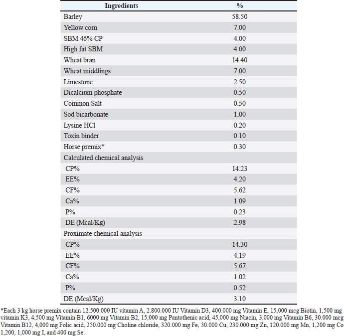
AbstractBackground: In equine practice, nutraceuticals are usually used to improve a horse’s performance. Aim: This study evaluated the influence of FERTISUPPORT M® given orally on follicular development in anestrous Arabian mares. Methods: Twenty anestrous straight Egyptian Arabian mares were used in the present study. These animals were divided randomly into two groups (ten mares each). Group 1 served as a control and was fed a basal diet only. Group 2 (supplemented group) was fed the basal diet supplemented with 50 ml of FERTISUPPORT M® according to manufacture recommendation that was given orally once daily for 21 days. Ovarian ultrasonography examination and assessment of blood indices, antioxidants, and hormonal status were conducted at days 0, 7, and 21 after dietary supplementation. Results: After 3 ± 0.5 days of treatment, ultrasound scanning revealed an increase in follicular size. On day 9 ± 1 of treatment, there was a large follicle depicted on the ovary with evidence of characteristic endometrial edema in the estrus phase. There were no significant differences between the treated and control groups regarding RBCs, HB, blood platelets, WBCs, eosinophils, segmented neutrophils, lymphocytes, monocytes, MCV, MCH, and MCHC (p > 0.05). While there were significant differences in hematocrit (p=0.046) and staff neutrophils count (p=0.011) between the treated and control groups. Moreover, the treated group had a statistically higher level of total antioxidants than the control group at different times (p=0.017). There were no significant differences between both groups at different times regarding T3, T4, progesterone, and estrogen levels (p > 0.05). Conclusion: FERTISUPPORT M® supplementation stimulates follicular development and increases the total antioxidant capacity in anestrous Arabian mares. Keywords: Antioxidants, Arabian mare, Follicular development, Nutraceuticals, Sex hormones. IntroductionArabian horses are known for their extraordinary beauty and scarcity; therefore, preserving their fertility is an important goal for preserving their lineages. (Marei, 2014; Derbala et al., 2025). Due to their seasonal polyestrous nature, most mares cycle during long daylight hours. Anestrus happens in the winter when there is less daylight. During anestrus, the ovaries are dormant with no large follicles >10 mm or corpora lutea; therefore, plasma levels of estrogen and progesterone remain low (Allen, 1985). Reproductive issues in horses are relatively prevalent, often resulting in low conception rates and financial setbacks. Both stallions and mares commonly experience infertility challenges (Giesecke et al., 2010). As mares age, their fertility declines; particularly in those aged 15 years and older, they may experience a reduction of 10%–20% in fertility each year (Koca et al., 2016). The decline in fertility in mares is influenced by multiple factors, including age, nutritional status, management practices, and reproductive disorders such as endometritis and anatomical abnormalities (Snider, 2015; Koca et al., 2016; Lopez, 2022; Derbala and Abu-Seida, 2024; Gil-Miranda et al., 2024). According to Schoster (2014) nutraceuticals are supplements added to horse feed with the goal of improving performance or health. Probiotics, vitamins, minerals, extra protein, energy, and herbal supplements are frequently among these options. In many cases, nutrition might help with fertility problems. As mentioned before, malnutrition is one of the main causes of horse infertility. In order to guarantee that every horse is fed a well-balanced diet that is high in nutrients and free of excesses or deficiencies, veterinarians and breeders should pay close attention to a horse's body weight and body condition score (BCS). Inadequate protein intake can cause irregular ovulation in mares (Martin-Rosset et al., 2006), and energy content is particularly important for mares that are ready to mate. Supplemented with vitamin E and vitamin C, the equine diet should be rich in macronutrients and micronutrients (Coenen, 2013). Significant advantages are also offered by antioxidants. Vitamins A and E are very important for fertility in mares (Parej et al., 2017). Nutritional status plays a crucial role in reproductive performance in mares. Adequate intake of energy, protein, vitamins, and minerals is essential for maintaining normal ovarian function and supporting fertility (Martin-Rosset et al., 2006; Coenen, 2013; Parej et al., 2017). FERTISUPPORT M® (Dana Vet Co., Egypt) is an innovative, first liquid nutraceutical supplement created especially to improve mare fertility in breeding programs through providing a combination of amino acids, vitamins, and fatty acids. According to the manufacturer, each liter of the product contains 309,000 mg L-carnitine, 101500 mg L-arginine, 150,000 mg omega 3 fatty acids, 123.76 mg chelated Cu, 8,180 mg chelated Se, 12,000 mg chelated zinc, 60 mg vitamin A, 5,559 mg vitamin E, 0.41 mg vitamin B12, 227 mg vitamin B6, 18,000 mg vitamin C, and 560 mg folic acid that enhance the oocyte’s metabolic process and the growth of the supplemented mare’s embryo (Zeyner and Harmeyer, 1999; Robles et al., 2019; Gobesso et al., 2020). Although several supplements are available to support equine fertility, their efficacy has not been fully validated under controlled conditions. Therefore, the present study aimed to evaluate the effect of oral supplementation with FERTISUPPORT M® on follicular development in anestrous Arabian mares. Materials and MethodsAnimalsThe current study was carried out in Giza province (Shubramant private equine stud farms), Egypt, during the period between 2023 and 2024. The study involved twenty straight Egyptian Arabian mares suffering from anestrum. The age of mares ranged between 3 and 18 years. Before the experiment, all mares were inspected twice, 10 days apart, using transrectal palpation and ultrasound (Sonoscape E1V adapted, China) to record the presence of ovarian structures and confirm anestrus. Mares were randomized to one of the two following groups (ten mares in each group) using a random number generator. Group 1 served as control was fed a basal diet formulated according to NRC (2007)as shown in Table 1. Table 1. Physical and chemical composition of the mare basal concentrated feed.
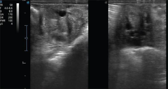
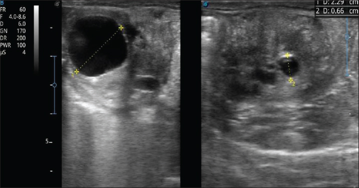
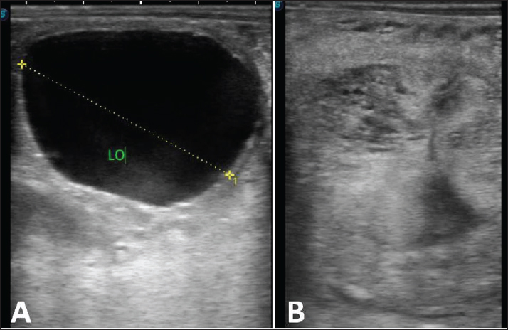
Group 2 was supplemented and was fed the basal diet supplemented with 50 ml of FERTISUPPORT M® (Dana Vet, Egypt) according to manufacturer recommendations given orally once daily for 21 days. Dietary manipulationAll mares were fed 2.0% of their body weight daily feed intake over a period of 21 days. Mares’ feeds were composed of 65% of daily feed intake of good-quality berseem hay and 35% concentrated mares’ feed. Animals were housed under sheds in semi-closed pens. Concentrate feed mixture was offered in two equal parts daily, and roughages were offered in between. Animals were given water free choice all day round. Ultrasonography assessmentTransrectal ultrasonography was performed three times per week (every 2–3 days) using a real-time ultrasound scanner (Sonoscape E1V, China). Follicular development was monitored by recording the number and diameter of ovarian follicles (Kwong and Klein, 2019). Examinations were performed by a blinded operator to avoid bias. Until follicular development resulted in the appearance of a follicle >35 mm in diameter along with mild to significant edema, therapy and blood sampling were continued for 14 days. To induce ovulation, 2500 IU of hCG (Chorulo; Merck, Toronto, Ontario) was given intramuscularly. About 500 million motile sperm from a single stallion with known fertility were used to artificially inseminate mares 24 hours after hCG was administered. The mares were then monitored every 24 hours until ovulation was detected. To determine if an embryonic vesicle was present or absent, transrectal ultrasonography was performed 12 and 14 days following ovulation (Derbala and Abu-Seida, 2024). Assessment of blood indicesAt days 0, 7, and 21 of the experiment, blood samples were taken from all mares. Complete blood count was performed according to standard hematological methods (Jain, 1993). Hemoglobin concentration was determined using standard spectrophotometric methods (Jain, 1993). A manual differential leukocyte count was performed by examining 100–200 leukocytes per blood smear and classifying them according to morphological characteristics. The percentage of each leukocyte type was determined and used to calculate absolute leukocyte counts. The hematocrit was performed manually by filling a capillary tube with blood, centrifuging it, and measuring the percentage of the blood that consisted of red blood cells. This was useful in some conditions that might cause automated hematocrit results to be incorrect. Assessment of antioxidant statusBlood was collected from the jugular vein and placed in a heparin tube at days 0, 7, and 21 of the experiment. Total antioxidant capacity mM/l (TAC), malondialdehyde nmol/ml (MDA), catalase U/l (CAT), and glutathione reductase U/l (GSH) levels were measured in plasma separated by centrifugation using the techniques of Koracevic et al. (2001); Ohkawa et al. (1979); Aebi (1984) and Beutler et al. (1963) respectively. Special, highly standardized kits from the bio-diagnostic kits in Egypt were used for all measurements. Assessment of hormones statusSerum T3, T4, progesterone, and estradiol were measured using an enzyme-linked fluorescent immunoassay (ELFA) with the VIDAS system (bioMérieux, France) according to the manufacturer’s instructions, as previously described (Derbala and Abu-Seida, 2024a,b). Statistical analysisAfter gathering data in an Excel spreadsheet, SPSS version 22 was used to analyze the impact of various intervention groups over three follow-up periods as independent variables on the quantitative variable, which was represented by mean ± SD. Pairwise comparisons for both study groups and study periods were then conducted after post hoc Bonferroni adjustment. p-value < 0.05 was considered significant. Ethical approvalAll experimental procedures in this study were approved by the Institutional Animal Care and Use Committee of the Faculty of Veterinary Medicine, Cairo University, Egypt (Approval Reference No.: Vet CU 08102051243). All methods were carried out in accordance with relevant guidelines and regulations, and the study was reported in compliance with the ARRIVE guidelines. ResultsUltrasonography findingsUltrasonography, the anestrus mares were detected. These mares showed no change in the follicular size, which was ≤10 mm in diameter after 10 days of the first examination, with absence of CL (Fig. 1). After the beginning of treatment, ultrasound scanning was carried out with a 3-day interval for detection of follicular changes. After 3 ± 0.5 days of treatment, there was an increase in the follicular size (Fig. 2). On day 9 ± 1 of treatment, there was a large follicle depicted on the ovary with evidence of characteristic endometrial edema in the estrus phase (Fig. 3). All treated mares in group 2 had an embryonic vesicle.
Fig. 1. Ultrasonographic images of both ovaries in a 7-year-old Arabian anestrous mare showing small follicles (<10 mm in diameter) with no CL.
Fig. 2. Ultrasonography images showing the beginning of the follicular growth (2.29 cm in diameter) in a 6-year-old Arabian mare after 3 days of treatment.
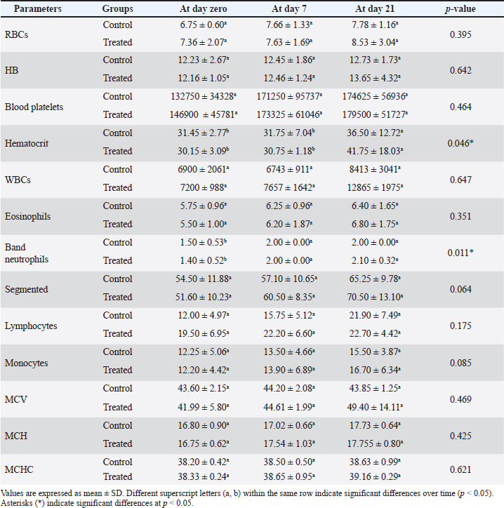
Fig. 3. (A) Ultrasonography image showing a large estrus follicle (>4 cm in diameter) in a 9-year-old Arabian mare after 10 days of treatment. (B) Ultrasonography image showing the characteristic rosette shape appearance of the endometrial folds of the uterine horn in the same mare. Results of blood indicesNo significant differences were observed between the treated and control groups in RBCs, hemoglobin (HB), platelets, total WBCs, eosinophils, segmented neutrophils, lymphocytes, monocytes, MCV, MCH, and MCHC at any time point (p > 0.05; Table 2). Table 2. Effect of FERTISUPPORT M® supplementation on hematology parameters (Mean ± SD) at different time intervals.
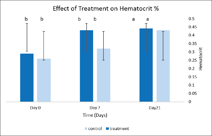
Hematocrit values significantly increased at day 21 compared to days 0 and 7 in both groups (p=0.046), with numerically higher values recorded in the treated group at day 21 compared to the control group (Table 2; Fig. 4).
Fig. 4. Effect of treatment on hematocrit percentage in control and treated groups at different time points (Day 0, 7, and 21). Data are presented as mean ± SD. Statistical analysis was performed using two-way ANOVA followed by Bonferroni post hoc test. Different superscripts indicate significant differences at p < 0.05
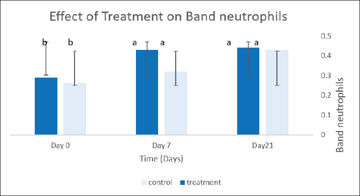
Fig. 5. Effect of treatment on band neutrophils in control and treated groups at different time points (Day 0, 7, and 21). Data are presented as mean ± SD. Statistical analysis was performed using two-way ANOVA followed by Bonferroni post hoc test. Different superscripts indicate significant differences at p < 0.05
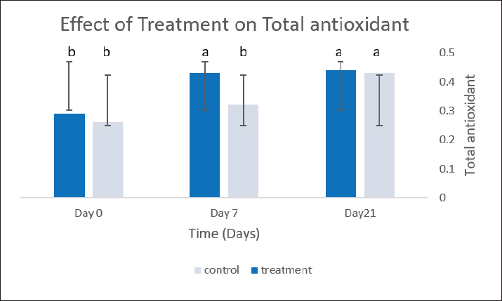
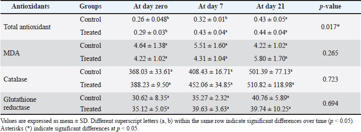
Fig. 6. Effect of treatment on total antioxidant (mM/l) in control and treated groups at different time points (Day 0, 7, and 21). Data are presented as mean ± SD. Statistical analysis was performed using two-way ANOVA followed by Bonferroni post hoc test. Different superscripts indicate significant differences at p < 0.05. Similarly, band neutrophils showed a significant increase at days 7 and 21 compared to day 0 in both groups (p=0.011; Table 2), with no significant differences between treated and control groups at corresponding time points. Regarding temporal changes, lymphocyte counts increased progressively over time, with a statistically significant effect of time (p=0.007), whereas all other hematological parameters remained unchanged throughout the experimental period (p > 0.05; Table 2). Results of antioxidant statusTotal antioxidant capacity (mM/l) was significantly higher in the treated group compared to the control group throughout the experimental period (p=0.017; Table 3). In addition, TAC levels increased over time in both groups, reaching the highest values at day 21 (p < 0.001). Table 3. Effect of FERTISUPPORT M ® supplementation on antioxidant parameters (Mean ± SD) at different time intervals.
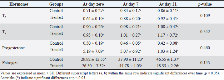
No significant differences were observed between treated and control groups in MDA, catalase (CAT), and glutathione reductase (GSH) levels at any time point (p > 0.05). With respect to temporal variation, catalase activity increased over time, showing a significant effect of time (p=0.010), whereas MDA and GSH levels did not show significant changes during the experimental period (p > 0.05; Table 3). Results of hormones statusNo significant differences were detected between treated and control groups in serum T3, T4, progesterone, or estrogen levels at any time point (p > 0.05; Table 4). Similarly, T3, T4, and progesterone levels did not show significant changes over time (p > 0.05). On the contrary, estrogen levels increased over time in both groups, with a significant effect of time (p=0.001), reaching higher values at day 21 compared to baseline (Table 4). Table 4. Effect of FERTISUPPORT M ® supplementation on hormone levels (Mean ± SD) at different time intervals.
DiscussionNutraceuticals are increasingly used in equine practice due to their potential to support physiological functions beyond basic nutrition, particularly in reproduction and athletic performance (Vervuert and Stratton-Phelps, 2021). In the present study, supplementation with FERTISUPPORT M® was associated with enhanced follicular development in anestrous Arabian mares, supporting its potential role as a reproductive modulator. The observed improvement in follicular growth may be attributed to the synergistic effects of its bioactive components. L-carnitine plays a crucial role in mitochondrial fatty acid transport and energy production, which are essential for oocyte maturation and follicular activity. In addition, its antioxidant properties may protect ovarian cells from oxidative stress, thereby improving follicular viability. Arginine, as a precursor of nitric oxide, likely enhances ovarian blood flow, facilitating nutrient and hormone delivery to developing follicles. Furthermore, omega-3 fatty acids are known to modulate inflammatory responses and improve endometrial and ovarian function, which could indirectly support follicular development. Interestingly, the stimulatory effect on follicular development was observed after approximately 9 days of supplementation, suggesting a relatively rapid physiological response. This may indicate that the mechanism of action is more closely related to improving metabolic and vascular support rather than direct endocrine modulation. Despite the improvement in follicular dynamics, no significant changes were detected in T3, T4, estrogen, or progesterone levels. This finding suggests that the effect of FERTISUPPORT M® is likely mediated through local ovarian mechanisms rather than systemic hormonal alterations. Similar observations were reported by Porto et al. (2023) indicating that such nutraceutical components may enhance reproductive performance without significantly altering circulating hormone levels. Hematological parameters remained within normal physiological ranges in both groups, confirming the general health status of the animals and the safety of the supplementation protocol. Although previous studies (Yessenbayeva et al., 2023) reported increases in hematological indices following dietary supplementation, the lack of significant changes in the present study may be attributed to differences in supplement composition, duration of administration, or baseline physiological status of the mares. The significant increase in total antioxidant capacity (TAC) in treated mares highlights the role of oxidative balance in reproductive efficiency. Enhanced antioxidant status may protect follicular cells from oxidative damage, thereby improving follicular development and function. This is consistent with previous findings indicating the importance of metabolic and oxidative support in equine reproduction (Fielding et al., 2018). ConclusionIt is important to note that the current study has some limitations, including the relatively small sample size and short duration of supplementation. Therefore, further studies involving larger populations and extended treatment periods are warranted to confirm these findings and to better elucidate the underlying mechanisms of action. FERTISUPPORT M® supplementation stimulates follicular development and increases the total antioxidant capacity in anestrous Arabian mares. AcknowledgmentsNone. FundingThis research did not receive any funding from any agency. Conflict of interestsAll authors declare no competing interests. Authors’ contributionsConceptualization, project management, resources, supervision, methodology, investigation, writing, review, and editing were all completed by K. N. F. Formal analysis, data curation, investigation, methodology, software, visualization, writing—original draft, writing—review, and editing were all done by M. A. The formal analysis, data curation, investigation, methodology, software, visualization, and writing of the first draft were completed by M. K.D. Formal analysis, data curation, research, technique, software, and writing—original draft—were created by E. S. H. Data availabilityAll data generated or analyzed during this study are included in this published article. ReferencesAebi, H. 1984. Catalase in vitro. Methods. Enzymology. 105(84), 121–126. Allen, W.R. 1985. Practical control of anoestrus in thoroughbred broodmares. In: Current Topics in Veterinary Medicine and Animal Science. Eds., Ellendorff, F. and Elsaesser, F. Dordrecht, The Netherlands: Springer, Vol. 31. Beutler, E., Duron, O. and Kelly, B.M. 1963. Improved method for the determination of blood glutathione. J. Lab. Clin. Med. 61, 882–888. Coenen, M. 2013. Macro and trace elements in equine nutrition. Equine Appl. Clin. Nutr. 190–228; doi: 10.1016/B978-0-7020-3422-0.00010-9 Derbala, M. and Abu-Seida, A. 2024. Endoscopic laser ablation of endometrial cysts in Straight Egyptian Arabian mares: outcome on embryo recovery and conception rate. Egypt. J. Vet. Sci. 55(2), 371–380; doi:10.21608/EJVS.2023.232922.1589 Derbala, M.K., Hassan, E.A. and Abu-Seida, A.M. 2025. Outcome following standing flank laparotomy for treatment of uterine torsion in 87 Arabian mares. J. Equine Vet. Educ. 37(11), 580–586; doi:10.1111/eve.14076 Fielding, R., Riede, L., Lugo, J. and Bellamine, A. 2018. L-carnitine supplementation in recovery after exercise. Nutrients 10(3), 349; doi:10.3390/nu10030349 Giesecke, K., Sieme, K.H. and Distl, O. 2010. Infertility and candidate gene markers for fertility in stallions: a review. Vet. J. 185(3), 265–271. Gil-Miranda, A., Macnicol, J., Orellana-Guerrero, D., Samper, J.C. and Gomez, D.E. 2024. Reproductive tract microbiota of mares. Vet. Sci. 11(7), 324. Gobesso, A.A.D.O., Mazzo, H.C., Bianconi, C., Freitas, F.V., Pombo, G.D.V., Pereira, Y.S., Centini, T.N., Françoso, R. and Balieiro, J.C.D.C. 2020. The effect of supplementation with omega-3 and 6 fatty acids to mares during late gestation and early lactation on the transfer of passive immunity in foals. Livest. Sci. 237, 104072; doi:10.1016/j.livsci.2020.104072 Jain, N.C. 1993. Essentials of veterinary hematology. Philadelphia, PA: Lea & Febiger. Koca, E., Özenç, E., Şeker, E., Sevimli, A., Vural, M.R., Uçar, M. and Küplülü, S. 2016. Evaluation of the reproductive and metabolic problems encountered during two breeding seasons in barren Arabian mares. Turkish J. Vet. Anim. Sci. 40(1), 60–67. Koracevic, D., Koracevic, G., Djordjevic, V., Andrejevic, S. and Cosic, V. 2001. Method for the measurement of antioxidant activity in human fluids. J. Clin. Pathol. 54(5), 356–361; doi:10.1136/jcp.54.5.356 Kwong, G.P.S. and Klein, C. 2019. Deslorelin and naltrexone stimulate follicular development in mares during autumn transition and early anestrus. Can. Vet. J. 60(8), 855–858. Lopez, J. 2022. Breeding the problem mare. PetersonSmith Equine Hospital + Complete Care. https://petersonsmith.com/breeding-the-problem-mare/Accessed at 6/9/2025. Marei, N., 2014. The Arabian Horse of Egypt. Liveright Publisher, 90-110. Martin-Rosset, W., Vervuert, I. and Austbo, D. 2006. Energy and protein requirements and recommended allowances in pregnant mares. Nutrition and Feeding of the Broodmare, 13–39. National Research Council. 2007. Division on Earth, Life Studies, Institute for Laboratory Animal Research, Committee for the Update of the Guide for the Care and Use of Laboratory Animals, 2024.. Ohkawa, H., Ohishi, N. and Yagi, K. 1979. Assay for lipid peroxides in animal tissues by thiobarbituric acid reaction. Analyt. Biochem. 95(2), 351–358; doi:10.1016/0003-2697(79)90738-3 Parej, J., Pongracz, L. and Papp, A.B. 2017. Study of vitamin A, vitamin E and selenium supply of Thoroughbred mares. Cabidigitallibrary.org. Porto, A.C.R.C., Redoan, M.A., Massoco, C.O., Furtado, P.V. and Oliveira, C.A. 2023. Additional effects using progestins in mares on levels of thyroid hormones and steroids in neonates. Anim. Reprod. 20(4), e20230029; doi:10.1590/1984-3143-AR2023-0029 Robles, M., Couturier-Tarrade, A., Derisoud, E., Geeverding, A., Dubois, C., Dahirel, M., Aioun, J., Prezelin, A., Calvez, J., Richard, C., Wimel, L. and Chavatte-Palmer, P. 2019. Effects of dietary arginine supplementation in pregnant mares on maternal metabolism, placental structure and function and foal growth. Sci. Rep. 9(1), 6461; doi:10.1038/s41598-019-42941-0 Schoster, A. 2014. Probiotics and dietary supplements in horses: useful or not? Zurich: University of Zurich. Snider. 2015. Reproductive disorders in horses. Vet. Clin. Eq. Pract. 31(2), 389–405. Vervuert, I. and Stratton-Phelps, M. 2021. The safety and efficacy in horses of certain nutraceuticals that claim to have health benefits. Vet. Clin. North Amer. Eq. Pract. 37(1), 207–222; doi:10.1016/j.cveq.2020.11.002 Yessenbayeva, Z., Akbasova, A.D. and Yuldashbek, D.H. 2023. Study of the effect of protein-vitamin-mineral feed additives on the increase in live weight of horses. Gylym Zane Bilim 4(73), 28–35. Zeyner, A. and Harmeyer, J. 1999. Metabolic functions of L‐Carnitine and its effects as feed additive in horses. A review. Archiv Für Tierernaehrung. 52(2), 115–138; doi:10.1080/17450399909386157 Values are expressed as mean ± SD. Different superscript letters (a, b) within the same row indicate significant differences over time (p < 0.05). Asterisks (*) indicate significant differences at p < 0.05. | ||
| How to Cite this Article |
| Pubmed Style Abdulredha MAH, Elmosalamy SH, Derbala MK, Fahmy KNE. Potential effects of nutraceutical supplementation on follicular development and antioxidant capacity in anestrous Arabian mares. Open Vet. J.. 2026; 16(7): 4952-4961. doi:10.5455/OVJ.2026.v16.i7.72 Web Style Abdulredha MAH, Elmosalamy SH, Derbala MK, Fahmy KNE. Potential effects of nutraceutical supplementation on follicular development and antioxidant capacity in anestrous Arabian mares. https://www.openveterinaryjournal.com/?mno=310788 [Access: July 27, 2026]. doi:10.5455/OVJ.2026.v16.i7.72 AMA (American Medical Association) Style Abdulredha MAH, Elmosalamy SH, Derbala MK, Fahmy KNE. Potential effects of nutraceutical supplementation on follicular development and antioxidant capacity in anestrous Arabian mares. Open Vet. J.. 2026; 16(7): 4952-4961. doi:10.5455/OVJ.2026.v16.i7.72 Vancouver/ICMJE Style Abdulredha MAH, Elmosalamy SH, Derbala MK, Fahmy KNE. Potential effects of nutraceutical supplementation on follicular development and antioxidant capacity in anestrous Arabian mares. Open Vet. J.. (2026), [cited July 27, 2026]; 16(7): 4952-4961. doi:10.5455/OVJ.2026.v16.i7.72 Harvard Style Abdulredha, M. A. H., Elmosalamy, . S. H., Derbala, . M. K. & Fahmy, . K. N. E. (2026) Potential effects of nutraceutical supplementation on follicular development and antioxidant capacity in anestrous Arabian mares. Open Vet. J., 16 (7), 4952-4961. doi:10.5455/OVJ.2026.v16.i7.72 Turabian Style Abdulredha, Mohammad A. H., Sherif Hamed Elmosalamy, Mohamed K. Derbala, and Khaled Nasr Eldin Fahmy. 2026. Potential effects of nutraceutical supplementation on follicular development and antioxidant capacity in anestrous Arabian mares. Open Veterinary Journal, 16 (7), 4952-4961. doi:10.5455/OVJ.2026.v16.i7.72 Chicago Style Abdulredha, Mohammad A. H., Sherif Hamed Elmosalamy, Mohamed K. Derbala, and Khaled Nasr Eldin Fahmy. "Potential effects of nutraceutical supplementation on follicular development and antioxidant capacity in anestrous Arabian mares." Open Veterinary Journal 16 (2026), 4952-4961. doi:10.5455/OVJ.2026.v16.i7.72 MLA (The Modern Language Association) Style Abdulredha, Mohammad A. H., Sherif Hamed Elmosalamy, Mohamed K. Derbala, and Khaled Nasr Eldin Fahmy. "Potential effects of nutraceutical supplementation on follicular development and antioxidant capacity in anestrous Arabian mares." Open Veterinary Journal 16.7 (2026), 4952-4961. Print. doi:10.5455/OVJ.2026.v16.i7.72 APA (American Psychological Association) Style Abdulredha, M. A. H., Elmosalamy, . S. H., Derbala, . M. K. & Fahmy, . K. N. E. (2026) Potential effects of nutraceutical supplementation on follicular development and antioxidant capacity in anestrous Arabian mares. Open Veterinary Journal, 16 (7), 4952-4961. doi:10.5455/OVJ.2026.v16.i7.72 |


