





| Research Article | ||
Open Vet. J.. 2026; 16(7): 4427-4440 Open Veterinary Journal, (2026), Vol. 16(7): 4427-4440 Research Article Molecular identification of Tribolium castaneum using SYBR green quantitative PCR and evaluation of Silybum marianum seed extract-mediated silver nanoparticles as a biopesticideBanaz Sdiq Abdulla*Department of Biology, College of Education, Salahaddin University-Erbil, Iraq *Corresponding Author: Banaz Sdiq Abdulla. Department of Biology, College of Education, Salahaddin University-Erbil, Iraq. Email: banaz.abdulla [at] su.edu.krd Submitted: 22/02/2026 Revised: 03/06/2026 Accepted: 16/06/2026 Published: 11/07/2026 © 2025 Open Veterinary Journal
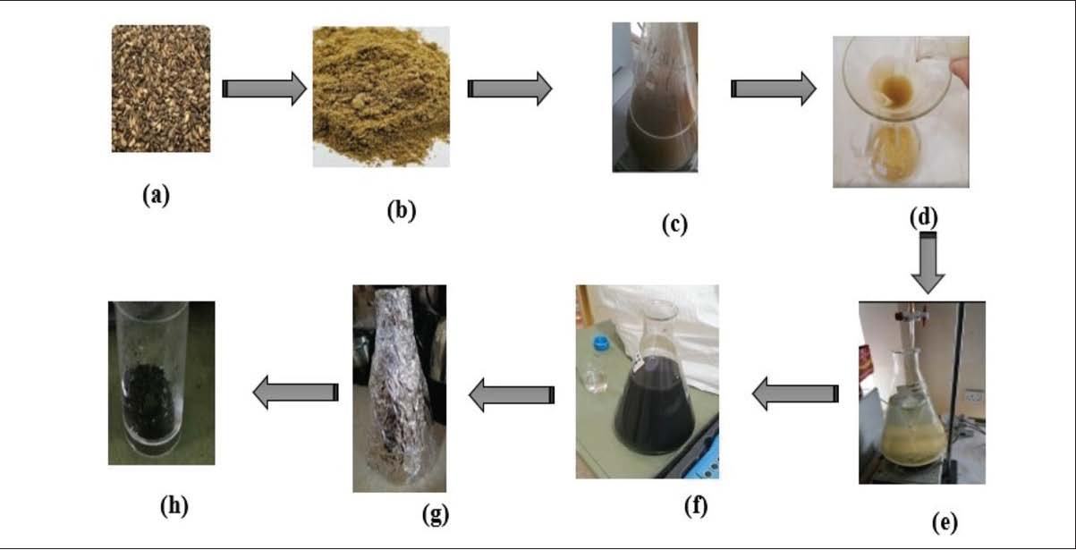
ABSTRACTBackground: Tribolium castaneum Herbst (Coleoptera: Tenebrionidae) causes economic damage to stored foods, especially wheat, and is controlled by synthetic insecticides. Synthetic pesticides work but are expensive, non-biodegradable, pesticide-resistant, and harmful to the environment. Green-synthesized nanoparticles may improve pesticides. Traditionally, T. castaneum is identified by adult morphology. Pest management programs still struggle to identify this species quickly and accurately. Aim: Using species-specific PCR and real-time quantitative PCR with SYBR Green dye. The second purpose was to produce milk thistle seed AgNPs and test their bioinsecticide efficacy against T. castaneum. Methods: To identify T. castaneum, real-time qPCR was used with specific primers for the mt-COI gene fragment. Production and evaluation of silver nanoparticles synthesized from aqueous extract of Silybum marianum seed (SM-AgNPs) against the red flour beetle. The synthesized AgNPs were characterized. In the bioassay of biopesticides, SM-AgNPs were added to flour at 0.75, 1.5, 3, and 6 mg/g to evaluate their larvicidal (2nd and 5th larval instar) and adulticidal activities on T. castaneum. Mortality was reported at 24, 48, 72, and 96 hours following application. Results: PCR amplified a 330-bp COI gene fragment, and mtCOI gene qPCR identified T. castaneum efficiently, quickly, and species-specifically. The synthesized SM-AgNPs were characterized. FTIR spectra showed milk thistle functional groups. SEM showed circular to oval particles. The UV-Vis of SM-AgNPs showed significant absorption bands at 408 nm. AgNP particles averaged 119 nm and had a zeta potential of −48 mV. The bioassay results demonstrated significant dose- and time-dependent larvicidal and adulticidal effects of SM-AgNPs (p < 0.05). Second-instar larvae were most responsive to SM-AgNPs, while adults were least. At a concentration of 0.75 mg/g, SM-AgNPs showed the lowest mortality rate (5.6%) in adult red flour beetles after 24 hours of exposure. On the contrary, the highest mortality rate (70%) was observed in second-instar larvae after the fourth day of exposure to the highest concentration of SM-AgNPs (6 mg/g). Conclusion: SYBR Green real-time qPCR identified T. castaneum rapidly and accurately. The mortality rates at different nanoparticle concentrations and exposure times showed that SM-AgNPs could be an eco-friendly and effective biological control approach for this pest species. Keywords: AgNPs, Bioinsecticide, Milk thistle, qPCR, Tribolium castaneum. IntroductionThe red flour beetle is a common and important insect pest that infests stored grain products globally. Adult and larval beetles can infest cereal grains, dried fruits, pulses, husk, germ, grain dust, and processed cereal products (Papanikolaou et al., 2022). The phenotypic resemblance of adult and non-adult flour beetles makes morphological identification difficult. Tribolium species share many physical characteristics, making typical methods unreliable for species identification. An alternative to taxonomy, this method is a DNA-based approach, which is the only technique that can achieve this result. Quantitative PCR (qPCR) uses a fluorescent dye that intercalates into double-stranded DNA to detect, identify, and quantify specific DNA sequences with high sensitivity. The utilization of qPCR for the detection of stored insect pests has been documented for numerous species. Real-time qPCR TaqMan probes targeting mitochondrial COI gene areas can rapidly recognize any pest, as demonstrated by Zhang et al. (2016). Specific reaction conditions now enable the identification of several species, including storage pests such as Tribolium, with high sensitivity, precision, and quantitative accuracy. Synthetic insecticides have been used to control these pests. However, they have major disadvantages, including damage to ecosystems, non-target species, environmental persistence, insect resistance, and health risks. Agriculture workers account for about 75% of pesticide poisonings owing to improper use (Sindhu et al., 2022). The use of synthetic pesticides without discrimination leads to the development of insect resistance, and therefore the necessity to discover other pest control strategies. There is a need for pest control with eco-friendly and cost-effective solutions such as the use of nanoparticles (Kannan et al., 2023). Nanoparticles have generated a lot of interest in the use of bio–nano insecticides in agriculture. They are 1–100 nm in size; they are around 103 nm smaller than bacterial cells and 100 nm smaller than viral cells. Materials with smaller sizes are more reactive and efficient than larger ones. Due to the high surface area to volume ratio of nanoscale materials, molecular collisions become more frequent (Karimi-Maleh et al., 2024). Nanomaterials provide significant progress in the control of agricultural pests. Biopesticides derived from plant extracts and nanomaterials are an effective and safe alternative to traditional insecticides (Qiu et al., 2023). Therefore, nanotechnology has evolved as an important tool to tackle this problem efficiently (Iqbal et al., 2024). The synthesis of nanomaterials using plant extracts has been extensively studied, using metals such as copper, zinc, titanium/nickel, magnesium, gold, and silver. Silver nanoparticles (AgNPs) have gained much attention owing to their minimal toxicity in humans (Jaswal and Gupta, 2023). Silver nanoparticles are used more in food, pharmaceuticals, healthcare, industry, and agriculture. Due to their large surface area, biocompatibility, simplicity of modification, and ability to penetrate target cells, silver nanoparticles are ideal for drug administration (Xu et al., 2020). Medicinal herb milk thistle (Silybum marianum L., Asterales: Asteraceae) assists in detoxification (Bjørklund et al., 2024). The flavonolignans found in extracts of milk thistle seeds, in particular silymarin, are physiologically active. The most pharmacologically active flavonolignan in silymarin is silybin, followed by isosilybin, silydianin, and silychristin. It is a small molecule and structurally resistant. It is highly functionalized. Silymarin provides various therapeutic effects such as immunomodulatory, anti-inflammatory, and antifibrotic activities (Erfanian et al., 2024). The primary aim of this study was to identify T. castaneum using a real-time qPCR method by amplifying a specific segment of the mtCOI gene. The secondary purpose was to produce silver nanoparticles (AgNPs) by a green synthesis method utilizing milk thistle seed extract. The third objective was to characterize the produced nanoparticles by UV-visible spectroscopy, XRD, FTIR, SEM, TEM, DLS, and zeta potential, and finally, to evaluate the biopesticide activity of these AgNPs against T. castaneum. Materials and MethodsInsect collecting and rearingRed flour beetles were collected from infested flour at some stores in the Erbil Governorate in the Kurdistan Region of Iraq. A mixture consisting of 95% wheat flour and 5% dry yeast was cultured on a wheat flour medium. It was preserved in a 300-ml glass jar, closed with a tulle cloth. Insect cultures were maintained in a controlled setting at 28°C ± 2°C and 70% ± 5% humidity. A study was performed to distinguish instars and acquire the necessary instar larvae and adults for each experiment. Adults were enclosed in a container with a camel-hair brush for two days; the containers were then removed, allowing the eggs to incubate. Larvae were maintained in dark settings at a temperature of 28°C ± 2°C in plastic Petri dishes measuring 9 × 1.6 cm, which were covered with tulle. Fresh flour was provided as required to nourish the larvae in the Petri plates. Experiments were conducted using adults, second- and fifth-instar larvae (Al-Fatlawy and Al-Zurfi, 2023). Molecular identification of T. castaneumDNA extraction, PCR amplification of the COI region, real-time qPCR, and COI sequencing: The Jena Bioscience animal DNA preparation kit was used to extract whole-genomic DNA from the heads and thoraxes of adult T. castaneum, according to the manufacturer’s protocol (Aslam et al., 2019). For COI amplification, a pair of targeted primers, forward Tca33F26 (5′- GAATAGTAGGCACTTCATTAAGACTC-3′) and reverse Tca346R24 (5′- CCATGTGCAATGTTTGATGAGAGG-3′), was used, as Zhang et al. (2016) had previously designed. The PCR amplification process required a 25-μl reaction volume, which comprised 0.5 μl of each primer (1 μM), 1.5 μl of extracted DNA (about 20 ng/μl), 10 μl of distilled water that had been sterilized, and 12.5 μl of master mix with loading dye. The polymerase chain reaction (PCR) process included an initial denaturation step of 3 minutes at 94°C, 35 cycles of 1 minute each at 94°C, 52°C, and 72°C, and a final extension step of 10 minutes at 72°C. During the reactions, a Veriti™ 96-well Thermal Cycler from ABI (USA) was used. A 1.0% (w/v) agarose gel in a 1× Tris acetate–EDTA solution was used to separate the PCR products. After that, the gel was stained with ethidium bromide and viewed under a UV lamp. PCR tests were carried out, and the SYBR Green technique was used to perform quantitative PCR on the COI gene in conformity with the manufacturer's instructions (Add Script qPCR SYBR Master (2× conc.), Korea). The same specific primers that go forward and backward for COI Tca33F26 (5′- GAATAGTAGGCACTTCATTAAGACTC-3′) and reverse Tca346R24 (5′- CCATGTGCAATGTTTGATGAGAGG-3′) were used in the qPCR reaction. The following components were used in each qPCR reaction: 13 µl of SYBR Green PCR master mix, 9 µl of PCR-grade water, 0.5 µl of each forward and reverse primer (100 pmol/µl), and 2 µl of the DNA template (RT+/RT−/water/ sample/control). The total volume of the mixture was 25 µl. At Zheen International Hospital/EXO-GENE for Genetic Diagnosis (Erbil, Iraq), an automated sequencer was utilized to retrieve the sample sequences. After the validation of PCR results on an agarose gel, gel extraction was conducted using the Zymoclean gel DNA recovery kit. Bidirectional sequencing of isolated fragments was performed using the Nimagen Brilliant Dye terminator cycle sequencing kit V3.1 (BRD3-100/1000) and subsequently purified with the Zymo Research ZR-96 DNA sequencing cleanup kit. The purified fragments were analyzed using the ABI 3500 ? l genetic analyzer for each reaction of every sequenced sample. Sequencing was performed in both forward and backward directions. Biosynthesis of the green-synthesized silver nanoparticlesSeeds of the S. marianum plant were bought from the local market. Preparation of milk thistle seed extracts was done according to the following method of Mohammadinejad et al. (2013) with modest adjustments. Twenty grams of dry seeds of S. marianum were washed several times with distilled water to remove dust. The seeds were ground. Distilled water was added to a final volume of 200 ml, maintaining the ratio of extract to water (w/v) at 1:10. After boiling for 2 hours at 100°C, the mixture was combined with 10 ml of milk thistle seed extract and 90 ml of AgNO3 solution (1.0 mM) It was incubated at room temperature for 24–48 hours. The filtered extract was then combined with seeds at 1:5 (v/v) containing 1-mM AgNO3. The mixture of AgNO3 solution and plant extract was then stored in an airtight bottle, away from direct sunlight. Absorbance was recorded by UV-visible spectrophotometry after 24 hours. The reaction mixtures were left at room temperature for 72 hours, and the complete reduction of all Ag ions into AgNPs was observed. The shift in color of the solution from pale brown to deep reddish-brown was observed due to the reduction of silver ions to generate silver nanoparticles. After the synthesis was completed, the mixture was centrifuged to extract the AgNPs. The AgNPs produced were purified using centrifugation at a speed of 15,000 rpm for 15 minutes for this. The air-dried semi-solid AgNPs were collected as powder (Fig. 1). The synthesized AgNPs were submitted to the Scientific Research Center at Soran University for different characterizations.
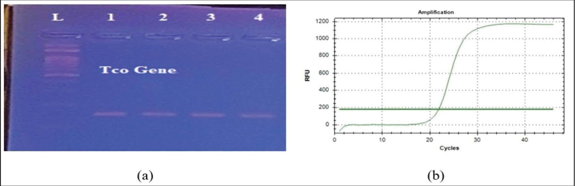
Fig. 1. Preparation of S. marianum milk thistle silver nanoparticles (SM-AgNPs): (a) Seeds of milk thistle; (b) Fine powder of milk thistle seeds; (c) Aqueous seed extract solution of milk thistle before filtration; (d) Aqueous seed extract of milk thistle after filtration; (e) Adding of milk thistle solution to AgNO3 solution by buriet; (f) Nanoparticle solution; (g) Nanoparticle solution incubation; (h) Nanoparticle powder. Characterization of the green synthesis silver nanoparticlesThe synthesis of AgNPs from S. marianum plant seed extract was initially seen by the naked eye as a color shift. Then, UV-Vis spectroscopy was again carried out to characterize the prepared AgNPs and to determine the absorbance peak related to their surface plasmon resonance. Fourier transform infrared spectroscopy (FTIR) was used to determine the organic components in the sample. Scanning electron microscopy (SEM) and transmission electron microscopy (TEM) were used to determine the size and form of AgNPs. The size and zeta potential of the AgNPs from milk thistle were characterized by dynamic light scattering (Shah et al., 2020). Biopesticidal effect of green-synthesized Ag-NPs on T. castaneumThe bioinsecticidal activity of synthetic AgNPs prepared from S. marianum seed extract was evaluated against the second and fifth larval instars and adult stages of red-flour beetles at the Department of Biology, Salaheddin University-Erbil. This evaluation was done at several concentrations (0.75, 1.5, 3, and 6 mg). The bioassay was conducted at a temperature of 28°C ± 2°C and relative humidity of 70% ± 5% (Jahan and Rasheed, 2023). Each jar contained 20.0 g of flour and was treated with varied concentrations of milk thistle nanoparticles. The jar was shaken for about 10 minutes to ensure that the nanoparticles were evenly distributed throughout the flour (Al-Fatlawy and Al-Zurfi, 2023). To evaluate the bioinsecticidal efficacy of synthesized AgNPs derived from S. marianum, for each treatment, ten insects of the corresponding stages were placed in Petri dishes containing flour that had been treated with nanocomposites and adequately covered. Although the control group did not receive any nanoparticles combined with flour, there were four distinct concentrations of milk thistle nanoparticles that were evaluated. Each of the treatments consisted of a total of thirty insects, with three replicates for each treatment. Following treatment, mortality was recorded at 1, 2, 3, and 4 days for both the adult and larval stages of T.castaneum. The mortality correction was performed utilizing Abbott's formula as follows: Mc=(Mo-Me)/(100-Me) X 100 Mo=Death rate of treated adults or larvae as observed (%) Me=Controlled mortality rate (%) Mc=Ratio of corrected deaths (%) Statistical data analysisData were analyzed using one-way analysis of variance (ANOVA). Duncan's multiple range test was employed to statistically compare the biological control treatments. Differences were statistically significant at p < 0.05 with a 95% confidence interval. The insecticidal activity was assessed using the lethal concentration 50 (LC50): the concentration that kills 50% of the tested population of T. castaneum adult stages and larval stages in the second and fifth instars. The LC50 values were calculated using GraphPad Prism version 10.4.1. Ethical approvalNot needed for this study. ResultsMolecular identification of T. castaneumMolecular biology techniques are increasingly being used to identify species. This work aims to use a sensitive, species-specific molecular technique for rapid and precise detection of the red flour beetle. A real-time qPCR test with SYBR Green was used to detect T. castaneum efficiently and rapidly. PCR amplification was performed using a particular primer against the target mitochondrial cytochrome oxidase I gene and specifically distinguished T. castaneum from T. confusum. The analysis of the PCR results on an agarose gel confirmed the existence of the specified target at 330 bp (Fig. 2a). The amplification plots of qPCR of T. castaneum were made utilizing the changes in fluorescence against the cycle number. The specific PCR exhibited one major product peak at 21 (Fig. 2b), whereas the negative control did not show any peak. In qPCR, the fluorescence is measured once each cycle after the product extension. Adult morphological traits are frequently employed to distinguish Tribolium species. However, it is almost impossible to rapidly distinguish between adult pieces and non-adult phases using external morphological characteristics. Particularly for pests of stored commodities, molecular techniques are required for rapid and accurate identification of flour-beetle species. Compared to physical characteristics, analyzing amplified DNA sequences from animals provides a more thorough means of identification.
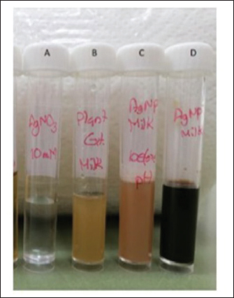
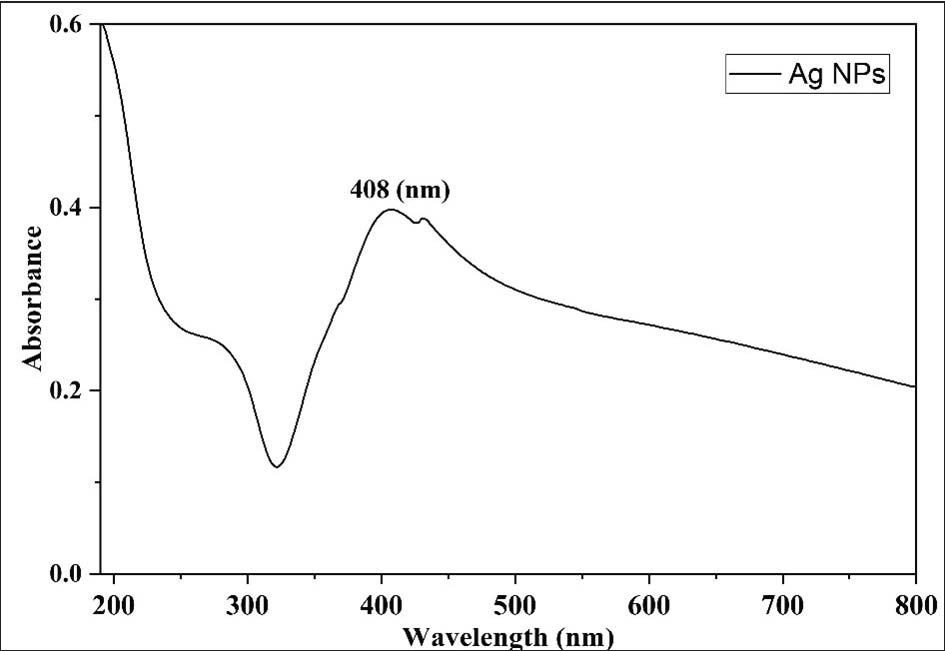
Fig. 2. This figure depicts (a) PCR amplification of T. castaneum with the mitochondrial cytochrome oxidase subunit 1 gene using a particular primer, L. ladder (DNA marker 100 bp). (b) qPCR showing the amplification curve of T. castaneum with mitochondrial cytochrome oxidase. Green synthesis of silver nanoparticles and UV anlysisThe synthesis of AgNPs from aqueous seed extracts of S. marianum was observed using color shift and UV-VIS spectra. The AgNPs were formed by the reduction of silver ions after 30 minutes of reaction within a time frame of 24–48 hours. When the silver nitrate and water-based extract solutions were combined, the hue changed quickly. The color change observed during synthesis utilizing milk thistle seed (Fig. 3) confirmed the creation of silver nanoparticles in the present investigation. Then, the silver nanoparticles were characterized by UV-Vis spectroscopy; the AgNPs solution absorption spectrum showed a sharp surface plasmon resonance peak at 408 nm (Fig. 4). The apparent peak shows silver reduction. The aqueous extract of S. marianum seeds displayed appreciable reducing activity in the reduction of Ag⁺ ions to Ag⁰. Bioreduction is the process in which metal ions or oxides are converted into nanoparticles of zero-valent metal.
Fig. 3. This figure depicts a change in color that occurs during the green production of silver nanoparticles utilizing seed extract from S. marianum. (a) AgNO3 alone; (b) S. marianum seed extract; and (c) combining silymarin seed extract with AgNO3. After 30 minutes, the white hue of AgNO3 instantly turns yellow. D: After 24 hours, a dark brownish hue indicates the production of S. marianum AgNPs.
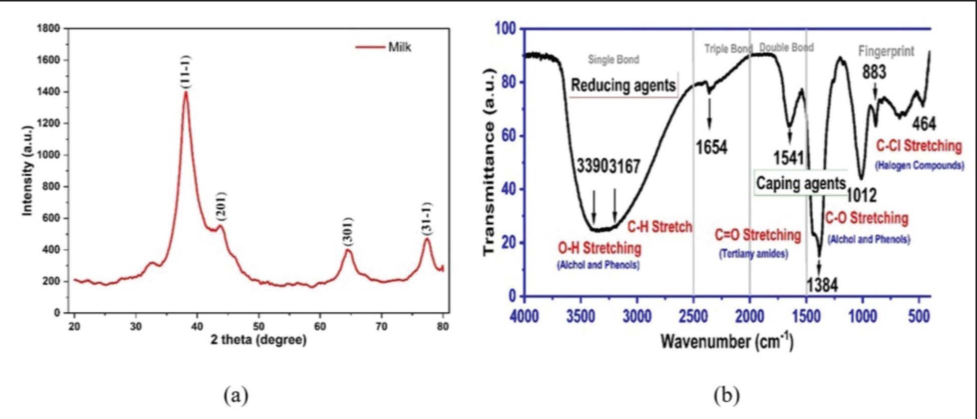
Fig. 4. This figure depicts UV-Vis absorption spectra of AgNPs produced using seed extract from Silybum marianum. Characterization of the green synthesis of silver nanoparticlesX-Ray diffraction (XRD)Nanoparticles composed of a crystalline material can be characterized using XRD, a rapid analytical method that also provides information on unit-cell dimensions. In order to confirm the crystalline nature of seed milk-thistle-mediated Ag-NPs, XRD analysis was performed. The diffraction pattern of the biosynthesized silver showed distinct peaks (Fig. 5a), confirming their crystalline structure. Prominent diffraction peaks were observed at 2θ values of 38.4°, 43.3°, 63.4°, and 77.0°, corresponding to the (111), (202), (220), and (311) crystallographic planes of face-centered cubic (FCC) silver, respectively.
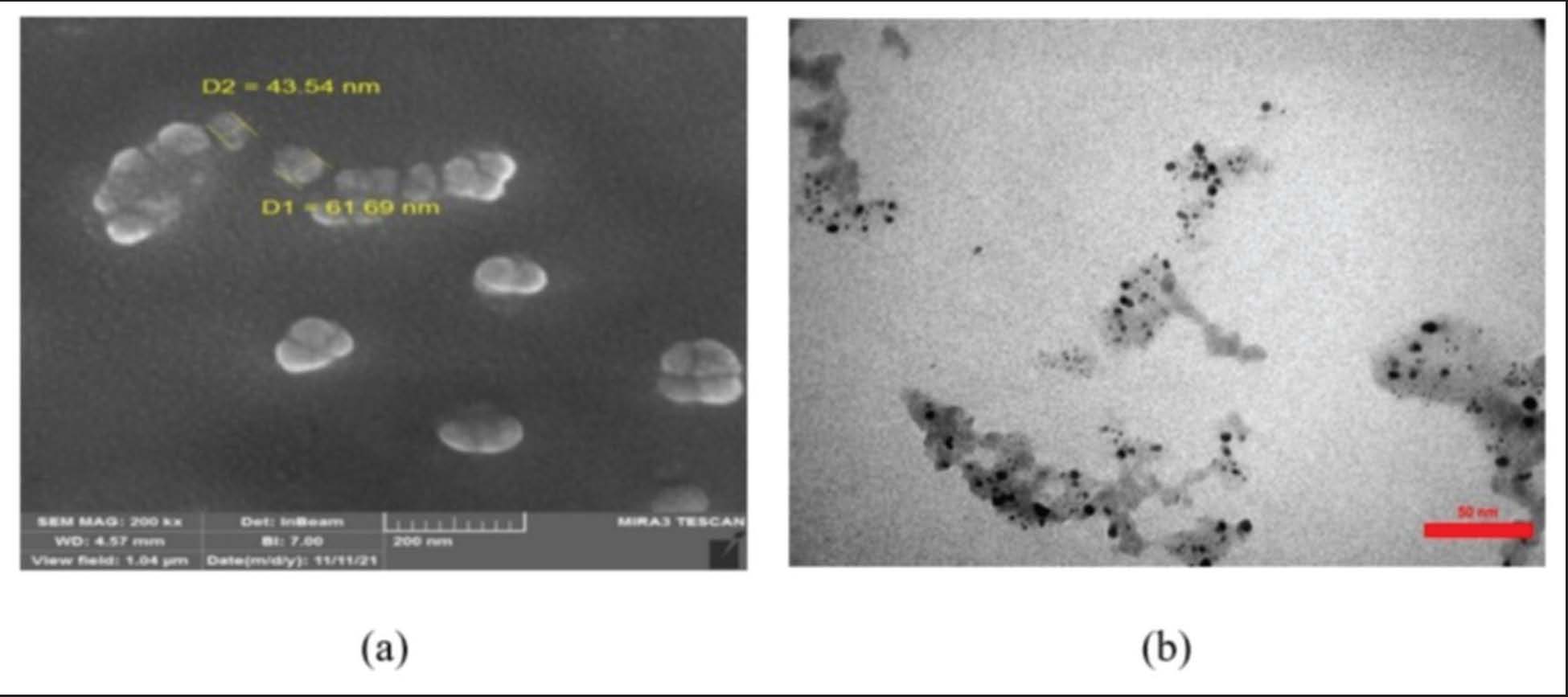
Fig. 5. This figure depicts (a) the XRD pattern of green Ag nanoparticles and (b) FTIR analysis of silver nanoparticles synthesized by S. marianum seed extract. Fourier transform infrared spectroscopy (FTIR)FTIR analysis was employed to identify biomolecules responsible for the reduction, stabilization, and capping of silver nanoparticles synthesized using milk thistle seed extract. This technique was also used to determine the functional groups present in both AgNO3 and the reducing agent (silymarin seed extract) and to infer their role in nanoparticle formation. The FTIR spectrum (Fig. 5b) exhibited characteristic peaks corresponding to various functional groups involved in the synthesis process. Prominent absorption bands were observed at 3,390, 3,167, 1,654, 1,541, 1,384, 1,012, 883, and 464 cm?1. These bands indicate the presence of functional groups such as O–H, C–H, C=O, C-O, and C-Cl, suggesting the bioactive compounds in the milk thistle seed extract contributed to the reduction and stabilization of synthesized AgNPs. Scanning electron microscopic analysis (SEM)SEM images were required to analyze the morphology and size of the synthesized silver nanoparticles. Scanning electron microscopy is a technique that uses electrons instead of light to determine the size, shape, morphology, and distribution of synthetically generated silver nanoparticles. Fig. 6a illustrates the SEM image of AgNPs synthesized by a green technique utilizing silymarin seed extract. The image shows that the particles are aggregated with different sizes and shapes, mostly spherical shapes with average diameters of 43.54 to 61.69 nm. The highly agglomerated nanoparticles generate massive aggregates that provide evidence of the aggregation of nanoparticles during the sample preparation, which results in the larger size of AgNPs.
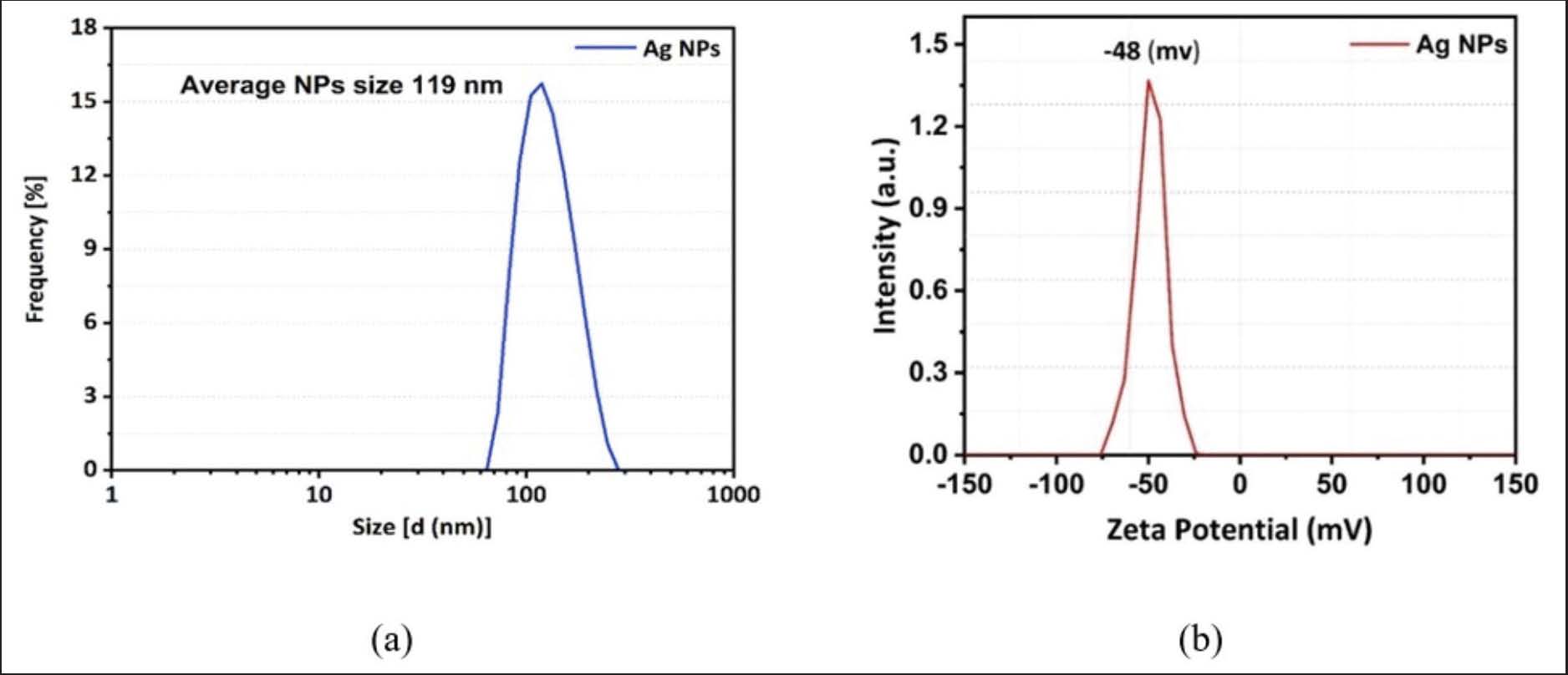
Fig. 6. This figure depicts (a) a scanning electron microscopy image of the green AgNPs and (b) a transmission electron microscopy image of the green AgNPs synthesized with S. marianum seed extract. Transmission electron microscope (TEM)The size, shape, and morphology of the green AgNPs synthesized using milk thistle seed extract were examined by transmission electron microscopy. The spherical nanoparticles had a diameter of around 50 nm and a spherical morphology, as shown in the transmission electron microscopy (Fig. 6b). The pattern of dispersion of the nanoparticles indicated the stabilization of the nanoparticles by the capping agents. Dynamic light scattering (DLS) analysisThe synthesized green AgNPs from the S. marianum seed extract were evaluated for average size and distribution using a dynamic light scattering technique. The mean size of AgNPs reported in the data was 119 nm (Fig. 7a). DLS measures the scattering intensity; the elevated DLS value is due to the existence of a significant solvation layer and the intensity-weighted characteristics of DLS, which is sensitive to the hydrodynamic volume rather than the physical core. Moreover, the larger DLS value indicates the presence of tiny particles in the aqueous phase.
Fig. 7. This figure depicts (a) dynamic light scattering analysis of the green AgNPs and (b) zeta potential (mV) of the green AgNPs synthesized with S. marianum seed extract. Zeta potential analysisZeta potential analysis was used to observe the surface charges accumulated by the AgNPs, which is a clear indication of the stability of the created colloidal AgNPs. The charge on the nanoparticle surface defines the electrostatic repulsion between the nanoparticles. The negative charge on the nanoparticles prevents aggregation and provides long-term stability. The zeta potential of AgNPs in this study was −48 mV, confirming the presence of monodisperse nanoparticles without any aggregates (Fig. 7b). Biopesticidal effect of green-synthesized Ag-NPs on T. castaneumThe contact toxicity method was used to evaluate the pesticidal effect of green-synthesized Ag-NPs from S. marianum seed extract against adult, second-instar larvae, and fifth-instar larval stages of T. castaneum. Each of the three insect stages was exposed to SM-AgNPs at doses of 0.75, 1.5, 3, and 6 mg/g for different exposure durations of 24 hours (p-value=0.024), 48 hours (p-value=0.01), 72 hours (p-value=0.0054), and 96 hours (p-value=0.0001). The analysis of variance showed significant differences (p ˂ 0.05) in mortality rates in adult stages and in the 2nd and 5th larval instars. Fig. 8a illustrates that the toxic effects of green-synthesized AgNPs infused with milk thistle seed extract at all dosages exhibited a substantial difference in impact on adult T. castaneum and larvae (2nd and 5th instars) after 24 hours post-application of the AgNPs. Red flour beetle larvae that are in their second instar had the maximum mortality rate of 32% at a concentration of 6 mg/g, but adult red flour beetles demonstrated the lowest mortality rate of 5.6% at a concentration of 0.75 mg/g. The control group showed a notable disparity compared with all concentrations. On the second day of the application process, the death rates of red flour beetle larvae and adults differed greatly by treatment concentration. After 48 hours, the second-instar T. castaneum larvae showed the highest mortality of 50.6% when exposed to SM-AgNPs at 6 mg/g and 50% at the rate of 3 mg/g. The minimum death rate for adults was found at 0.75 mg/g, resulting in 9.3% mortality (Fig. 8b). A statistically significant difference was observed between the control group and each concentration, as the control group had a lower mortality rate than all other concentrations. As shown in Fig. 8c, all concentrations of SM-AgNPs resulted in considerable mortality of T. castaneum larvae and adults at 72 hours after treatment. The highest mortality was found in the 2nd and 5th instar larvae of T. castaneum at 6 mg/g, with 62% and 56.6%, respectively. The control revealed minimal mortality on the third day post-application, but all other concentrations were substantially different from the control. Fig. 8d demonstrated a substantial difference among all SM-AgNP concentrations after 96 hours of treatment. The SM-AgNPs at 0.75 mg/g had a lower larval and adult mortality rate as compared to all other concentrations. The highest mortality rate for the second-instar larvae of T. castaneum was 70% on the fourth day at the highest dosage of AgNP. There was no significant mortality seen in the control group.
Fig. 8. This figure depicts the percentage mortality of T. castaneum subjected to diverse concentrations (0.75, 1.5, 3, and 6 mg/g) of silver nanoparticles synthesized using S. marianum seed extracts after 24 hours (p-value=0.024) (a), 48 hours (p-value=0.01) (b), 72 hours (p-value=0.0054) (c), and (d) 96 hours of exposure (p-value=0.0001). The bars represent mean mortalities (%) ± SEM. Bars labeled with different letters indicate statistically significant differences (p < 0.05). The LC50 value for larvae in their second instar decreased from 42.37 mg/g at 24 hours to 4.54 mg/g at 48 hours, then to 1.83 mg/g at 72 hours and 0.80 mg/g at 96 hours. The larvae in their fifth instar show a similar pattern, with LC50 values decreasing from 223.23 mg/g (24 hours) to 6.84 mg/g (48 hours), 3.26 mg/g (72 hours), and 1.79 mg/g (96 hours). At the adult stage, the LC50 value drops from a significantly higher 257.55 mg/g at 24 hours to 10.50 mg/g at 48 hours, 7.63 mg/g at 72 hours, and 5.15 mg/g at 96 hours. The latest findings on the biopesticidal efficacy of AgNPs indicate that the maximum pesticide activity recorded against the red flour beetle in its 2nd larval instar at a concentration of 6 mg/g of AgNP seed extract from S. marianum after 96 hours reached 70%. On the contrary, the minimum pesticide activity (5.6%) was recorded against adult stages of the red flour beetle at 0.75 mg/g after 24 hours of treatment. The mortality rate is notably low in the first few days following the application of the bioassay, despite the fact that increased death rates are recorded once the experiment has been finally concluded. Factors such as dose concentration, duration, and nanoparticle dispersion are among those that could affect the outcomes of a bioassay. The percentage of mortality increased in direct correlation with the concentration and exposure time. DiscussionInfestations of T. castaneum in stored cereal grains result in considerable postharvest damage and food contamination. Therefore, it is very important to rapidly and precisely identify this pest and implement effective biological management practices at any stage of its life cycle. Quantitative PCR is a rapid and precise detection method for T. castaneum. Tribolium species are commonly distinguished based on morphological characteristics. However, rapid differentiation of fragmented adult specimens and immature developmental stages using external morphology is often difficult or impossible. On the contrary, amplified DNA sequences can be compared among species, providing a more reliable method of identification than morphological traits. qPCR is a useful tool to identify sympatric species, overcoming the limitations related to taxonomy and morphological identification. In addition, it is particularly suitable for detecting pests at immature life stages, including eggs, larvae, and pupae, making it a practical tool for rapid diagnostic application. The results highlighted the importance of species-specific PCR and qPCR, as well as the use of short, standardized DNA segments from the cytochrome oxidase I mitochondrial region, for rapid and accurate identification of T. castaneum. This result is consistent with what has been found in earlier studies. The cytochrome b and cytochrome c oxidase subunit I regions among species of the flour beetle were sequenced. The results showed that T. castaneum and T. confusum are genetically different despite their similar size and shape (Ming et al., 2015). Zhang et al. (2016) separated six species of Tribolium: T. castaneum, T. confusum, T. destructor, T. madens, T. freemani, and T. brevicornis from stored products using species-specific polymerase chain reaction and qPCR techniques based on mtCOI barcodes. Similarly, Negi et al. (2021) used qPCR with specific primers designed from the mtCOI gene to successfully detect 0.001 T. castaneum individuals per 5g of wheat flour, which was intentionally infested with adults. Our results also indicated that real-time quantitative PCR with SYBR Green and species-specific PCR were rapid and more effective methods for identifying T. castaneum. In this study, we also prepared silver nanoparticles from aqueous seed extracts of S. marianum, which were characterized using UV-visible spectroscopy, FTIR, SEM, TEM, XRD, DLS, and zeta potential. The synthesis of AgNPs from S. marianum aqueous seed extracts was detected by UV-VIS spectra and color shift. After 24–48 hours of mixing, the hue changes from light yellow to deep brown due to the AgNPs’ activation of surface plasmon vibrations (Mohammadinejad et al., 2013). The presence of AgNPs is indicated by a gradual increase in the extract’s color following the addition of 1-mM AgNO3. After nearly 48 hours, the solution’s dark brown hue indicates the presence of AgNPs. The main indication of the formation of silver nanoparticles was the color change of the reaction mixture; variations among plants occur in the period of color shift and color density. The presence of H+ ions may be responsible for the reduction of silver or for quantitative fluctuations in the synthesis of silver nanoparticles. Our results are consistent with those of Kumar et al. (2014). The absorbance spectra of the AgNPs solution show a noticeable surface plasmon resonance band (SPR) at 408 nm after the silver nanoparticles were screened using a UV-visible spectrometer. The reported results were in agreement with Gopalakrishnan and Raghu (2014), who observed a surface plasmon resonance band in the 450–400 nm range. On the contrary, Noreen et al. (2024) observed separate absorption peaks at two wavelengths, with a strong SPR at 200 and 400 nm. UV-Vis spectroscopy investigation revealed changes due to increased surface energy and tension. Minor aggregation in the micrograph may be due to these features (Aouf et al., 2024). The distinct peak indicated that silver had decreased; there was a notable reduction in the conversion of Ag⁺ ions to Ag⁰ in the aqueous extract of S. marianum seeds. In the bioreduction process, metal ions or oxides are converted into zero-valent metal nanoparticles. Many phytochemicals can reduce the ions independently, but the most prevalent ones are amides, aldehydes, quinones, carboxylic acids, and flavonoids. The plant S. marianum has been found to contain flavonoids, phenols, and tannins, which collectively make up silymarin and are effective bioreduction agents (Shah et al., 2020). XRD investigation was performed to confirm the crystallinity of milk thistle-mediated AgNPs. The XRD pattern of the silver crystals showed distinct peaks at 2θ values of 38.4°, 43.3°, 63.4°, and 77.0° corresponding to (111), (202), (2201), and (311) planes, respectively, confirming the crystalline nature of the silver crystals. The significant peaks in the XRD pattern were due to the presence of organic substances, such as flavonoids and phenols, in plants that reduce and stabilize the silver ions in the produced AgNPs and the high crystallinity of the AgNPs. Silver nanoparticles synthesized using milk thistle seed extract were stabilized and capped by biomolecules that were identified by FTIR analysis. There were absorption peaks at 3,390, 3,167, 1,654, 1,541, 1,384, 1,012, 883, and 464 cm-¹ in the FTIR spectrum (Fig. 5b), which corresponded to functional groups including O–H, C–H, C=O, C–O, and C–Cl. These molecules indicate the existence of flavonoids and phenols. The existence of flavonoids, polyphenols, and protein amide groups, in addition to other phytochemicals, which are primarily in charge of lowering the AgNO3 to create AgNPs. Shah et al. (2020) reported similar results. As a result, silymarin seed extract is a potentially useful source for the development of AgNPs. The XRD results corroborated the conclusions of prior investigations (Kumar et al., 2014; Shah 2019). SEM images of silymarin seed extract-green-synthesized AgNPs showed aggregated particles of various sizes and shapes, primarily spherical, with average diameters ranging from 43.54 to 61.69 nm. Our findings are consistent with earlier research (Salah et al., 2023(Noreen et al., 2024). The SEM analysis showed a spherical morphology with an average particle size of 52.63 nm, which indicates the stability of the produced silver nanoparticles, attributed to the active constituents in milk thistle seeds (Abdel-Rahman et al., 2022). The insecticidal or larvicidal properties of nanoparticles can be attributed to their structure, size, and significant coverage capacity, facilitating their infiltration into the insect's body. Transmission electron microscopy revealed that the spherical NPs had a spherical morphology and a diameter of approximately 50 nanometers. The nanoparticles’ dispersion pattern indicates that capping agents have stabilized them. These findings concur with those published by Sedighi et al. (2019). A dynamic light scattering approach was used to evaluate the green AgNPs generated from S. marianum seed extract to determine their average size and distribution; the average particle size was 119 nm. The large size observed in DLS (119 nm) compared to TEM (~50 nm) is attributed to DLS measuring the hydrodynamic diameter, which includes the metallic core, the surrounding hydration layer, and organic capping agents from the milk thistle extract in the aqueous solution. In order to provide more clarity regarding the homogeneity of the sample, we have supplied the polydispersity index (PDI) value of 0.32, which shows a size distribution that is either narrow or broad. The zeta potential of the AgNPs in this study was −48 mV, which indicated that they were not aggregated and were monodispersed. The current research indicates that silymarin fruit extract may serve as a viable source for the efficient production of stable silver nanoparticles through a simple, economical, and environmentally sustainable synthesis process. Ayad et al. (2019) and El-Saadony et al. (2020) both supported our findings. The bioinsecticidal activity of green-synthesized S. marianum AgNPs against the adults and second and fifth-instar larvae of T. castaneum was significant. It was observed that higher doses and longer exposure times of milk thistle AgNPs increased the insecticidal activity against T. castaneum adults and larvae (2nd and 5th instars). Larval mortality in the second instar revealed variable responses to concentrations and time of exposure. The second instar showed the highest mortality (70%) after 96 hours of exposure to 6 mg/g SM-AgNPs treatment, which was higher than the mortality rates in the fifth larval instar and adult stages (61% and 54%, respectively). In the fifth-instar larvae, mortality escalated with higher concentrations and extended exposure periods. However, the mortality rate was lower than that observed in second-instar larvae. The adult stage of the red flour beetle showed a lower mortality response compared with both larval stages (2nd and 5th instars). Mortality of larvae and adults could be due to several variables such as absorption and interaction, mode of action, and cumulative toxicity of NPs (Tuncsoy and Tuncsoy, 2023). Larvae can efficiently absorb SM-AgNPs via surface contact, resulting in internal accumulation and subsequent disruption of essential physiological processes required for development and survival. In addition, AgNPs possess antimicrobial properties that may disrupt cellular integrity and cause cellular toxicity, perhaps leading to larval mortality (El-Samad et al., 2022). Furthermore, AgNPs can inhibit acetylcholinesterase activity, thereby impairing neuronal function. Collectively, these effects interfere with larval development, reproduction, and survival (Shahzad and Manzoor, 2021). The LC50 values decreased with increasing nanoparticle concentrations and extended treatment duration. The findings indicate that prolonged exposure durations and elevated SM-AgNPs concentrations correlated with heightened mortality rates in both adults and larvae (2nd and 5th instars). The toxicity of SM-AgNPs increased with time, as shown by the lowering LC50 values. Possible causes of this pattern include bioaccumulation, developmental stage sensitivity, and duration of exposure to AgNPs (Wāng et al., 2024). The larvae have the potential to accumulate increased levels of SM-AgNPs over time. Increasingly, this accumulation surpasses the levels that are lethal (El-Samad et al., 2024). Metabolic rates vary with age and surface-to-volume ratios in different instars of larvae (2nd and 5th), as well as in adult stages, as stated by Pathipati and Kanuparthi (2021). Because of this, their susceptibility to the SM-AgNP concentrations used in experiments varied. The effectiveness of SM-AgNPs decreases when the larvae reach the fifth instar and eventually adults, and they are most toxic to younger larvae (2nd instar). This variation is evident from the higher mortality observed in second-instar larvae compared with adult and fifth-instar stages of red flour beetles. The ability of insects to tolerate insecticides is largely influenced by stage-specific cuticular proteins. Zhu et al. (2021) reported that the thicker proteinaceous cuticle of adult red flour beetles provides greater protection than that of the larval stage. In addition, red flour beetles have detoxification enzymes capable of degrading toxic compounds, thereby enhancing their survival following pesticide exposure. The lower susceptibility in the adult stage could be related to increased activity of detoxifying enzymes involved in pesticide degradation, as well as reduced sensitivity of target enzymes, such as acetylcholinesterase, to pesticides (Ranganathan et al., 2022). The LC50 value of the synthesized SM-AgNPs biopesticide was increased with respect to the aged red flour beetles. This indicates that biopesticide concentrations have to be lower to kill larvae compared to adults. Results showed that red flour beetles' tolerance levels increased as they matured and gained weight (Fareed et al., 2023). Our results confirmed that increased concentrations and longer exposure times to SM-AgNPs significantly enhanced insecticidal efficacy against adult and second and fifth-instar larvae of T. castaneum. The second larval instar was most sensitive to the toxicity of the synthesized AgNPs from milk thistle. These results were in line with previous studies. Salah et al. (2023) reported that AgNPs had no significant effect on T. castaneum adults during the first day of exposure at all tested concentrations (100, 200, 300, and 400 ppm). However, at 400 ppm, mortality reached 40% by the fourth day. Noreen et al. (2024) also reported that maximal mortality was recorded in the 4th instar of the fall armyworm after 72 hours of exposure to a 500-ppm MOL-AgNPs treatment, which was higher than that reported in the 5th and 6th larval instars. The effectiveness of milk thistle as a bioinsecticide indicates that this plant has toxic, repellent, and feeding–inhibitory properties against the red flour beetle. Silver nanoparticles synthesized using a 4% acetone concentration of the clove, Syzygium aromaticum, essential oil achieved 100% larvicidal mortality against the red flour beetle (Selvaraj et al., 2019) . AgNPs derived from the leaf extract of Nerium oleander exhibited greater larvicidal effectiveness against T. castaneum and Callosobruchus maculatus compared with the plant extract alone (Annon and Jafar, 2020). In another study, adult T. confusum were exposed to silver nanoparticles synthesized from Citrus sinensis peel extract. Mortality rates of 77%–83% in filter-paper residue and feeding assays, whereas the peel extract alone exhibited no insecticidal activity (Sedighi et al., 2019). Likewise, green silver nanoparticles synthesized using Spirogyra hyalina extract were used as both capping and reducing agents, caused 30% mortality in T. castaneum at a concentration of 500 ppm (Al-Radadi et al., 2022). Al-Hamdani and Alebady (2023) observed that combining milk thistle oil with Aktara insecticide resulted in the highest average mortality rates for flour beetle larvae and adults, reaching 40%. These findings are consistent with the present result. Jahan et al. (2023) reported that the biosynthesized ZnONPs derived from Silybum marianum seed extracts caused a mortality rate of 78% against T. castaneum after 72 hours of exposure. Similarly, Solorzano Toala et al. (2024) demonstrated that copper nanoparticles synthesized from Acacia cornigera and Annona purpurea exhibited significant insecticidal activity against T. castaneum. Iqbal et al. (2024) also reported that M. oleifera M3 nanosuspension significantly increased insecticidal efficiency, achieving an 83.00% mortality rate against T. castaneum after 72 hours of exposure. On the contrary, the M. oleifera extract alone exhibited significantly lower insecticidal activity. Agriculture and the environment have the priority of sustainable pest management. The use of typical chemicals to produce nanoparticles is expensive, poisonous, and environmentally harmful. Green synthesis, which uses plants, microbes, and biopolymers, is a sustainable solution with little environmental impact. The biocidal potential of green-produced silver nanoparticles offers a bridge between the traditional approach of pest control and modern nanotechnology, as they have the ability to offer better pest control while reducing environmental impact (Salomi et al., 2023). In modern agriculture and pest management, nanotechnology has emerged as a tool that has the potential to bring about substantial advances, especially in managing insect pests. It has gained much attention because of its ability to provide more effective, precise, and environmentally friendly alternatives to conventional pesticides. A major advantage of nano-biopesticides is the ability to reduce pollution and pesticide waste by better pesticide delivery through focused action and controlled release mechanisms (Liu et al., 2024). The results indicate that silver nanoparticles synthesized from S. marianum seed extracts provide an environmentally sustainable technique for the management of T. castaneum. Biosynthesized AgNPs have the potential to be a powerful bioinsecticide, but further research is required to evaluate large-scale field application, their environmental safety, long-term stability, impacts on non-target organisms, and effectiveness compared to commercially available alternatives. Furthermore, better methods of fast molecular detection for use in integrated pest management and real-time pest monitoring should be the subject of future research. ConclusionThe present study indicated that qPCR was a sensitive, effective, quick, and species-specific method for T. castaneum detection. This method permits the rapid and reliable identification of stored product pests. In addition, silver nanoparticles produced from Silybum marianum seed extract were found to be effective against second and fifth instars and adults of T. castaneum. The bioinsecticidal efficacy of SM-AgNPs exhibited considerable variation with the developmental stage of the red flour beetle, concentration levels, and duration of exposure. Throughout all developmental stages, including larvae (2nd and 5th instars) and adults, prolonged exposure to SM-AgNPs reduced LC50 values. The results reveal that considerable bioinsecticidal activity of SM-AgNPs was seen against red flour beetles. The production of silver nanoparticles is safe, cost-effective, and eco-friendly for the control of important economic pests like T. castaneum. AcknowledgmentsThe authors acknowledge the continuous support of Salahaddin University-Erbil throughout this work. FundingThere is no funding for this project. Authors’ contributionsThe author solely contributed to the conception, study design, data collection, data analysis, interpretation of results, and manuscript. Conflicts of interestThe author declares no conflict of interest. Data AvailabilityAll the data generated or analyzed during this inquiry are included in this paper. ReferencesAbdel-Rahman, L.H., Al-Farhan, B.S., Abou El-ezz, D., Abd–el Sayed, M.A., Zikry, M.M. and Abu-Dief, A.M. 2022. Green biogenic synthesis of silver nanoparticles using aqueous extract of Moringa oleifera: access to a powerful antimicrobial, anticancer, pesticidal and catalytic agents. J. Inorganic Organometallic Polym. Mater. 32(4), 1422–1435. Abdullah., Al-Radadi, N.S., Hussain, T., Faisal, S. and Ali Raza Shah, S. 2022. Novel biosynthesis, characterization and bio-catalytic potential of green algae (Spirogyra hyalina) mediated silver nanomaterials. Saudi. J. Biol. Sci. 29(1), 411–419. Al-Fatlawy, N.C.H. and Al-Zurfi, S. 2023. Effectiveness of three Commercially Nanoparticles against Tribolium castaneum (Herbst)(Coleoptera: tenebrionidae) under the Lab. conditions. Bionatura. Latin. Am. J. Biotechnol. Life. Sci. 8(1), 1–9. Al-Hamdani, O.U.A. and Mohammed Alebady, E.Q. 2023. The stimulatory effect of mixtures milk thistle and wild safflower oil with some insecticides on the adults and larvae of flour beetles. NTU J. Agriculture Vet. Sci. 3(2):51–57. Annon, M.R. and Jafar, F.S. 2020. The effectiveness of silver and silica nanoparticles on productivity and adult emergence of T. castaneum and C. maculatus. Journal of Physics: Conference Series. IOP Publishing, Bristol, United Kingdom, p 012110. Aouf, D., Khane, Y., Fenniche, F., Albukhaty, S., Sulaiman, G.M., Khane, S., Henni, A., Zoukel, A., Dizge, N., Mohammed, H.A. and Abomughaid, M.M. 2024. Biogenic silver nanoparticles of Moringa oleifera leaf extract: characterization and photocatalytic application. Nanotechnol. Rev. 13(1), 20240002. Aslam, A.F., Sultana, S., Rain, F.F., Das, S.R., Siddika, A. and Howlader, A.J. 2019. Molecular characterization and identification of three stored grain pests based on mitochondrial cytochrome C oxidase subunit I (COI) gene sequences. Bangladesh. J. Zool. 47(1), 1–11. Ayad, Z.M., Ibrahim, O.M.S. and Omar, L.W. 2019. Biosynthesis and characterization of silver nanoparticles by Silybum marianum (silymarin) fruit extract. Adv. Anim. Vet. Sci. 7(2), 122–130. Bjørklund, G., Cruz-Martins, N., Goh, B.H., Mykhailenko, O., Lysiuk, R., Shanaida, M., Lenchyk, L., Upyr, T., Rusu, M.E., Pryshlyak, A., Shanaida, V. and Chirumbolo, S. 2024. Medicinal plant-derived phytochemicals in detoxification. Curr. Pharm. Design 30(13), 988–1015. El-Saadony, M.T., Abd El-hack, M.E., Taha, A.E., Fouda, M.M.G., Ajarem, J.S., N. Maodaa, S., Allam, A.A. and Elshaer, N. 2020. Ecofriendly synthesis and insecticidal application of copper nanoparticles against the storage pest Tribolium castaneum. Nanomaterials 10(3), 587. El-Samad, L.M., Bakr, N.R., Abouzid, M., Shedid, E.S., Giesy, J.P., Khalifa, S.A., El-Seedi, H.R., El Wakil, A. and Al Naggar, Y. 2024. Nanoparticles—mediated entomotoxicology: lessons from biologicals. Ecotoxicology 33(3), 305–324. El-Samad, L.M., Bakr, N.R., El-Ashram, S., Radwan, E.H., Abdul Aziz, K.K., Hussein, H.K., El Wakil, A. and Hassan, M.A. 2022. Silver nanoparticles instigate physiological, genotoxicity, and ultrastructural anomalies in midgut tissues of beetles. Chemico-Biological Interact. 367, 110166. Erfanian, S.S., Ansari, H., Javanmard, S.H., Amini, Z. and Hajigholami, A. 2024. The hepatorenal protective effects of silymarin in cancer patients receiving chemotherapy: a randomized, placeholder-controlled trial. BMC. Complementary. Med. Therapies. 24(1), 329. Fareed, N., Nisa, S., Bibi, Y., Fareed, A., Ahmed, W., Sabir, M., Alam, S., Sajjad, A., Kumar, S., Hussain, M., Syed, A., Bahkali, A.H., Elgorban, A.M. and Qayyum, A. 2023. Green synthesized silver nanoparticles using carrot extract exhibited strong antibacterial activity against multidrug resistant bacteria. J. King. Saud. University-Science. 35(2), 102477. Gopalakrishnan, R. and Raghu, K. 2014. Biosynthesis and characterization of gold and silver nanoparticles using milk thistle (Silybum marianum) seed extract. J. Nanoscience 1(1), 905404. Iqbal, H., Jahan, N., Ali, S., Shahzad, A. and Iqbal, R. 2024. Formulation of Moringa oleifera nanobiopesticides and their evaluation against Tribolium castaneum and Rhyzopertha dominica. J. Plant Dis. Prot. 131(1), 133–142. Jahan, N. and Rasheed, K. 2023. Green inspired synthesis of zinc oxide nanoparticles using Silybum marianum (milk thistle) extract and evaluation of their potential pesticidal and phytopathogens activities. PeerJ 11, e15743. Jaswal, T. and Gupta, J. 2023. A review on the toxicity of silver nanoparticles on human health. Mater. Today. Proc. 81, 859–863. Kannan, M., Bojan, N., Swaminathan, J., Zicarelli, G., Hemalatha, D., Zhang, Y., Ramesh, M. and Faggio, C. 2023. Nanopesticides in agricultural pest management and their environmental risks: a review. Int. J. Environ. Sci. Technol. 20(9), 10507–10532. Karimi-Maleh, H., Ghalkhani, M., Saberi Dehkordi, Z., Mohsenpour Tehran, M., Singh, J., Wen, Y., Baghayeri, M., Rouhi, J., Fu, L. and Rajendran, S. 2024. MOF-enabled pesticides as developing approach for sustainable agriculture and reducing environmental hazards. J. Ind. Eng. Chem. 129, 105–123. Kumar, D.A., Palanichamy, V. and Roopan, S.M. 2014. Green synthesis of silver nanoparticles using Alternanthera dentata leaf extract at room temperature and their antimicrobial activity. Spectrochimica. Acta. Part. A. Mol. BioMol. Spectrosc. 127, 168–171. Liu, T., Xu, C., Ma, D., Cao, L., Ge, S. and Li, Y. 2024. Glycine-doped metal–organic frameworks as intelligent nanocarriers to enhance pesticide delivery and provide micronutrient in plants. Chem. Eng. J. 495, 153287. Ming, Q., Wang, A. and Cheng, C. 2015. Molecular identification of Tribolium castaneum and T. confusum (Coleoptera: tenebrionidae) using PCR-RFLP analysis. J. Genet. 94(Suppl 1), 17–21. Mohammadinejad, R., Pourseyedi, S., Baghizadeh, A., Ranjbar, S. and Mansoori, G.A. 2013. Synthesis of silver nanoparticles using Silybum marianum seed extract. Int. J. Nanoscience. Nanotechnol. 9(4), 221–226. Negi, A., Anandharaj, A., Kalakandan, S. and Rajamani, M. 2021. A molecular approach for the detection and quantification of Tribolium castaneum (Herbst) infestation in stored wheat flour. Food Technol. Biotechnol. 59(1), 112–121. Noreen, A., Hussain, M., Malik, M.F., Iftikhar, A., Zeb, U., Farid, A., Alarfaj, A.A. and Ansari, M.J. 2024. Moringa oleifera based silver nanoparticles: synthesis and insecticidal toxicity against fall armyworm. Notulae Bot. Horti Agrobotanici Cluj-Napoca 52(4), 14066. Papanikolaou, N.E., Kavallieratos, N.G., Iliopoulos, V., Evergetis, E., Skourti, A., Nika, E.P. and Haroutounian, S.A. 2022. Essential oil coating: mediterranean culinary plants as grain protectants against larvae and adults of Tribolium castaneum and Trogoderma granarium. Insects 13(2), 165. Pathipati, U.R. and Kanuparthi, P.L. 2021. Silver nanoparticles for insect control: bioassays and mechanisms. In Silver nanomaterials for agri-food applications. Ed., Abd-Elsalam, K. Amsterdam, The Netherlands: Elsevier, pp:471–494. Qiu, C., Zeng, J., Tang, Y., Gao, Q., Xiao, W. and Lou, Y. 2023. The fall armyworm, Spodoptera frugiperda (lepidoptera: noctuidae), influences Nilaparvata lugens population growth directly, by preying on its eggs, and indirectly, by inducing defenses in rice. Int. J. Mol. Sci. 24(10), 8754. Ranganathan, M., Narayanan, M. and Kumarasamy, S. 2022. Importance of metabolic enzymes and their role in insecticide resistance. In New and Future Development in Biopesticide Research: Biotechnological Exploration. Springer, Singapore, Singapore, pp: 243–260. Salah, S., Alyousuf, A.A. and Abass, M.H. 2023. Efficiency of silicon and silver nanoparticles against the infestation of Tribolium castanium (Herbst)(Coleoptera: tenebrionidae) on wheat grains under laboratory conditions. Basrah. J. Agricult. Sci. 36(2), 175–184. Salomi, V., Alfaris, A.A., Saranraj, P., Swetha, M. and Gayathri, K. 2023. Extraction of fungal pigment melanin from Aspergillus niger and analysis of its antimicrobial activity. Eur. Chem. Bull. 12(11), 431–438. Sedighi, A., Imani, S., Moshtaghi Kashanian, G., Najafi, H. and Fathipour, Y. 2019. Efficiency of green synthesized silver nanoparticles with sweet orange, Citrus sinensis (L.)(Rutaceae, Sapindales) against Tribolium confusum Duval.(Coleoptera, Tenebrionidae). J. Agricult. Sci. Technol. 21(6), 1485–1494. Selvaraj, P., Angelin, P.C. and Jayaseeli, J.P.R. 2019. Insecticidal and antibacterial potential of Syzygium aromaticum (L.) Merrill and Perry. J. Biopestic. 12(2), 191–196. Shah, A. 2019. Biomedical Potential of Silver and Gold Nanoparticles Synthesized from Daphne mucronata and Monotheca buxifolia as New Precursors. University of Peshawar, Peshawar. Shah, M., Nawaz, S., Jan, H., Uddin, N., Ali, A., Anjum, S., Giglioli-Guivarc'H, N., Hano, C. and Abbasi, B.H. 2020. Synthesis of bio-mediated silver nanoparticles from Silybum marianum and their biological and clinical activities. Mater. Sci. Eng. C 112, 110889. Shahzad, K. and Manzoor, F. 2021. Nanoformulations and their mode of action in insects: a review of biological interactions. Drug Chem. Toxicol. 44(1), 1–11. Sindhu, R. K., Yadav, S. K., Kaur, A., Kumar, M. and Kumar, P., 2022. X-Ray fluorescence: current trends and future scope. X-Ray Fluorescence in biological sciences: principles, instrumentation, and applications, pp. 623–646. Solorzano Toala, R., Gutierrez-Miceli, F., Valdez-Salas, B., Beltran-Partida, E., Gonzalez-Mendoza, D., Tzintzun-Camacho, O., Grimaldo-Juarez, O. and Basilio-Cortes, A. 2024. Biosynthesis of copper nanoparticles from Acacia cornigera and Annona purpurea and their insecticidal effect against Tribolium castaneum. Reactions 5(2), 274–284. Tuncsoy, B. and Tuncsoy, M. 2023. Toxicological effects of nanomaterials in terrestrial and aquatic insects. In Handbook of Green and Sustainable Nanotechnology: Fundamentals, Developments and Applications. Eds., Shanker, U., Hussain, C.M. and M. Rani. Cham, Switzerland: Springer, pp: 1–15. Wāng, Y., Han, Y. and Xu, D.X. 2024. Developmental impacts and toxicological hallmarks of silver nanoparticles across diverse biological models. Environ. Sci. Ecotechnology. 19, 100325. Xu, L., Wang, Y.Y., Huang, J., Chen, C.Y., Wang, Z.X. and Xie, H. 2020. Silver nanoparticles: synthesis, medical applications and biosafety. Theranostics 10(20), 8996. Zhang, T., Wang, Y.J., Guo, W., Luo, D., Wu, Y., Kučerová, Z., Stejskal, V., Opit, G., Cao, Y., Li, F.J. and Li, Z.H. 2016. DNA barcoding, species-specific PCR and real-time PCR techniques for the identification of six Tribolium pests of stored products. Scientific Rep. 6(1), 28494. Zhu, J.Y., Li, L., Xiao, K.R., He, S.Q. and Gui, F.R. 2021. Genomic and transcriptomic analysis reveals cuticular protein genes responding to different insecticides in fall armyworm Spodoptera frugiperda. Insects 12(11), 997. | ||

| How to Cite this Article |
| Pubmed Style Banaz Sdiq Abdulla. Molecular identification of Tribolium castaneum using SYBR green quantitative PCR and evaluation of Silybum marianum seed extract-mediated silver nanoparticles as a biopesticide. doi:10.5455/OVJ.2026.v16.i7.28 Web Style Banaz Sdiq Abdulla. Molecular identification of Tribolium castaneum using SYBR green quantitative PCR and evaluation of Silybum marianum seed extract-mediated silver nanoparticles as a biopesticide. https://www.openveterinaryjournal.com/?mno=311376 [Access: July 11, 2026]. doi:10.5455/OVJ.2026.v16.i7.28 AMA (American Medical Association) Style Banaz Sdiq Abdulla. Molecular identification of Tribolium castaneum using SYBR green quantitative PCR and evaluation of Silybum marianum seed extract-mediated silver nanoparticles as a biopesticide. doi:10.5455/OVJ.2026.v16.i7.28 Vancouver/ICMJE Style Banaz Sdiq Abdulla. Molecular identification of Tribolium castaneum using SYBR green quantitative PCR and evaluation of Silybum marianum seed extract-mediated silver nanoparticles as a biopesticide. doi:10.5455/OVJ.2026.v16.i7.28 Harvard Style Banaz Sdiq Abdulla (2026) Molecular identification of Tribolium castaneum using SYBR green quantitative PCR and evaluation of Silybum marianum seed extract-mediated silver nanoparticles as a biopesticide. doi:10.5455/OVJ.2026.v16.i7.28 Turabian Style Banaz Sdiq Abdulla. 2026. Molecular identification of Tribolium castaneum using SYBR green quantitative PCR and evaluation of Silybum marianum seed extract-mediated silver nanoparticles as a biopesticide. doi:10.5455/OVJ.2026.v16.i7.28 Chicago Style Banaz Sdiq Abdulla. "Molecular identification of Tribolium castaneum using SYBR green quantitative PCR and evaluation of Silybum marianum seed extract-mediated silver nanoparticles as a biopesticide." doi:10.5455/OVJ.2026.v16.i7.28 MLA (The Modern Language Association) Style Banaz Sdiq Abdulla. "Molecular identification of Tribolium castaneum using SYBR green quantitative PCR and evaluation of Silybum marianum seed extract-mediated silver nanoparticles as a biopesticide." doi:10.5455/OVJ.2026.v16.i7.28 APA (American Psychological Association) Style Banaz Sdiq Abdulla (2026) Molecular identification of Tribolium castaneum using SYBR green quantitative PCR and evaluation of Silybum marianum seed extract-mediated silver nanoparticles as a biopesticide. doi:10.5455/OVJ.2026.v16.i7.28 |


