Open Veterinary Journal, (2026), Vol. 16(7): 4566-4576
Research Article
10.5455/OVJ.2026.v16.i7.40
Effect of Cinnamomum burmannii bioactive fraction DLBS2411 on renal histopathology, serum malondialdehyde, and TNF-α in unilateral ureteral obstruction-induced renal fibrosis in male rats (Rattus norvegicus)
Novadian Novadian1*, Mgs Irsan Saleh2, Zulkhair Ali1, Evi Lusiana2, Elfiani Elfiani1,
Novandra Abdillah1, Rery Tri Ferri Yuniarti1 , Debby Hadiyanti Harahap2, Nur Riviati3
and Raymond Rubianto Tjandrawinata4
1Division of Nephrology and Hypertension, Department of Internal Medicine, Faculty of Medicine, Universitas Sriwijaya/Mohammad Hoesin Hospital, Palembang, Indonesia
2Department of Pharmacology, Faculty of Medicine, Universitas Sriwijaya, Palembang, Indonesia
3Division of Geriatrics, Department of Internal Medicine, Faculty of Medicine, Universitas Sriwijaya/Mohammad Hoesin Hospital, Palembang, Indonesia
4School of Bioscience, Technology and Innovation, Atma Jaya Catholic University of Indonesia, Jakarta, Indonesia
*Corresponding Author: Novadian Novadian. Division of Nephrology and Hypertension, Department of Internal Medicine, Faculty of Medicine, Universitas Sriwijaya/Mohammad Hoesin Hospital, Palembang, Indonesia.
Email: novadian [at] fk.unsri.ac.id
Submitted: 02/03/2026 Revised: 08/06/2026 Accepted: 19/06/2026 Published: 17/07/2026
© 2025 Open Veterinary Journal
This is an Open Access article distributed under the terms of the Creative Commons Attribution-Non Commercial-No Derivatives License (http://creativecommons.org/licenses/by-nc-nd/4.0/), which permits non-commercial re-use, distribution, and reproduction in any medium, provided the original work is properly cited, and is not altered, transformed, or built upon in any way.
ABSTRACT
Background: Renal tubulointerstitial fibrosis is a major pathological endpoint of progressive kidney disease and is closely associated with oxidative stress and inflammation. DLBS2411, a standardized bioactive fraction derived from Cinnamomum burmannii, has been investigated mainly for gastroprotective and mucoprotective effects, but its potential activity in renal fibrosis remains unclear.
Aim: This study evaluated the effect of C. burmannii bioactive fraction DLBS2411 (CB) on renal histopathology, serum malondialdehyde (MDA), and tumor necrosis factor-alpha (TNF-α) in a unilateral ureteral obstruction (UUO)-induced renal fibrosis model in male rats.
Methods: Thirty male Sprague–Dawley rats underwent UUO and were randomized on Day 7 into five groups (n=6/group): UUO + vehicle, UUO + methylprednisolone 1 mg/kgBW, and UUO + CB at 50, 100, or 200 mg/kgBW. Treatments were administered orally once daily for 14 days. Serum MDA and TNF-α were measured at baseline, Day 7, and Day 21. At Day 21, obstructed kidneys were collected for hematoxylin–eosin staining and blinded semiquantitative histopathological scoring.
Results: UUO increased serum MDA and TNF-α by Day 7. The UUO vehicle group showed severe tubular dilatation and interstitial inflammation, moderate protein casts, and mild interstitial fibrosis. CB treatment improved renal histopathology, particularly at 100 and 200 mg/kgBW. The CB200 group showed the most favorable renal morphology, with no detectable interstitial fibrosis. Serum MDA decreased significantly after CB treatment at 50 mg/kgBW (p=0.017), 100 mg/kgBW (p=0.007), and 200 mg/kgBW (p=0.009), with the greatest reduction in the CB100 group. Between-group ΔMDA analysis showed greater MDA reduction in CB-treated groups than in the UUO vehicle group. Serum TNF-α decreased significantly in the CB100 group (p=0.033) and methylprednisolone group (p=0.011). Between-group ΔTNF-α analysis showed a significantly greater reduction in CB100 than in the UUO vehicle group.
Conclusion: DLBS2411 improved renal histopathological injury and reduced systemic oxidative stress in UUO-induced renal fibrosis. CB200 showed the most favorable histological profile, whereas CB100 showed the most consistent biochemical response.
Keywords: Cinnamomum burmannii, DLBS2411, Malondialdehyde, Renal fibrosis, Unilateral ureteral obstruction.
Introduction
Kidney fibrosis is a final common pathway of most progressive kidney diseases and is characterized by excessive extracellular matrix accumulation, tubular atrophy, interstitial inflammation, and progressive loss of functional nephrons (Panizo et al., 2021; Reiss et al., 2024). Chronic kidney disease (CKD) remains a major global health problem and is associated with substantial morbidity, mortality, and healthcare burden (Deng et al., 2025). Current clinical strategies, particularly renin–angiotensin–aldosterone system blockade and sodium–glucose cotransporter-2 inhibitors, offer meaningful protection but do not fully halt fibrotic progression (Forouzanmehr et al., 2024). Therefore, there remains a need to identify adjunctive therapeutic candidates that target key mechanisms of renal fibrosis, particularly oxidative stress and inflammation.
The unilateral ureteral obstruction (UUO) model is widely used to investigate mechanisms and candidate treatments for tubulointerstitial fibrosis. UUO reproducibly induces tubular dilatation, inflammatory cell infiltration, tubular injury, oxidative stress, and extracellular matrix accumulation in the obstructed kidney (Martínez-Klimova et al., 2019). This model is valuable in experimental veterinary and biomedical research because it allows controlled assessment of histopathological renal remodeling and treatment-related responses in vivo. Although the UUO model does not fully replicate the multifactorial nature of naturally occurring CKD, it provides a reproducible and well-established experimental platform for evaluating tubulointerstitial injury, inflammation, oxidative stress, and early fibrotic remodeling. Therefore, findings from this model are valuable for preclinical assessment of candidate renoprotective agents.
Cinnamomum burmannii is an Indonesian medicinal plant traditionally used as a culinary spice and herbal remedy. Its bioactive constituents, including cinnamaldehyde, flavonoids, and polyphenols, have been associated with antioxidant and anti-inflammatory properties (Rahayu et al., 2022). DLBS2411 is a standardized bioactive fraction derived from C. burmannii. Previous studies have mainly focused on its gastroprotective and mucoprotective effects, including stimulation of mucus-related pathways and protection against ethanol-induced gastric damage (Wulandari et al., 2016; Tjandrawinata and Nailufar, 2020). DLBS2411 has also been reported to contain not less than 15% total phenolic compounds in prior gastroprotective work, supporting its biological plausibility as an antioxidant-rich fraction (Tjandrawinata and Nailufar, 2020).
Oxidative stress and inflammation are central drivers of renal fibrosis. Malondialdehyde (MDA), a lipid peroxidation product, is commonly used as an index of oxidative stress, whereas tumor necrosis factor-alpha (TNF-α) is a pro-inflammatory cytokine involved in tubular injury, inflammatory cell recruitment, fibroblast activation, and extracellular matrix accumulation (Mehaffey and Majid, 2017; Sagar et al., 2023; Stefania et al., 2024). Experimental suppression of TNF-α activity has been shown to reduce renal inflammation and fibrosis in kidney injury models, although fibrosis may persist through other pathways such as TGF-β/Smad signaling and sustained immune activation (Meldrum et al., 2007; Taguchi et al., 2021). Although DLBS2411 has been investigated mainly for its gastroprotective and mucoprotective activities, evidence regarding its potential role in renal fibrotic injury remains limited. Considering the involvement of oxidative stress and inflammation in UUO-induced renal fibrosis, evaluation of DLBS2411 in this model may provide further insight into its possible renoprotective activity beyond previously studied gastrointestinal effects. Therefore, this study aimed to evaluate the effect of C. burmannii bioactive fraction DLBS2411 on renal histopathology, serum MDA, and serum TNF-α in UUO-induced renal fibrosis in male Sprague–Dawley rats.
Materials and Methods
Study design
This experimental study used a randomized, controlled, pre-test and post-test design to investigate the effect of C. burmannii bioactive fraction DLBS2411 (CB) on renal histopathology and serum biomarkers in a rat model of UUO-induced renal fibrosis. The study was conducted from April to October 2025 at the iRATco Animal House, Dramaga, Bogor, Indonesia.
Experimental animals
Thirty healthy male Sprague–Dawley rats (Rattus norvegicus), aged 8–12 weeks and weighing 170–200 g, were obtained from iRATco Animal House, Dramaga, Bogor, Indonesia. All animals underwent a 7-day acclimatization period before the experiment and were maintained under standardized laboratory conditions, including a 12-hour light/12-hour dark cycle, room temperature of 22°C ± 2°C, relative humidity of 55%–65%, and ad libitum access to standard chow and water. During acclimatization and throughout the study period, animals were monitored daily for general health, behavior, food and water intake, and signs of distress. Only animals that appeared clinically healthy after acclimatization were included in the experiment. The sample size of six animals per group was selected as an exploratory animal study sample size for preliminary therapeutic evaluation in a rodent disease model. No formal a priori power calculation was performed.
Chemicals, reagents, and test substances
DLBS2411 was obtained as a standardized bioactive fraction of C. burmannii (Dexa Laboratories of Biomolecular Sciences, PT Dexa Medica, Indonesia). In the present study, DLBS2411 was used as supplied by the manufacturer. DLBS2411 is a water-dispersible bioactive fraction. The CB suspension was freshly prepared each day by dispersing the required dose in distilled water immediately before oral gavage administration. No co-solvent, surfactant, or suspending agent was used. Methylprednisolone was obtained as a pharmaceutical-grade lyophilized preparation (Methylprednisolone Sodium Succinate for Injection, PT Dexa Medica, Indonesia) and was reconstituted with sterile water for injection according to the manufacturer’s instructions, then further diluted with sterile 0.9% sodium chloride (NaCl) solution to a final gavage volume of 5 ml/kgBW. The negative control group received an equivalent volume of sterile 0.9% NaCl as a vehicle. All oral treatments, including CB suspensions, were administered at a gavage volume of 5 ml/kgBW, adjusted to each animal’s most recent body weight. Serum MDA was measured using a thiobarbituric acid reactive substances assay kit (BT Lab, China). Serum TNF-α was measured using a commercial rat TNF-α enzyme-linked immunosorbent assay kit (BT Lab, China). All biochemical assays were performed according to the manufacturer’s instructions.
Dose selection and treatment rationale
The CB doses of 50, 100, and 200 mg/kgBW were selected to evaluate dose-related biological activity in the UUO model. The lowest dose was chosen based on prior experimental evidence showing biological activity of DLBS2411 in rat gastroprotective models (Tjandrawinata and Nailufar, 2020), while the 100 and 200 mg/kgBW doses were selected as higher exploratory doses to assess whether increased exposure would enhance renal histological or biochemical responses in a fibrosis model. Because no previous study has clearly established an effective renal-fibrosis dose of DLBS2411, these doses were used as an exploratory dose range. Methylprednisolone at 1 mg/kgBW was used as an anti-inflammatory positive control to provide a reference comparator for inflammatory biomarker suppression. This dose was selected as a low-dose anti-inflammatory comparator rather than as a high-dose antifibrotic regimen.
Induction of unilateral ureteral obstruction
Renal fibrosis was induced using the UUO procedure. Rats were anesthetized with ketamine 80 mg/kgBW and xylazine 10 mg/kgBW administered intraperitoneally. After adequate anesthesia was confirmed by loss of pedal withdrawal reflex, the abdominal area was shaved and disinfected. A midline laparotomy was performed to expose the left ureter. The left ureter was ligated at two points using sterile 4–0 silk sutures, and the segment between ligatures was excised. The abdominal wall and skin were closed using standard suturing techniques. Animals were returned to their cages and monitored until full recovery from anesthesia. Postoperative analgesia was provided using buprenorphine 0.05 mg/kgBW administered subcutaneously after surgery and every 12 hours for 48 hours. Additional monitoring was performed to assess postoperative pain, wound condition, mobility, food and water intake, and general behavior. A sham-operated biochemical control group was not included. Renal tissue from non-UUO rats was used only as a descriptive histological reference for normal renal architecture and was not included in statistical comparisons.
Randomization, allocation, and treatment groups
On Day 7 after UUO surgery, rats were randomly allocated into five groups using a computer-generated randomization list, with six rats in each group. The negative control group received UUO + 0.9% NaCl vehicle, the positive control group received UUO + methylprednisolone 1 mg/kgBW, and the treatment groups received UUO + CB at doses of 50 mg/kgBW (CB50), 100 mg/kgBW (CB100), or 200 mg/kgBW (CB200). All treatments were administered once daily by oral gavage from Day 7 to Day 21 after UUO. The individual dose volume was adjusted according to the most recent body weight of each animal.
Blood sampling, euthanasia, and kidney collection
Blood sampling was performed at three time points: before UUO surgery (baseline), Day 7 after UUO, before treatment initiation, and Day 21 after completion of treatment. For blood collection, light sedation was induced using ketamine at 40 mg/kgBW administered intraperitoneally. No adjunct anesthetic agent was used for this procedure. Samples were transferred into plain tubes and allowed to clot for 30 minutes at room temperature, followed by centrifugation at 3,000 rpm for 10 minutes. The serum was carefully separated and stored at −80°C until analysis. Animals were monitored after each blood collection for signs of weakness, pallor, reduced activity, or other clinical signs suggestive of anemia. No clinical signs of anemia were observed during the study period.
On Day 21, animals were euthanized under deep anesthesia induced by intraperitoneal ketamine 80 mg/kgBW and xylazine 10 mg/kgBW. After surgical-plane anesthesia and absence of reflexes were confirmed, euthanasia was completed by exsanguination via cardiac puncture, in accordance with institutional animal care guidelines and the approved ethical protocol. The left obstructed kidneys were immediately harvested, rinsed with normal saline to remove residual blood, and fixed in 10% neutral buffered formalin for 24–48 hours at room temperature.
Histopathological processing and scoring
Fixed kidney tissues were processed using standard paraffin-embedding procedures, sectioned at 3–5 µm thickness, and stained with hematoxylin and eosin. Histopathological evaluation was performed using a semiquantitative scoring system based on INHAND criteria (Frazier et al., 2012). Renal lesions were assessed for tubular dilatation, interstitial inflammation, protein casts, and interstitial fibrosis using a 0–3 scale: 0=normal, 1=mild, 2=moderate, and 3=severe. Scoring was performed by an experienced pathologist who was blinded to group allocation. At least five non-overlapping high-power fields per section were evaluated using light microscopy. The median score for each animal was used for statistical analysis.
Outcome and blinding
The primary outcome was renal histopathological injury score at Day 21. Secondary outcomes were serum MDA and TNF-α levels measured at baseline, Day 7, and Day 21. All outcome assessments were performed under blinded conditions. The pathologist who performed histopathological scoring was blinded to group allocation. Laboratory personnel who measured serum MDA and TNF-α levels were also blinded to treatment groups, and statistical analysis was conducted using coded group labels before group identities were revealed.
Statistical analysis
Statistical analysis was performed using SPSS version 26.0 (IBM Corp., Armonk, NY, USA). Normality of continuous biomarker data was assessed using the Shapiro–Wilk test. Normally distributed serum biomarker data were presented as mean ± standard deviation, whereas non-normally distributed data were presented as median and interquartile range. Histopathological scores were presented as median with minimum–maximum values. Within-group changes in serum MDA and TNF-α between Day 7 and Day 21 were analyzed using paired t-tests for normally distributed data or Wilcoxon signed-rank tests for non-normally distributed data. Between-group comparisons of ΔMDA and ΔTNF-α were performed using one-way analysis of variance followed by Tukey’s post hoc test for normally distributed data, or Kruskal–Wallis test followed by Dunn–Bonferroni post hoc analysis for non-normally distributed data. These between-group Δ analyses are presented in Figures 3 and 4. Histopathological scores were compared among groups using the Kruskal–Wallis test. When a significant overall difference was detected, pairwise comparisons were performed using the Mann–Whitney U test with Bonferroni correction. Pairwise comparisons with the negative control group were reported where appropriate. A p-value < 0.05 was considered statistically significant. Exact p-values were reported whenever possible.
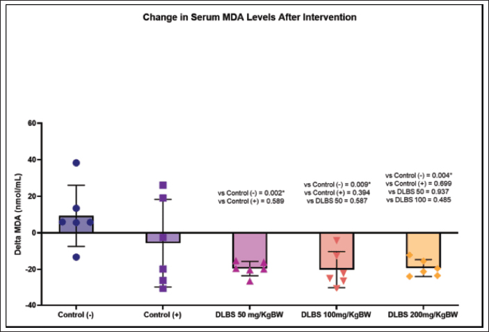
Fig. 3. Between-group comparison of changes in serum MDA levels from Day 7 to Day 21. ΔMDA was calculated as Day 21 − Day 7; negative values indicate a reduction after treatment. Data were normally distributed and analyzed using one-way ANOVA, followed by Tukey’s post hoc test for pairwise comparisons. Overall, ANOVA showed a significant between-group difference (p < 0.001). P-values displayed in the figure represent Tukey’s post hoc pairwise comparisons. NC: negative control; PC: positive control; CB50: DLBS2411 50 mg/kgBW; CB100: DLBS2411 100 mg/kgBW; CB200: DLBS2411 200 mg/kgBW. *Significant, p < 0.05.
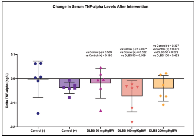
Fig. 4. Between-group comparison of changes in serum TNF-α levels from Day 7 to Day 21. ΔTNF-α was calculated as Day 21 − Day 7; negative values indicate a reduction after treatment. Data were normally distributed and analyzed using one-way ANOVA, followed by Tukey’s post hoc test for pairwise comparisons. Overall, ANOVA showed a significant between-group difference (p < 0.001). P-values displayed in the figure represent Tukey’s post hoc pairwise comparisons. NC: negative control; PC: positive control; CB50: DLBS2411 50 mg/kgBW; CB100: DLBS2411 100 mg/kgBW; CB200: DLBS2411 200 mg/kgBW. *Significant, p < 0.05.
Ethical approval
This study was approved by the Animal Ethics Committee of the School of Veterinary Medicine and Biomedical Sciences, IPB University (Approval No. 287/KEH/SKE/I/2025). The ethical approval date is January 25, 2025. The study was reported with consideration of the ARRIVE 2.0 recommendations for animal research.
Results
A total of 30 male Sprague–Dawley rats were enrolled, and all animals completed the experimental protocol without mortality. No animals met predefined exclusion criteria. UUO-induced obstructive renal injury, as shown by increased serum MDA and TNF-α levels by Day 7 compared with baseline values. At Day 21, histopathological examination of the obstructed kidneys confirmed tubulointerstitial injury characterized by tubular dilatation, interstitial inflammation, protein cast formation, and interstitial fibrosis in the negative control group.
Renal histopathology
The semiquantitative renal histopathological scores are illustrated in Figure 1, and representative hematoxylin and eosin stain (H&E)-stained renal histopathological findings are shown in Figure 2. The non-UUO renal tissue used as a descriptive normal reference showed preserved renal architecture, regularly arranged proximal and distal tubules, and absence of tubular dilatation, intraluminal protein casts, inflammatory cell infiltration, or interstitial fibrosis. On the contrary, the UUO negative control group showed marked tubulointerstitial injury, with severe tubular dilatation, severe interstitial inflammation, moderate protein cast formation, and mild interstitial fibrosis. No necrosis was observed. Methylprednisolone treatment improved the histopathological profile compared with the negative control group, with lower tubular dilatation and interstitial inflammation scores and absence of detectable interstitial fibrosis.
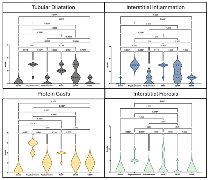
Fig. 1. Semiquantitative renal histopathological scores across treatment groups in UUO-induced renal fibrosis. Violin plots show the distribution of tubular dilatation, interstitial inflammation, protein casts, and interstitial fibrosis scores in the normal reference, negative control, positive control, and DLBS2411-treated groups. Histopathological lesions were scored using a 0–3 semiquantitative scale based on INHAND criteria, where 0=normal, 1=mild, 2=moderate, and 3=severe. Pairwise comparisons were performed using the Mann–Whitney U test after Kruskal–Wallis analysis, with Bonferroni adjustment for multiple comparisons. Adjusted p-values are shown above the plots; adjusted p < 0.05 was considered statistically significant. NC: negative control (UUO + NaCl vehicle); PC: positive control (UUO + methylprednisolone 1 mg/kgBW); CB50, CB100, and CB200: DLBS2411 at 50, 100, and 200 mg/kgBW, respectively. *Significant, p < 0.05.
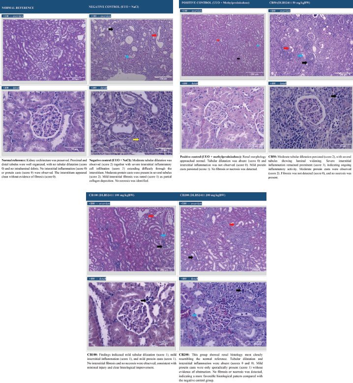
Fig. 2. Representative renal histopathology across treatment groups. Hematoxylin and Eosin (H&E) staining. Upper panel: ×100 magnification (overview of tissue architecture); lower panel: ×400 magnification (detail of lesions). Scale bar=200 µm. Black arrows: tubular dilatation. Red arrows: interstitial inflammatory cell infiltration. Blue arrows: intraluminal protein casts. Yellow arrowheads: areas interpreted as interstitial fibrosis on H&E staining. Semiquantitative scores based on INHAND criteria: 0=normal, 1=mild, 2=moderate, 3=severe. NC: negative control (UUO + NaCl vehicle); PC: positive control (UUO + methylprednisolone 1 mg/kgBW); CB50, CB100, CB200: DLBS2411 at 50, 100, and 200 mg/kgBW, respectively.
CB-treated groups showed lower histopathological lesion scores than the negative control group, particularly at 100 and 200 mg/kgBW. The CB50 group showed persistent moderate tubular dilatation and severe interstitial inflammation, although interstitial fibrosis was not detected in most animals. The CB100 group showed reduced tubular dilatation, interstitial inflammation, and protein cast formation, with minimal-to-absent fibrosis. The CB200 group demonstrated the most favorable histological profile among the CB-treated groups, with renal morphology closest to the normal reference. Pairwise comparisons of semiquantitative histopathological scores are shown in Figure 1, whereas the corresponding representative microscopic findings across groups are shown in Figure 2.
Serum MDA
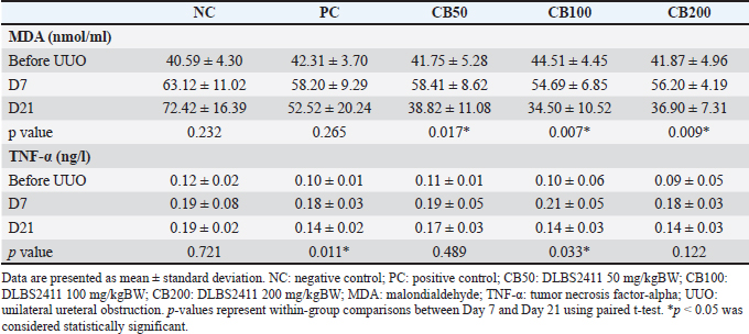
Serum MDA levels are presented in Table 1, and between-group comparisons of ΔMDA are shown in Figure 3. Serum MDA increased by Day 7 after UUO induction in all groups compared with baseline values. In the negative control group, MDA remained elevated at Day 21, with no significant reduction between Day 7 and Day 21 (p=0.232). In the methylprednisolone group, MDA showed a numerical decrease between Day 7 and Day 21, but this change was not statistically significant (p=0.265). On the contrary, CB treatment significantly reduced serum MDA after 14 days of treatment, with significant within-group reductions observed in the CB50 group (p=0.017), CB100 group (p=0.007), and CB200 group (p=0.009). The greatest reduction was observed in the CB100 group. Between-group analysis of ΔMDA, calculated as Day 21 − Day 7, showed a significant difference among groups by one-way ANOVA (p < 0.001). Tukey’s post hoc analysis demonstrated that CB-treated groups showed greater reductions in ΔMDA than the negative control group, with the pairwise p-values shown in Figure 3.
Table 1. Serum MDA and TNF-α levels before UUO, at Day 7 after UUO, and at Day 21 after treatment.
Serum TNF-α
Serum TNF-α levels are presented in Table 1, and between-group comparisons of ΔTNF-α are shown in Figure 4. Serum TNF-α increased by Day 7 after UUO induction in all groups compared with baseline values. The negative control group showed no significant change between Day 7 and Day 21 (p=0.721). Methylprednisolone significantly reduced TNF-α between Day 7 and Day 21 (p=0.011). In the CB-treated groups, TNF-α levels decreased after treatment, with a significant within-group reduction observed in the CB100 group (p=0.033). Reductions in the CB50 and CB200 groups did not reach statistical significance in within-group analysis.
Between-group analysis of ΔTNF-α, calculated as the Day 21 − the Day 7 value, showed a significant difference among groups by one-way ANOVA (p < 0.001). Tukey’s post hoc analysis showed that the CB100 group had a significantly greater reduction in ΔTNF-α than the negative control group, while pairwise comparisons among the remaining groups are shown in Figure 4. Among the CB-treated groups, CB100 showed the greatest reduction in ΔTNF-α. Pairwise comparisons with the methylprednisolone group are shown in Figure 4.
Overall, the CB200 group showed the lowest histopathological lesion scores among the CB-treated groups, whereas the CB100 group showed the greatest reduction in serum MDA and a significant reduction in serum TNF-α.
Discussion
This study applied the UUO model to reproduce key features of obstructive nephropathy. The rise of serum MDA and TNF-α by Day 7 indicates that UUO was accompanied by systemic oxidative stress and inflammatory responses, which are known contributors to obstructive renal injury (Aranda-Rivera et al., 2021). The renal histopathology in the negative control group further showed prominent tubular dilatation, diffuse interstitial inflammation, protein cast formation, and interstitial fibrosis, which are characteristic changes during progressive tubulointerstitial injury (Frazier et al., 2012). These findings are consistent with the concept that renal fibrosis evolves through persistent tubular epithelial injury, inflammatory infiltration, and maladaptive repair that ultimately promotes extracellular matrix accumulation and nephron loss (Panizo et al., 2021; Reiss et al., 2024).
Methylprednisolone was included as a positive control because corticosteroids are widely used in experimental settings to suppress inflammation-driven tissue injury (Flombaum, 1986). In UUO, inflammation and immune-cell recruitment amplify tubular damage and can accelerate fibrotic remodeling; therefore, an anti-inflammatory comparator is useful for assessing whether CB produces a treatment effect in the expected direction (Klahr and Morrissey, 2000; Meldrum et al., 2007). In this study, methylprednisolone improved histopathology compared with the negative control, with lower scores for tubular dilatation and interstitial inflammation and absence of detectable fibrosis. It also significantly reduced TNF-α between Day 7 and Day 21, consistent with the known capacity of corticosteroids to suppress pro-inflammatory cytokine signaling. However, methylprednisolone was used as a low-dose anti-inflammatory comparator rather than a high-dose antifibrotic regimen, and this should be considered when interpreting comparisons with CB.
CB treatment improved renal histopathology, particularly at 100 and 200 mg/kgBW, where injury scores were lower than in the negative control group, and fibrosis was minimal to absent. Among the CB-treated groups, CB200 showed renal morphology closest to the normal reference, suggesting a more favorable histological profile at this dose. This finding is relevant because histopathological injury is a direct structural outcome in renal fibrosis. Previous work using C. burmannii preparations has also reported protective effects on kidney histology in experimental metabolic injury models, supporting the possibility that this plant-derived preparation may influence renal tissue injury under pathological conditions (Budiastuti et al., 2025). Nevertheless, the present histopathological assessment was based on semiquantitative H&E scoring, so the antifibrotic interpretation should be supported in future work by fibrosis-specific staining and molecular markers.
In parallel with the histopathological findings, CB significantly reduced serum MDA at all tested doses. MDA reflects lipid peroxidation and systemic oxidative stress, and its reduction is biologically relevant because oxidative stress contributes to mitochondrial dysfunction, tubular epithelial injury, inflammation, and activation of fibrogenic pathways (Su et al., 2019; Tejchman et al., 2021). The greatest MDA reduction was observed at 100 mg/kgBW, whereas the 200 mg/kgBW dose did not show further biochemical improvement. This finding suggests that the biochemical response was not strictly dose-linear. However, because this study did not include pharmacokinetic analysis, tissue concentration measurement, or dose–response modeling, the non-linear MDA pattern should be interpreted cautiously. It may reflect biological variability or differences between systemic oxidative stress markers and intrarenal tissue responses rather than a definitive plateau effect.
TNF-α results were also favorable, with the clearest reduction observed in the CB100 group. TNF-α is an important mediator in renal fibrosis because it drives inflammatory cell recruitment, promotes tubular epithelial injury, and supports fibroblast activation and extracellular matrix accumulation (Klahr and Morrissey, 2000; Meldrum et al., 2007). Persistent elevation of TNF-α has been associated with inflammatory and fibrotic progression in kidney disease (Ortega and Fornoni, 2010; Toda et al., 2018) . In this study, within-group analysis showed a significant reduction in TNF-α in the CB100 group, while reductions in CB50 and CB200 did not reach statistical significance. Between-group analysis of ΔTNF-α also showed that CB100 had a significantly greater reduction than the negative control group. These findings suggest that CB100 produced the most consistent anti-inflammatory biomarker response among the tested CB doses. The lack of a uniform TNF-α response across all CB doses may reflect biological variability, the limited sample size, and the dynamic nature of inflammatory mediator expression during the injury and repair phases of UUO (Mavrogonatou et al., 2018; Stefania et al., 2024; Aripova et al., 2025).
The combined biomarker pattern suggests that CB exerted a more consistent effect on oxidative stress than on systemic TNF-α within the 14-day treatment window. This is plausible considering the phytochemical profile of C. burmannii, which contains polyphenols, flavonoids, and cinnamaldehyde, compounds commonly associated with antioxidant and anti-inflammatory activity (Tjandrawinata and Nailufar, 2020; Lusiana et al., 2022). DLBS2411 has also been reported to contain not less than 15% total phenolic compounds, which supports its biological plausibility as an antioxidant-rich bioactive fraction (Tjandrawinata and Nailufar, 2020). Anti-inflammatory effects may also be related to the modulation of inflammatory signaling pathways such as nuclear factor kappa-light-chain-enhancer of activated B cells, although this mechanism was not directly evaluated in the present study. Because oxidative stress and inflammation can reinforce each other during renal fibrosis, the concurrent reduction in MDA and TNF-α, particularly at 100 mg/kgBW, supports further investigation of CB as a candidate renoprotective agent.
Overall, the findings suggest that CB may have potential renoprotective activity in this exploratory UUO model. CB improved structural kidney injury and reduced systemic oxidative stress, with supportive evidence of anti-inflammatory activity based on TNF-α reduction. The 200 mg/kgBW dose showed the most favorable histopathological profile, whereas the 100 mg/kgBW dose showed the most consistent biochemical response. This difference between histological and biochemical outcomes suggests that tissue-level and systemic biomarker responses may not follow identical dose-response patterns.
Limitations and future perspectives
Several limitations should be acknowledged. First, a sham-operated biochemical control group was not included. Although non-UUO renal tissue was used as a descriptive normal histological reference, the absence of a sham group limits the interpretation of surgery-related systemic biomarker changes. Second, the observation period was limited to 21 days; therefore, the long-term antifibrotic efficacy of CB and its impact on sustained renal remodeling remain uncertain. Third, fibrosis assessment relied on semiquantitative H&E-based histopathological scoring without fibrosis-specific staining or quantitative molecular markers such as α-smooth muscle actin, collagen I/III, fibronectin, or evaluation of profibrotic signaling pathways such as TGF-β/Smad. Fourth, biomarker analysis was restricted to serum MDA and TNF-α and did not include tissue-specific oxidative stress or inflammatory markers that could more directly reflect intrarenal processes. Fifth, the relatively small sample size and absence of formal a priori power calculation may have limited statistical power, particularly for detecting dose-dependent differences in TNF-α. Sixth, only male rats were used, so possible sex-related differences were not evaluated. Finally, pharmacokinetic analysis and tissue-level measurement of DLBS2411 constituents were not performed. Future studies should include sham-operated controls, larger sample sizes with power calculation, longer follow-up, fibrosis-specific staining, molecular pathway analysis, renal tissue biomarkers, renal functional assessment, and pharmacokinetic evaluation to strengthen mechanistic interpretation and translational relevance.
Conclusion
In this exploratory UUO model, CB administration was associated with improved semiquantitative renal histopathological injury scores, particularly at 100 and 200 mg/kgBW. The CB200 group showed the most favorable renal morphology among the CB-treated groups, with lesion scores closest to the normal histological reference and no detectable interstitial fibrosis. CB also reduced systemic oxidative stress, as shown by significant decreases in serum MDA at all tested doses, with the greatest reduction observed at 100 mg/kgBW. For the inflammatory biomarker, serum TNF-α was significantly reduced in the CB100 group, while CB50 and CB200 showed non-significant reductions. Overall, DLBS2411 was associated with improved structural kidney injury scores and reduced systemic oxidative stress in this exploratory UUO model, with CB200 showing the most favorable histological profile and CB100 showing the most consistent biochemical response.
Acknowledgments
This study is part of a dissertation submitted in partial fulfillment of the requirements for the Doctoral Program in Biomedical Science at Universitas Sriwijaya. The authors thank the laboratory staff of iRATco Animal House, Dramaga, Bogor, Indonesia, and the School of Veterinary Medicine and Biomedical Sciences, IPB University, for their technical assistance and support during the animal experiments.
Conflict of interest
The authors declare that this study was funded by Dexa Medica. The authors declare no other competing interests.
Funding
This research was funded by Dexa Medica. The article processing charge was funded by the author(s). The funder had no role in data analysis, interpretation of results, or manuscript preparation.
Authors’ contributions
Conceptualization, N and MIS; methodology, N; software, NA; validation, N, MIS, and NA; formal analysis, EL; investigation, N and EL; resources, DHH; data curation, N; writing—original draft preparation, N, NR; writing—review and editing, MIS, ZA, RTFY and NA; visualization, ZA; supervision, RTFY and NA; project administration, N; funding acquisition, RRT. All authors have read and agreed to the published version of the manuscript.
Data availability
All data supporting the findings of this study are available within the manuscript.
References
Aranda-Rivera, A.K., Cruz-Gregorio, A., Aparicio-Trejo, O.E., Ortega-Lozano, A.J. and Pedraza-Chaverri, J. 2021. Redox signaling pathways in unilateral ureteral obstruction (UUO)-induced renal fibrosis. Free Radic. Biol. Med. 172, 65–81.
Aripova, N., Duryee, M.J., Zhou, W., England, B.R., Hunter, C.D., Klingemann, L.E., Aripova, N., Nelson, A.J., Katafiasz, D., Bailey, K.L., Poole, J.A., Thiele, G.M. and Mikuls, T.R. 2025. Citrullinated and malondialdehyde-acetaldehyde-modified fibrinogen activates macrophages and promotes profibrotic responses in human lung fibroblasts. Am. J. Physiol. Lung Cell. Mol. Physiol. 328, L134–L147.
Budiastuti, B., Noviana Thesia, N.L., Plumeriastuti, H., Effendi, M.H., Ugbo, E.N., Khairullah, A.R., Moses, I.B., Priscilia Riwu, K.H., Ahmad, R.Z., Yanestria, S.M., Wasito, W., Pratama, B.P., Awwanah, M. and Prasetyo, A. 2025. Effect of cinnamon oil (Cinnamomum burmannii) on the histological kidney of male diabetic rats (Rattus norvegicus). Open. Vet. J. 15, 923–930.
Deng, L., Guo, S., Liu, Y., Zhou, Y., Liu, Y., Zheng, X., Yu, X. and Shuai, P. 2025. Global, regional, and national burden of chronic kidney disease and its underlying etiologies from 1990 to 2021: a systematic analysis for the Global Burden of Disease Study 2021. BMC. Public Health 25, 636.
Flombaum, C.D. 1986. Treatment of acute obstructive renal failure with high-dose methylprednisolone. Arch. Intern. Med. 146, 58–61.
Forouzanmehr, B., Hedayati, A.H., Gholami, E., Hemmati, M.A., Maleki, M., Butler, A.E., Jamialahmadi, T., Kesharwani, P., Yaribeygi, H. and Sahebkar, A. 2024. Sodium-glucose cotransporter 2 inhibitors and renin-angiotensin-aldosterone system, possible cellular interactions and benefits. Cell Signal. 122, 111335.
Frazier, K.S., Seely, J.C., Hard, G.C., Betton, G., Burnett, R., Nakatsuji, S., Nishikawa, A., Durchfeld-Meyer, B. and Bube, A. 2012. Proliferative and nonproliferative lesions of the rat and mouse urinary system. Toxicol. Pathol. 40, 14S–86S.
Klahr, S. and Morrissey, J.J. 2000. The role of vasoactive compounds, growth factors and cytokines in the progression of renal disease. Kidney. Int. 57, S7–S14.
Lusiana, E., Tamzil, N.S., Oktariana, D. and Septadina, I.S. 2022. Effectivity of cinnamon (Cinnamomum burmanii) to decrease urea levels. Int. J. Islam. Complement. Med. 3, 41–48.
Martínez-Klimova, E., Aparicio-Trejo, O.E., Tapia, E. and Pedraza-Chaverri, J. 2019. Unilateral ureteral obstruction as a model to investigate fibrosis-attenuating treatments. Biomolecules 9, 141.
Mavrogonatou, E., Konstantinou, A. and Kletsas, D. 2018. Long-term exposure to TNF-α leads human skin fibroblasts to a p38 MAPK- and ROS-mediated premature senescence. Biogerontology 19, 237–249.
Mehaffey, E. and Majid, D.S.A. 2017. Tumor necrosis factor-α, kidney function, and hypertension. Am. J. Physiol. Renal Physiol. 313, F1005–F1008.
Meldrum, K.K., Misseri, R., Metcalfe, P., Dinarello, C.A., Hile, K.L. and Meldrum, D.R. 2007. TNF-α neutralization ameliorates obstruction-induced renal fibrosis and dysfunction. Am. J. Physiol. Regul. Integr. Comp. Physiol. 292, R1456–R1464.
Ortega, L.M. and Fornoni, A. 2010. Role of cytokines in the pathogenesis of acute and chronic kidney disease, glomerulonephritis, and end-stage kidney disease. Int. J. Interferon Cytokine Mediat. Res. 2, 49–62; doi: 10.2147/IJICMR.S10111
Panizo, S., Martínez-Arias, L., Alonso-Montes, C., Cannata, P., Martín-Carro, B., Fernández-Martín, J.L., Naves-Díaz, M., Carrillo-López, N. and Cannata-Andía, J.B. 2021. Fibrosis in chronic kidney disease: pathogenesis and consequences. Int. J. Mol. Sci. 22, 408.
Rahayu, D.U.C., Hakim, R.A., Mawarni, S.A. and Satriani, A.R. 2022. Indonesian cinnamon (Cinnamomum burmannii): extraction, flavonoid content, antioxidant activity, and stability in the presence of ascorbic acid. Cosmetics 9, 57.
Reiss, A.B., Jacob, B., Zubair, A., Srivastava, A., Johnson, M. and De Leon, J. 2024. Fibrosis in chronic kidney disease: pathophysiology and therapeutic targets. J. Clin. Med. 13, 1881.
Sagar, P., Pranay, K., Shekhar, R., Prit Pal Singh. and Kumar, P. 2023. Serum malondialdehyde in different stages of chronic renal disorder. Asian. J. Med. Sci. 14, 113–118.
Stefania, K., Ashok, K.K., Geena, P.V., Katarina, P. and Isak, D. 2024. TMAO enhances TNF-α mediated fibrosis and release of inflammatory mediators from renal fibroblasts. Sci. Rep. 14, 9070.
Su, H., Wan, C., Song, A., Qiu, Y., Xiong, W. and Zhang, C. 2019. Oxidative stress and renal fibrosis: mechanisms and therapies.In Renal Fibrosis: mechanisms and Therapies. Eds., Liu, B.C., Lan, H.Y. and Lv, L.L Singapore: Springer, pp: 585–604.
Taguchi, S., Azushima, K., Yamaji, T. and et al. 2021. Effects of tumor necrosis factor-α inhibition on kidney fibrosis and inflammation in a mouse model of aristolochic acid nephropathy. Sci. Rep. 11, 23587.
Tejchman, K., Kotfis, K. and Sieńko, J. 2021. Biomarkers and mechanisms of oxidative stress — last 20 years of research with an emphasis on kidney damage and renal transplantation. Int. J. Mol. Sci. 22, 8010.
Tjandrawinata, R.R. and Nailufar, F. 2020. Gastroprotective effect of DLBS2411 bioactive fraction from Cinnamomum burmannii against ethanol-induced gastric damage in rats. J. Exp. Pharmacol. 12, 87–95.
Toda, N., Mukoyama, M., Yanagita, M. and Yokoi, H. 2018. CTGF in kidney fibrosis and glomerulonephritis. Inflammation Regeneration 38, 14.
Wulandari, A.S., Tandrasasmita, O.M. and Tjandrawinata, R.R. 2016. Bioactive fraction DLBS2411 from Cinnamomum burmannii (Nees and T. Nees) Blume as colon and gastroprotector by stimulating MUC5AC and cyclooxygenase-2 gene expression. Int. J. Pharm. Pharm. Sci. 8, 202–207.








