





| Research Article | ||
Open Vet. J.. 2026; 16(7): 4848-4861
Open Veterinary Journal, (2026), Vol. 16(7): 4848-4861 Research Article Automatic detection of animal disease using histogram and multi-scale multi-orientation Gabor Wavelet features with deep convolutional neural networksIthaya Rani Panneer Selvam1*, Saziya Tabbassum1, Surabhi Saxena2, Nikhat Parveen3, Sanasam Chanu Inunganbi4, Rathnamala Sankaranarayanan5 and Hari Prasath Thinakaran61Department of Computer Science and Engineering, Koneru Lakshmaiah Education Foundation, Vaddeswaram, India 2Department of Computer Science, CHRIST University, Bengaluru, India 3Department of Computer Science and Artificial Intelligence, College of Computing and Information Technology, University of Bisha, Bisha, Saudi Arabia 4Department of Computer Science and Engineering, IIIT Manipur, Imphal, India 5Department of AI & DS, Sethu Institute of Technology, Virudhunagar, India 6Department of EEE, Kamaraj College of Engineering and Technology, Virudhunagar, India *Corresponding Author: Ithaya Rani Panneer Selvam. Department of Computer Science and Engineering, Koneru Lakshmaiah Education Foundation, Vaddeswaram, Andhra Pradesh, India. Email: muhilrani [at] gmail.com Submitted: 05/03/2026 Revised: 01/06/2026 Accepted: 12/06/2026 Published: 20/07/2026 © 2025 Open Veterinary Journal
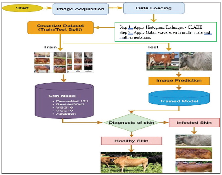
AbstractBackground: Lumpy skin disease (LSD) is an infectious cattle disease that poses a serious threat to animal welfare and the livestock industry. Conventional diagnostic approaches rely heavily on clinical examinations, laboratory testing, and veterinary expertise, making the detection process costly, labor-intensive, and time-consuming. Aim: This study aims to develop an automated framework for the early identification of LSD in cattle using histogram-based image enhancement, Gabor wavelet feature extraction, and deep learning techniques. Methods: Initially, histogram enhancement techniques were applied to cattle images to improve image quality and emphasize lesion regions. Subsequently, Gabor wavelet features with multiple scales and multi-orientations were extracted to capture significant texture patterns related to skin abnormalities and disease characteristics. The extracted features were then classified into healthy and infected categories using various deep learning architectures, including convolutional neural network (CNN), DenseNet121, ResNet50V2, InceptionV3, VGG16, VGG19, and Xception. The proposed framework was evaluated using three benchmark datasets: Mendeley, Kaggle, and Veterinary Research LSD datasets. Results: The experimental evaluation demonstrated that the proposed framework achieved high classification accuracies of 97.5% on the Mendeley dataset, 97% on the Kaggle dataset, and 97% on the Veterinary Research LSD dataset. The integration of histogram enhancement and Gabor texture analysis with CNN-based models significantly improved detection performance compared to existing state-of-the-art methods. Conclusion: The proposed automated LSD identification framework provides a reliable, scalable, and cost-effective solution for early disease detection in cattle. The integration of image enhancement, texture feature extraction, and deep learning techniques can effectively support veterinarians and livestock farmers in disease monitoring and management. Keywords: Gabor wavelet, Histogram, Convolutional neural network, DenseNet121, ResNet50V2. IntroductionFood systems and rural economies in more than a few areas of the world have been built on cattle production, providing a significant contributions to nutrition, employment, and income. Regardless of its significance, the livestock industry has been plagued by the problem of infectious diseases, especially the lumpy skin disease (LSD). Viral diseases are highly contagious among cattle populations and are characterized by severe clinical manifestations, production losses, movement limitations, and massive economic implications at the farm and national levels. Such diseases should be identified correctly and within the required time to contain and treat them. Nevertheless, the practices that are in use in the diagnosis are rely on visual examination, laboratory verification, and experienced veterinary knowledge. Such methods are also expensive, need special infrastructure, and can be impractical in isolated or underdeveloped areas. Thus, the time lag in the diagnosis might lead to uncontrolled infection and mortality of livestock. A considerable amount of recent research has focused on transfer learning using pretrained convolutional neural network (CNN) architectures for LSD detection. Abdullah et al. (2025) evaluated several deep learning models, including DenseNet121, InceptionV3, MobileNetV2, and Xception, and reported that DenseNet121 achieved superior performance because of its efficient feature-reuse capability. DenseNet121 is particularly effective for LSD detection, as its dense connectivity structure preserves low-level texture details while simultaneously learning high-level semantic features, which are essential for identifying subtle lesion characteristics. Although deeper architectures improve classification accuracy, their computational complexity restricts practical deployment in resource-constrained environments. This creates an important tradeoff between accuracy and efficiency that remains a challenge in current research. To enhance model robustness, (Shakeel et al., 2024) applied data augmentation techniques to improve performance under varying illumination and background conditions. While augmentation methods increase dataset diversity, they mainly rely on synthetic variations and may not completely represent real-world scenarios, thereby affecting generalization capability. Likewise, comparative analyses conducted by (Senthilkumar et al., 2024) highlighted the significance of lightweight architectures such as MobileNetV2, which provide lower computational cost and are suitable for real-time applications. However, the study by Saha (2024) indicated that the effectiveness of lightweight models is highly dependent on the application context. These architectures prioritize efficiency over deeper feature representation and may struggle to capture the complex textures, irregular lesion boundaries, and multi-scale patterns associated with LSD. In contrast, (Abdullah et al., 2025) demonstrated that deeper architectures such as DenseNet121 can achieve higher classification accuracy by effectively learning fine-grained lesion features and multi-scale representations. Therefore, lightweight models are advantageous for real-time implementation, whereas deeper architectures remain more suitable when diagnostic accuracy is the primary objective. Hybrid feature extraction approaches have also gained attention for improving deep learning performance. (Saha et al., 2024) achieved 99% accuracy in multimodal activity recognition by integrating thermal, RGB, and 3-D skeletal data for exercise assessment. Similarly, (Zaytoon et al., 2024) introduced the AMINA dataset containing 1.85 million Arabic news articles collected from nine newspapers for multilingual news analysis. Their study utilized models such as AraBERT, CAMeL-BERT, ResNet, EfficientNet, ViT, and RegNet for text and image classification tasks. Experimental findings showed that AraBERT produced the best text classification results, whereas RegNet achieved the highest image classification accuracy of 68.9%. These studies demonstrate the effectiveness of combining multiple feature representations and deep learning strategies for improved classification performance. For LSD detection specifically, (Reza et al., 2026) developed an image classification framework using cattle mouth and hoof images based on CNN architectures. Their results confirmed that deep learning models can effectively distinguish infected cattle from healthy animals without requiring manual feature extraction. In another study, (Olorunshola and Lakan, 2025) enhanced LSD classification performance by combining CNN-based features with Haralick texture descriptors, achieving better results than CNN-only approaches. Extending this concept, multi-stage feature extraction frameworks have shown promising potential by integrating histogram-based enhancement techniques for contrast improvement with patch-based Gabor wavelet features for localized texture and frequency analysis. Such integration improves feature representation by combining global intensity information with detailed lesion texture characteristics, thereby strengthening classification capability. In addition to these recent developments, earlier foundational studies conducted between 2015 and 2020 established the effectiveness of Gabor wavelets in image analysis applications. For example, (Jain et al., 1999) demonstrated that Gabor filters are highly effective in capturing texture, orientation, and frequency information for classification tasks. These findings provide a strong theoretical foundation for applying Gabor wavelet features to texture-rich skin lesion detection problems such as LSD. Despite their potential advantages, integrated frameworks that combine handcrafted texture descriptors with deep learning approaches remain relatively underexplored in the existing literature. Ensemble and hybrid learning methods have further been investigated to improve robustness and generalization performance. (Khandait et al., 2025) reported that combining multiple pretrained CNN architectures enhances classification accuracy across diverse datasets. Similarly, (Goddeti and Narayana, 2024) proposed CNN-based feature extraction integrated with extreme learning machines, achieving faster training and high diagnostic accuracy. Although these approaches improve reliability and predictive performance, their increased architectural complexity may limit scalability and real-time deployment capabilities. Recent studies also emphasize ensemble deep learning and object detection approaches for LSD diagnosis. Saqib et al. (2024) demonstrated that MobileNetV2 provides an effective balance between accuracy and computational efficiency for field-level applications, while (Sentoso et al., 2025) observed that VGG16 performs effectively on moderate-sized datasets, suggesting that model performance is highly influenced by dataset characteristics. Architectures such as Inception and Xception are particularly beneficial for LSD detection because of their ability to capture multi-scale lesion patterns. However, existing studies still do not provide a clear consensus regarding the most suitable architecture for LSD classification. Advanced approaches, including the ensemble CNN framework proposed by Ayon et al. (2026) and the hybrid YOLO-EfficientNet model developed by Ubaidullah et al. (2026) achieved high classification accuracy but mainly focused on specific architectures without integrating handcrafted and deep feature representations into a unified framework. Overall, current literature indicates that CNN-based approaches, especially when combined with transfer learning, augmentation strategies, and hybrid feature extraction techniques, offer significant potential for automated cattle disease diagnosis.However, key research gaps remain: 1. Limited integration of histogram enhancement and texture feature extraction (e.g., Gabor wavelets) with deep CNN models. 2. Lack of a systematic comparison of multiple CNN architectures within a unified framework. 3. Insufficient focus on balancing accuracy, computational efficiency, and real-world applicability. To address these limitations, and considering the texture-rich and multi-scale nature of LSD lesions, the present study proposes a histogram, Gabor wavelet feature extraction integrated with a deep learning framework. This approach is designed to enhance texture sensitivity and multi-scale representation, thereby improving classification performance. Materials and MethodsFigure 1 presents the overall architecture of the proposed framework, which combines histogram enhancement, Gabor wavelet feature extraction, and deep learning models for the automated identification of cattle skin disease. The process begins with image acquisition, where both healthy and LSD-infected cattle skin images are collected and resized to a uniform dimension to ensure consistent processing. The dataset is then separated into training (80%) and testing (20%) sets. During preprocessing, contrast-limited adaptive histogram equalization (CLAHE) is applied to improve image contrast and highlight disease-affected regions more clearly. After enhancement, Gabor wavelet transformation is employed to extract texture-related characteristics associated with lesions, skin irregularities, and abnormal patterns. These features capture important spatial frequency and orientation information, helping the system identify subtle differences between healthy and infected skin areas. The extracted features are then supplied to multiple deep learning architectures for automated feature learning and classification, and the learned representations are stored within the training database. In the final stage, the features obtained from testing images are analyzed against the patterns learned during training, enabling the proposed system to accurately categorize the images as either healthy cattle skin or LSD-infected skin.
Fig. 1. Histogram and Gabor wavelet-based deep learning framework for LSD detection. In this study, multiple pre-trained CNNs are utilized, including DenseNet121, InceptionV3, VGG16, VGG19, Xception, and ResNet50V2. Each architecture contributes unique strengths to the framework. CNNs are deep learning models that identify hierarchical patterns in images for tasks such as detection and classification. Their main components—convolutional, pooling, flattening, and fully connected layers—work together: convolutional layers extract features from low to high levels, pooling layers reduce computation, and fully connected layers perform classification. Advanced CNN architectures, such as ResNet50V2, InceptionV3, VGG16, VGG19, and Xception, enhance performance through techniques like residual connections, extraction of feature at multi-scale, convolutions which are separable on the basis of depth. ResNet50V2 improves gradient flow, InceptionV3 captures multi-scale features, VGG16/19 use simple filters with deep extraction, and Xception reduces computation while preserving rich features, making these models effective for complex image classification tasks. Pre-processing using histogram techniquesPre-processing is essential for improving image quality and optimizing model performance. Raw images may contain noise, low contrast, and uneven illumination, which can reduce the efficiency of feature extraction. To address these issues, adaptive histogram techniques, such as CLAHE, are applied, which redistribute the intensity values of pixels locally to enhance contrast across different regions of the image. Unlike standard global histogram equalization, CLAHE operates on small image tiles, ensuring that regions with varying illumination are enhanced without over-amplifying noise, thereby improving the visibility of skin lesions. The transformation can be expressed as follows: 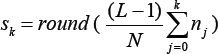
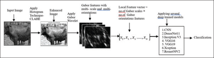
where L is the number of gray levels, nj is the number of pixels at intensity j, and N is the total number of pixels. This enhancement improves the visibility of features and supports better model accuracy, as shown in Figure 2.
Fig. 2. System diagram of the proposed enhancement: Gabor features with different scales and orientations. Feature extraction using Gabor waveletAfter histogram-based enhancement, the improved cattle images S are subjected to Gabor filtering for texture analysis (Jain et al., 1999). Each enhanced cattle image is convolved with a set of Gabor kernels at different orientations and wavelengths. This operation enhances directional textures such as wrinkles, spots, and lesion boundaries on the animal’s skin. The resulting output, commonly referred to as the Gabor magnitude response, emphasizes high-frequency components such as edges and irregular patterns while suppressing uniform regions. Mathematically, the filtered output can be expressed as: 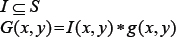
Where I(x,y) represents the enhanced cattle image and g(x,y) is the 2-D Gabor kernel. The symbol * denotes convolution. The magnitude is then computed as: 
This magnitude image provides a clearer representation of texture variations, making it highly effective for identifying abnormal skin patterns in cattle. Features extracted from the cattle image using Gabor wavelet and represented as IG. This process yields a feature vector of size (number of scales × number of orientations). This magnitude image provides a clearer representation of texture variations, making it highly effective for identifying abnormal skin patterns in cattle, as shown in Figure 2. Multimodal architectureConvolutional neural network (CNN)A CNN is a deep learning model used for image analysis, as proposed by Saha (2024). It automatically learns the spatial hierarchies of features from the input images through convolution layers that use filters for extracting patterns like edges, textures, and shapes. These features are downsampled using pooling layers to reduce computational complexity and prevent overfitting. For final classification, the mapping of features that are extracted is done by flattening and passing them to fully connected layers. CNNs use activation functions like ReLU to introduce nonlinearity and softmax for binary-class prediction. Due to weight sharing and local connectivity, CNNs efficiently manage large image data and are widely applied in medical imaging, object detection, and disease diagnosis. Hence, cow disease classification using convolution neural networks. Improve model input quality; design and train a CNN model on a diverse training set; and evaluate it to assess generalization and prevent overfitting on a separate test set. Using distinct training and test datasets ensured the model learned meaningful features and could reliably classify unseen images, as given in equation [3]. For input layer: 
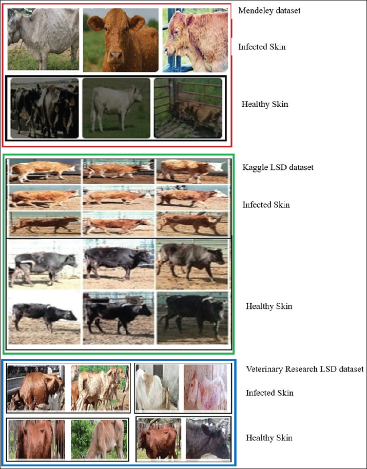
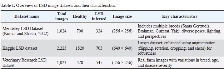
Convolutional layer 1: H1=Relu (W1 X + b1)(3) Where W1 and B1 represent weights and biases for the first convolutional layer, which represent the convolution operation. H1 represents the output feature map after applying 32 filters of size (3 × 3). The CNN was realized on the basis of the Keras Sequential of TensorFlow. It consisted of three consecutive convolutional networks with the number of filters slowly varying (32, 64, 128) and 2 × 2 max-pooling operations that followed them to extract features that were discriminative and downsize the spatial features. The ensuing feature maps were flattened and input to a fully connected layer that had 128 neurons that were activated by ReLU. To reduce overfitting, a dropout of 50% was used in front of the output. The last layer utilized the softmax activation function to produce class probability distributions among the two target categories, namely, healthy and lumpy skin disease, which made it possible to use the method for binary classification. Max Pooling Layer 1: P1=MaxPool (H1, 2)(4) In this case, P1 reduces the spatial resolution of H1 by a factor of 2. The Adam algorithm was used to optimize the network, where the loss function was categorical cross-entropy. The training was done for 20 epochs, and the classification accuracy was monitored during that period to evaluate the convergence behavior and possible overfitting. Convolutional Layer 2: H2=ReLU (W2 P1 + b2)(5) DenseNet121DenseNet121 is a deep convolutional architecture proposed by (Huang and Liu, 2017) that contains 121 layers organized through densely connected blocks. In this structure, each layer obtains inputs from all previous layers within the same block, enabling continuous feature reuse and improved information flow. This connectivity pattern strengthens gradient propagation, mitigates vanishing gradient issues, and reduces redundant parameters compared to conventional CNN models. The conventional convolution and pooling operations are performed using a network made up of a number of dense blocks, followed by a final classification stage. Owing to its compact design and strong feature extraction capability, DenseNet121 is extensively applied in image analysis and medical diagnosis tasks. ResNet50V2ResNet50V2 is a 50-layer residual network introduced by (Goceri, 2019). It employs identity shortcut connections that bypass intermediate layers, enabling improved gradient flow and preventing degradation in deep networks. The V2 variant adopts a pre-activation structure, where batch normalization and activation precede convolution, enhancing training stability. Owing to its strong feature extraction and efficient optimization, ResNet50V2 is widely used in image classification and medical imaging tasks. InceptionV3InceptionV3 is proposed by Rony (2024). The model utilizes inception modules that perform parallel convolutions with different filter sizes to extract multi-scale features efficiently. It also incorporates factorized convolutions, batch normalization, and auxiliary classifiers to reduce computational cost and enhance training convergence. Owing to its optimized structure and strong feature learning capability, this preparation helps the model learn distinct features and reduces overfitting for more accurate classification. VGG16VGGNet, introduced by Simonyan and Zisserman (2015) is a deep CNN known for its uniform architecture. It stacks small 3×3 convolution layers with periodic max-pooling, followed by fully connected layers for classification. The straightforward design enables effective hierarchical feature extraction, making VGG widely used in image analysis tasks. Hence, the VGG16-based classifier is used to sort cattle images into healthy and infected skin. VGG19VGG19 is a deep convolutional architecture containing 19 weight layers, developed by Simonyan and Zisserman (2015). The network is built using repeated 3 × 3 convolution operations combined with ReLU activation, after that periodic max-pooling and finally a dense layer for classification. Its greater depth enhances hierarchical feature representation, making it suitable for complex image analysis and medical diagnostic tasks. XceptionXception, proposed by Chollet (2017), is a deep convolutional architecture that relies entirely on depthwise separable convolutions. This approach divides standard convolution into channel-wise spatial filtering followed by pointwise convolution for feature integration, which reduces computational overhead while maintaining strong representational power. The network also incorporates residual connections to enhance gradient flow and training stability. Xception is extensively applied in image classification to classify cattle images into healthy and LSD categories because it is designed efficiently and achieves higher accuracy. Model.evaluate( ) is used to measure the trained network’s performance on previously unseen test samples by calculating the loss value and classification accuracy, thereby assessing its generalization capability. For further validation, randomly selected test images are visualized with their predicted categories and associated probability scores, which assists in spotting incorrect predictions. The saved model can also be loaded again to perform inference on new input images, enabling its effective application in cow disease diagnosis. Ethical approvalNot needed for this study. ResultsAn overview is provided in this section to introduce the findings and discussion of the proposed scheme. This study employs three publicly available datasets for experimental evaluation: the Mendeley LSD image dataset proposed by Kumar and Shastri (2022), the Kaggle lumpy skin disease image dataset, and the Veterinary Research LSD dataset (collected from publicly available veterinary sources and prior published studies). These datasets are used to evaluate the performance of the proposed framework. Sample images from the three datasets are illustrated in Figure 3, and their detailed descriptions are presented in Table 1. For computational efficiency, the image resolutions in all datasets were uniformly resized to (256 × 256) pixels. The proposed research was conducted on a personal computer equipped with an Intel Core i5 processor running at 3.20 GHz and 8 GB of RAM. All experiments and test cases were implemented using MATLAB R2013a.
Fig. 3. Sample images from three LSD datasets. Table 1. Overview of LSD image datasets and their characteristics.
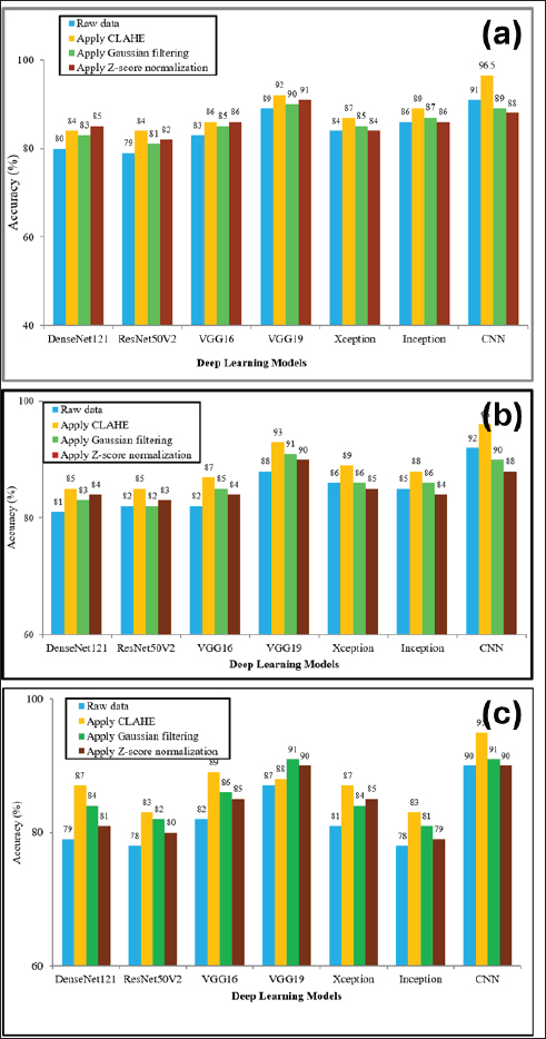
The models were trained using the Adam optimizer with a learning rate of 0.001 and a batch size of 32. Training was conducted for 50–100 epochs, with early stopping applied based on validation loss (patience=10), using an 80:20 training–testing split. Overfitting was controlled through the use of dropout and data augmentation techniques, including rotation, flipping, and scaling. As shown in Table 1, class imbalance was addressed by applying data augmentation to the minority (infected) class, along with class weighting during training to balance the loss contribution. The use of these three datasets, Mendeley, Kaggle, and the Veterinary Research LSD dataset, is significant for several reasons. They provide diversity in data sources, which helps reduce dataset bias and improves the reliability of the model. The datasets include variations in image conditions such as resolution, lighting, pose, and background, thereby simulating real-world scenarios. Each dataset also exhibits differences in class distribution between healthy and infected cattle, enabling evaluation of model stability under imbalanced conditions. Performance analysis of preprocessing methods for LSD identificationIn this experiment, raw cattle images were initially enhanced using the histogram-based CLAHE technique, along with Gaussian filtering and Z-score normalization, to improve image quality and feature representation. The processed images were then provided as input to several pre-trained architectures for the classification of healthy and infected cattle skin. The models were independently evaluated on the three datasets, and the results are presented in Figure 4. The findings indicate that CLAHE consistently improved classification accuracy across all models when compared to other preprocessing methods, while models trained on raw images exhibited lower performance. Among the evaluated architectures, the CNN achieved the highest accuracies of 96.5%, 96%, and 95% on the three datasets when combined with CLAHE. Overall, the results demonstrate that histogram-based enhancement significantly improves model performance for cattle image classification across different architectures.
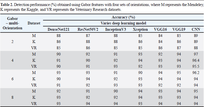
Fig. 4. Comparison of various preprocessing techniques with deep learning models. (a) Mendeley (b) Kaggle (c) Veterinary Research LSD dataset. Performance analysis of Gabor Wavelet with multi-scale and multi-orientationsIn this experiment, Gabor wavelet features were extracted from cattle images using multi-scale and multi-orientation parameters to capture discriminative texture information. These extracted features were then provided as input to various deep learning architectures for the classification of healthy and infected cattle skin. The orientation and scale parameters of the Gabor filter played a significant role in determining the effectiveness of texture-based feature extraction. A comparative evaluation was conducted using multi-orientation sets, including 2 (0°, 90°), 4 (−45°, 0°, 45°, 90°), 6 (−45°, −22.5°, 0°, 22.5°, 45°, 90°), and 8 (−67.5°, −45°, −22.5°, 0°, 22.5°, 45°, 67.5°, 90°), while maintaining a fixed scale value of eight. Experimental results obtained on the three datasets demonstrated that Gabor features combined with CNN achieved superior performance compared with other architectures, as presented in Table 2. In particular, the configuration with 4 orientations achieved the highest accuracies of 97%, 96.4%, and 95.5% on the Mendeley, Kaggle, and Veterinary Research datasets, respectively. Table 2. Detection performance (%) obtained using Gabor features with four sets of orientations, where M represents the Mendeley, K represents the Kaggle, and VR represents the Veterinary Research datasets.
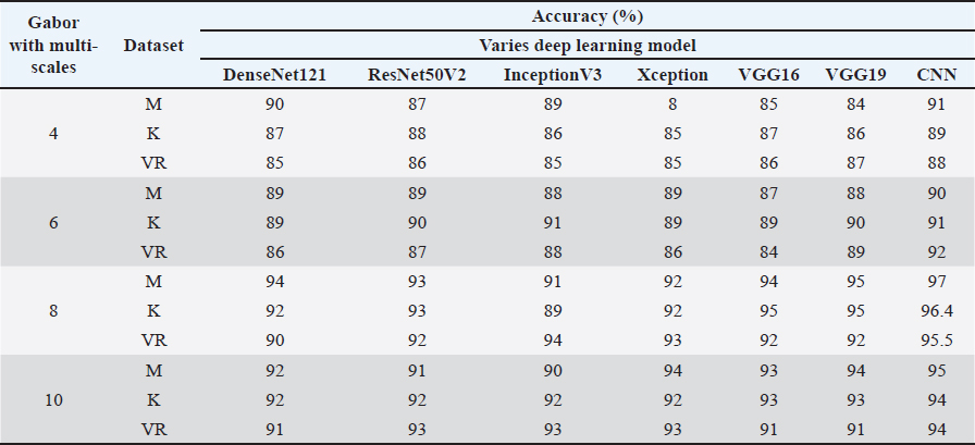
Furthermore, the impact of varying scale parameters was analyzed by fixing the number of orientations at four and evaluating scale ranges of (5:2:11), (5:2:15), (5:2:19), and (5:2:23), corresponding to 4, 6, 8, and 10 scales, respectively. The experimental findings indicate that the optimal balance between computational efficiency and classification accuracy was achieved using 4 orientations and 8 scales in combination with CNN. This configuration consistently outperformed the other settings across all three datasets, achieving accuracies of 97%, 96.4%, and 95.5% on the Mendeley, Kaggle, and Veterinary Research datasets, respectively, as shown in Table 3. Overall, the results confirm that the appropriate selection of Gabor orientation and scale parameters significantly enhances feature representation and improves the classification performance of deep learning models for cattle skin disease detection. The final Gabor feature extraction process generated a feature vector of size 32, derived from 8 scales and 4 orientations. Table 3. Detection performance (%) obtained using Gabor feature of four sets of scales, where M represents the Mendeley, K represents the Kaggle and VR represents the Veterinary Research datasets.
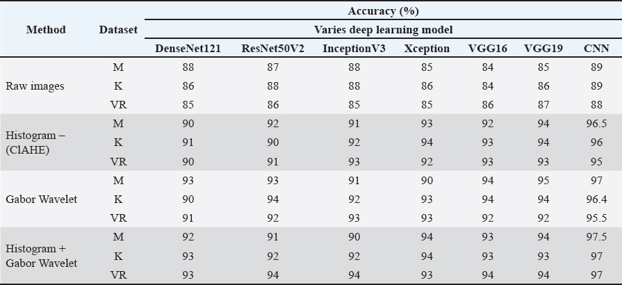
Performance analysis of histogram and Gabor wavelet with various deep learning modelsIn this experiment, raw cattle images, histogram-based CLAHE, Gabor wavelet features, and a hybrid combination of histogram enhancement with Gabor wavelet features were evaluated to improve image quality and feature representation for cattle skin disease classification. The processed images and extracted features were provided as input to several deep learning architectures. The models were independently evaluated on the three datasets, and the results are presented in Table 4 for the classification of healthy and infected cattle skin. The experimental findings indicate that CLAHE techniques significantly improved classification performance compared with raw images. The histogram-based CLAHE technique enhanced the visibility of important skin features and consistently improved the classification accuracy of all models. Table 4 Comparison of the accuracy of deep learning models with raw pixels, histogram-based CLAHE, Gabor wavelet, where M represents the Mendeley, K represents the Kaggle and VR represents the Veterinary Research datasets.
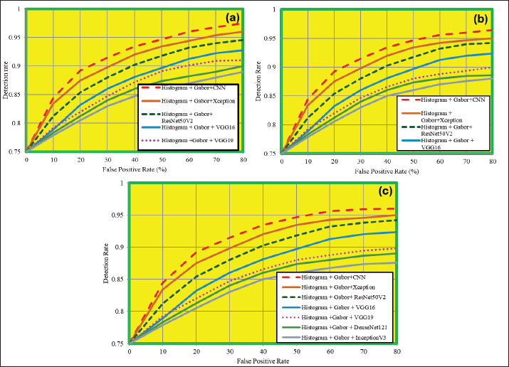
Furthermore, improvement was observed when Gabor wavelet features were utilized, demonstrating the effectiveness of texture-based feature extraction for identifying infected skin patterns. The CNN model with Gabor wavelet features achieved accuracies of 97%, 96.4%, and 95.5% across the three datasets. The best overall performance was obtained by integrating histogram-based enhancement with Gabor wavelet features. This combined approach effectively improved image contrast while simultaneously extracting discriminative texture information, leading to superior classification performance. Using the histogram + Gabor approach, the CNN achieved the highest accuracies of 97.5%, 97%, and 97% on the Mendeley, Kaggle, and Veterinary Research datasets, respectively. Overall, the results demonstrate that the integration of histogram enhancement and Gabor wavelet features significantly improves the feature representation capability of deep learning models and enhances the automated classification of healthy and infected cattle skin images across different datasets. The receiver operating characteristic (ROC) curves were plotted based on the results obtained using histogram + Gabor features combined with several pre-trained deep learning architectures. The evaluations were performed on the Mendeley, Kaggle, and Veterinary Research LSD datasets, and the corresponding results are presented in Figure 5. The ROC curve illustrates the tradeoff between the true positive rate (recognition rate) and the false positive rate. The experimental results indicate that the proposed histogram + Gabor features combined with the CNN model achieved superior performance in LSD detection compared with the other architectures. In particular, this combination achieved a high recognition rate while maintaining a low false positive rate across all datasets, demonstrating its effectiveness in accurately classifying cattle images as healthy or infected.
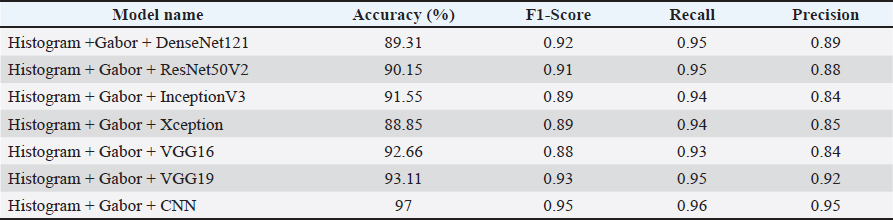
Fig. 5. ROC curve: False positive rate versus detection rate (a) Mendeley dataset (b) Kaggle dataset (c) Veterinary Research LSD dataset. Performance evaluation of deep learning models on the Veterinary Research LSD datasetThe Veterinary Research LSD dataset includes 1023 images (478 healthy, 545 infected), split 80:20 for training and testing. Performance evaluation of the models showed varied results, as shown in Table 5. Among all models, histogram + Gabor + CNN achieved the highest performance with 97% accuracy, 0.95 F1-score, 0.96 recall, and 0.95 precision. VGG19 also showed strong results with 93.11% accuracy and 0.93 F1-score, followed by VGG16 and InceptionV3. DenseNet121 and ResNet50V2 provided competitive performance, while Xception achieved the lowest accuracy of 88.85%. Based on these results, the corresponding confusion matrix is presented in Table 6. Out of the 545 infected images, 529 were correctly identified (true positives), while 16 were misclassified as healthy (false negatives). Similarly, among the 478 healthy images, 463 were correctly classified (true negatives), and 15 were incorrectly predicted as infected (false positives). Table 5. Performance of proposed method in the Veterinary Research dataset for LSD.
Table 6. Confusion matrix for LSD detection on the Veterinary Research LSD dataset.
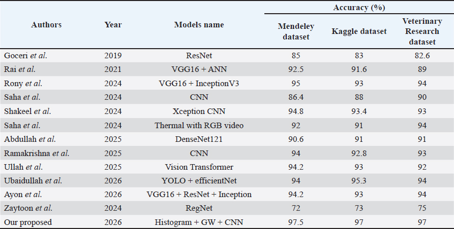
The model evaluation was conducted using an 80:20 training–testing split, and the following statistical measures further validated the robustness of the model: Cohen’s kappa coefficient was computed to assess the agreement between the predicted and actual classifications beyond chance. The obtained value of κ=0.94 indicates an almost perfect agreement. A 95% confidence interval (CI) for the kappa statistic was estimated as 0.91–0.97, demonstrating high reliability and consistency of model performance. McNemar’s test was performed to evaluate the statistical significance of misclassification differences. The results indicated no significant discrepancy (p > 0.05), confirming that the classification was balanced. A paired t-test was conducted using multiple experimental runs under the same 80:20 data split to compare the proposed model with baseline approaches, confirming a statistically significant improvement (p < 0.05). In addition, one-way ANOVA analysis was performed to compare multiple models, and the results showed statistically significant differences among them (p < 0.05). Performance comparison of state-of-the-art models for LSD detectionThe performance comparison of LSD detection methods using an 80:20 training–testing split demonstrates a clear improvement in model effectiveness. The comparative analysis in Table 7 shows that earlier methods, including ResNet, VGG16 + ANN, (Rai et al., 2021) CNN, (Ramakrishna, 2025) Xception CNN, Thermal with RGB video, DenseNet121, Vision Transformer, (Ullah, 2025) and EfficientNet-based models, achieved accuracies ranging from 82% to 95% across the Mendeley, Kaggle, and Veterinary Research datasets. In contrast, the proposed histogram + Gabor wavelet + CNN framework achieved the highest accuracies of 97.5%, 97%, and 97%, respectively. The improved performance is mainly due to the combination of histogram-based enhancement and Gabor wavelet texture extraction, which effectively highlights lesion regions and captures discriminative skin texture patterns. In addition, the CNN model provides efficient feature learning and classification, resulting in more accurate and robust LSD detection compared with existing state-of-the-art methods. Table 7. Comparison of existing LSD detection approaches with proposed methodology in three datasets.
Moreover, a ten-fold cross-validation strategy was adopted to ensure robust evaluation of the proposed model. The model achieved accuracies of 98.5%, 97.34%, and 98% on the Mendeley, Kaggle, and Veterinary Research LSD datasets, respectively. These results demonstrate the consistency and effectiveness of the proposed approach across multiple datasets, highlighting its strong generalization capability for LSD detection. DiscussionThe effectiveness of integrating histogram-based image enhancement, Gabor wavelet feature extraction, and deep learning models has been demonstrated for automated cattle disease detection, particularly LSD. Histogram enhancement techniques significantly improved image clarity and emphasized lesion regions, enabling better visual representation of disease-related abnormalities. In addition, Gabor wavelet features successfully captured discriminative texture patterns, including skin lesions, nodules, and irregular surface structures associated with LSD infection. The integration of these enhanced texture representations with transfer learning-based deep learning architectures, such as CNN, DenseNet121, ResNet50V2, InceptionV3, VGG16, VGG19, and Xception, resulted in strong classification performance across multiple benchmark datasets. The experimental findings indicate that combining histogram enhancement and Gabor texture analysis with CNN models improves feature representation capability and increases disease detection accuracy compared with existing state-of-the-art approaches. Furthermore, the framework reduces dependency on manual inspection and provides a more efficient and consistent diagnostic process for livestock disease monitoring. ConclusionIn conclusion, the proposed automated framework provides a reliable, scalable, and cost-effective solution for early cattle disease diagnosis, particularly for LSD detection. The integration of histogram-based preprocessing, Gabor wavelet texture analysis, and deep learning techniques achieved high classification accuracy across benchmark datasets, demonstrating its robustness and practical applicability. The developed system can assist veterinarians and livestock farmers in making timely and informed decisions, thereby improving livestock health management and reducing economic losses caused by disease outbreaks. AcknowledgmentsThe authors thank all the farm owners and technical staff who assisted in data collection and provided valuable insights. We also acknowledge the support of the Koneru Lakshmaiah Education Foundation for providing resources for this research. Moreover, the authors are thankful to the Deanship of Graduate Studies and Scientific Research at the University of Bisha for supporting this work through the fast-track research support program. FundingThis research received no specific grant. Authors' contributionsAll authors participated in data analysis, manuscript drafting, and revisions. Consent to assume responsibility for all aspects of this study. Conflict of interestThe authors declare that there is no conflict of interest. Data availabilityAll data were provided in the manuscript. ReferencesAbdullah, W., Tanwar, S. and Abouhawwash, M. 2025. Deep learning-based detection of lumpy skin disease in Livestock using CNNs. Sustain. Mach. Intell. J. 11(1), 1–10. Ayon, N.B., Rahman, M.S. and Islam, M.R. 2026. Ensemble deep learning approach for the automated detection of lumpy skin disease and foot-and-mouth disease in cattle. Artif. Intell. Agriculture 12, 2345–2353. Chollet, F. 2017. Xception: Deep learning with depthwise separable convolutions. Proceedings of the IEEE Conference on Computer Vision and Pattern Recognition (CVPR), Honolulu, HI, pp. 1800–1807. Goceri, E. 2019. Deep learning-based classification of skin lesions using ResNet architectures. Biomed. Signal Process. Control 51(1), 1–8. Goddeti, S. and Narayana, V.L. 2024. Lumpy skin disease diagnosis using deep feature extraction and extreme learning machines. Inter. J. Eng. Trends Technol. 72(9), 106–113. Huang, G. and Liu, Z. Densely connected convolutional networks. In IEEE Conf. Computer Vision and Pattern Recognition,2017 , pp 4700–4708. Jain, A.K. and Farrokhnia, F. 1991. Unsupervised texture segmentation using Gabor filters. Pattern. Recognit. 24(12), 1167–1186. Khandait, P.D., Lanjewar, K.K., Patle, S. and Meshram, R. 2025. Deep Learning-Based cattle disease detection: a CNN approach for Identifying lumpy skin disease and foot-and-mouth disease. Int. J. Innov. Res. Technol. (IJIRT). 11(10), 11. Kumar, S. and Shastri, S. 2022. Lumpy skin images dataset. Mendeley Data. Available from: https://universe.roboflow.com/operating-room/cows-mien3/dataset/1 Olorunshola, E. and Lakan, N. 2025. Hybrid convolutional neural network-haralick framework for foot-and-mouth disease classification in cattle. Asian. J. Res. CompSci. 18(6), 390–402. Rai Latha, S. and Sravanthi, S. 2021. Hybrid VGG16 and ANN-based classification of animal diseases. Procedia. Comput. Sci. 192, 2950–2959. Ramakrishna, P. 2025. Convolutional neural network-based classification of cattle diseases. InterJ. Advan. Comp. Sci. Appl. 16(2), 345–354. Reza, M.R., Sahriani, P. and Mukjizat, S. 2026. Clinical image based classification of Foot and Mouth disease in cattle using convolutional neural networks. JurnalSistem. Dan TeknologiInformasi. Indonesia 6(1), 22–31. Rony, M.A.T. 2024. Comparative analysis of vgg16 and inception v3 for livestock disease detection. Multimedia. Tools. Applications. 83(4), 10141–10163. Saha, D.K. 2024. Extensive investigation of convolutional neural network architectures for the diagnosis of Lumpy Skin Disease in dairy cattle. Halcyon 10(4), e27893. Saqib, S., Khan, M., Sharif, A. and Rehman, A. 2024. Lightweight CNN based detection of lumpy skin disease in cattle images. Comput. Biol. Med. 168, 107692. Senthilkumar, C., C, S., Vadivu, G. and Neethirajan, S. 2024. Early detection of lumpy skin disease in cattle using deep learning—A Comparative Analysis of Pretrained Models. Vet. Sci. 11(10), 510. Sentoso, F., Ardiansyah, R. and Wangsa, S. 2025. Performance evaluation of VGG and ResNet architectures for cattle disease image classification. Sistemasi. J. Syst. Inf. 14(1), 89–98. Shakeel, M.Z., Tauheed, N., Javaid, M.T. and Aslam, T. 2024. A deep learning tool for early detection and control of lumpy skin disease using convolutional neural networks. J. Comp. Biomed. Informat. 5(1), 1–14. Simonyan, K. and Zisserman, A. 2015. Very deep convolutional networks for large-scale image recognition. arXiv preprint arXiv:1409.1556. Ubaidullah, A., Hussain, H. and Jafri, M. 2026. LUMPNet: a hybrid YOLO–EfficientNet framework for lumpy skin disease detection. Comput. Biol. Med. 180, 107694. Ullah. 2025. Vision transformer-based automated classification of cattle skin diseases. Expert Syst. Appl. 2025, 235. Zaytoon, M. and Bashar, M 2024. Amina: An Arabic multi-purpose integral news articles dataset. Neural Comput. Appl. 36, 22149–22169; doi: 10.1007/s00521-024-10277-0 | ||

| How to Cite this Article |
| Pubmed Style Selvam IRP, Tabbassum S, Saxena S, Parveen N, Inunganbi SC, Sankaranarayanan R, Thinakaran HP. Automatic detection of animal disease using histogram and multi-scale multi-orientation Gabor Wavelet features with deep convolutional neural networks. Open Vet. J.. 2026; 16(7): 4848-4861. doi:10.5455/OVJ.2026.v16.i7.63 Web Style Selvam IRP, Tabbassum S, Saxena S, Parveen N, Inunganbi SC, Sankaranarayanan R, Thinakaran HP. Automatic detection of animal disease using histogram and multi-scale multi-orientation Gabor Wavelet features with deep convolutional neural networks. https://www.openveterinaryjournal.com/?mno=312737 [Access: July 20, 2026]. doi:10.5455/OVJ.2026.v16.i7.63 AMA (American Medical Association) Style Selvam IRP, Tabbassum S, Saxena S, Parveen N, Inunganbi SC, Sankaranarayanan R, Thinakaran HP. Automatic detection of animal disease using histogram and multi-scale multi-orientation Gabor Wavelet features with deep convolutional neural networks. Open Vet. J.. 2026; 16(7): 4848-4861. doi:10.5455/OVJ.2026.v16.i7.63 Vancouver/ICMJE Style Selvam IRP, Tabbassum S, Saxena S, Parveen N, Inunganbi SC, Sankaranarayanan R, Thinakaran HP. Automatic detection of animal disease using histogram and multi-scale multi-orientation Gabor Wavelet features with deep convolutional neural networks. Open Vet. J.. (2026), [cited July 20, 2026]; 16(7): 4848-4861. doi:10.5455/OVJ.2026.v16.i7.63 Harvard Style Selvam, I. R. P., Tabbassum, . S., Saxena, . S., Parveen, . N., Inunganbi, . S. C., Sankaranarayanan, . R. & Thinakaran, . H. P. (2026) Automatic detection of animal disease using histogram and multi-scale multi-orientation Gabor Wavelet features with deep convolutional neural networks. Open Vet. J., 16 (7), 4848-4861. doi:10.5455/OVJ.2026.v16.i7.63 Turabian Style Selvam, Ithaya Rani Panneer, Saziya Tabbassum, Surabhi Saxena, Nikhat Parveen, Sanasam Chanu Inunganbi, Rathnamala Sankaranarayanan, and Hari Prasath Thinakaran. 2026. Automatic detection of animal disease using histogram and multi-scale multi-orientation Gabor Wavelet features with deep convolutional neural networks. Open Veterinary Journal, 16 (7), 4848-4861. doi:10.5455/OVJ.2026.v16.i7.63 Chicago Style Selvam, Ithaya Rani Panneer, Saziya Tabbassum, Surabhi Saxena, Nikhat Parveen, Sanasam Chanu Inunganbi, Rathnamala Sankaranarayanan, and Hari Prasath Thinakaran. "Automatic detection of animal disease using histogram and multi-scale multi-orientation Gabor Wavelet features with deep convolutional neural networks." Open Veterinary Journal 16 (2026), 4848-4861. doi:10.5455/OVJ.2026.v16.i7.63 MLA (The Modern Language Association) Style Selvam, Ithaya Rani Panneer, Saziya Tabbassum, Surabhi Saxena, Nikhat Parveen, Sanasam Chanu Inunganbi, Rathnamala Sankaranarayanan, and Hari Prasath Thinakaran. "Automatic detection of animal disease using histogram and multi-scale multi-orientation Gabor Wavelet features with deep convolutional neural networks." Open Veterinary Journal 16.7 (2026), 4848-4861. Print. doi:10.5455/OVJ.2026.v16.i7.63 APA (American Psychological Association) Style Selvam, I. R. P., Tabbassum, . S., Saxena, . S., Parveen, . N., Inunganbi, . S. C., Sankaranarayanan, . R. & Thinakaran, . H. P. (2026) Automatic detection of animal disease using histogram and multi-scale multi-orientation Gabor Wavelet features with deep convolutional neural networks. Open Veterinary Journal, 16 (7), 4848-4861. doi:10.5455/OVJ.2026.v16.i7.63 |


