Open Veterinary Journal, (2026), Vol. 16(7): 4373-4387
Research Article
10.5455/OVJ.2026.v16.i7.24
Comprehensive anthocyanin profiling of Hibiscus sabdariffa flower extract and its bioactive potential as a natural poultry feed additive
Miftahul Jannah1, Armita Harahap2, Valdy Filando Sardi2, Yukine Moda3, Akira Nakatsuka3,4,
Kaeoko Murota3,4, Yose Rizal5, Friardi Ismed6 and Maria Endo Mahata5*
1Doctoral Program, Faculty of Animal Science, Universitas Andalas, Limau Manis Campus, Padang, Indonesia
2Doctoral Program, Faculty of Pharmacy, Universitas Andalas, Limau Manis Campus, Padang, Indonesia
3The United Graduate School of Agricultural Sciences (Shimane University section), Tottori University, Tottori, Japan
4Faculty of Life and Environmental Sciences, Shimane University, Matsue, Japan
5*Department of Animal Nutrition, Faculty of Animal Science, Universitas Andalas, Limau Manis Campus, Padang, Indonesia
6Department of Pharmacy, Faculty of Pharmacy, Universitas Andalas, Limau Manis Campus, Padang, Indonesia
*Corresponding Author: Maria Endo Mahata. Department of Animal Nutrition, Faculty of Animal Science, Universitas Andalas, Limau Manis Campus, Padang, Indonesia. Email: maria [at] ansci.unand.ac.id
Submitted: 06/03/2026 Revised: 11/06/2026 Accepted: 21/06/2026 Published: 11/07/2026
© 2025 Open Veterinary Journal
This is an Open Access article distributed under the terms of the Creative Commons Attribution-Non Commercial-No Derivatives License (http://creativecommons.org/licenses/by-nc-nd/4.0/), which permits non-commercial re-use, distribution, and reproduction in any medium, provided the original work is properly cited, and is not altered, transformed, or built upon in any way.
ABSTRACT
Background: Anthocyanins from plant sources have attracted considerable attention as natural bioactive compounds with antioxidant and hypocholesterolemic properties. Hibiscus sabdariffa L. flowers are rich in anthocyanins, which may serve as potential natural additives in poultry feed to reduce cholesterol levels.
Aim: This study aimed to determine the optimal solvent type and maceration duration for extracting anthocyanins from Hibiscus sabdariffa L. flowers and to characterize their anthocyanin profile as a potential natural hypocholesterolemic agent for poultry feed additives.
Methods: A factorial experiment with a completely randomized design was conducted. The first factor was the solvent type, and the second factor was the maceration duration. The evaluated parameters included color measurement, anthocyanin extract yield, total anthocyanin content (TAC), thin-layer chromatography (TLC), LC–MS/MS anthocyanin profiling, total phenolic content, and antioxidant activity using the DPPH radical scavenging assay.
Results: A significant interaction (P < 0.05) between solvent type and maceration duration was observed for color measurement, anthocyanin extract yield, and TAC. TLC analysis revealed five distinct anthocyanin spots. LC–MS/MS analysis identified three major anthocyanin compounds: delphinidin-3-sambubioside, cyanidin-3-sambubioside, and delphinidin. The extract also exhibited considerable total phenolic content, DPPH radical scavenging activity, and specific antioxidant activity. Variations in ΔE color change, anthocyanin extract yield, and TAC were observed among different solvent types and maceration durations.
Conclusion: Anthocyanins from H. sabdariffa L. contain delphinidin-3-sambubioside, cyanidin-3-sambubioside, and phenolics that may inhibit HMG-CoA reductase in the synthesis of cholesterol. DPPH antioxidant activity indicates free radical scavenging ability.
Keywords: Anthocyanin, Cholesterol,Feed additive, Hibiscus sabdariffa L, Poultry.
Introduction
The escalating global demand for poultry meat has established broilers as a cornerstone of the animal protein supply worldwide. Broiler meat remains highly accessible, particularly within emerging and middle-income economies such as Indonesia, characterized by rapid growth rates, superior feed conversion efficiency, and competitive production costs (Nugroho, 2020). However, intensive production systems frequently yield meat with elevated lipid and cholesterol concentrations. In contrast, extensive rearing systems are associated with higher PUFA profiles and reduced cholesterol levels, aligning more closely with contemporary nutritional guidelines (Giampietro-Ganeco et al., 2020).
In addition to red meat, chicken meat contains a high amount of cholesterol, which increases the risk of atherosclerosis and other related diseases, such as diabetes, cancer, and brain diseases, in humans (Falowo, 2022). Strategic interventions have been developed to enhance the lipid profile of poultry meat without compromising zootechnical performance. Nutritional intervention using functional feed additives has emerged as a promising research frontier. Specifically, phytogenic additives derived from plant bioactive compounds have garnered significant attention owing to their safety, sustainability, and multifaceted biological activities (Zhang et al., 2024). Anthocyanins, a class of flavonoid pigments responsible for the cyanic hues in various flora, possess potent hypolipidemic and antioxidant properties (Hina Farheen et al., 2025). Hibiscus sabdariffa L. is a prolific source of anthocyanins, particularly delphinidin- and cyanidin-based derivatives (Adisakwattana et al., 2020; Amer et al., 2022). Anthocyanins modulate lipid metabolism by inhibiting cholesterol synthesis, promoting lipid excretion, and attenuating oxidative stress (Xu et al., 2021). Furthermore, these compounds activate AMP-activated protein kinase (AMPK), which subsequently downregulates the activity of 3-hydroxy-3-methylglutaryl-coenzyme A HMG-CoA reductase, the rate-limiting enzyme in cholesterol biosynthesis (Chen et al., 2021; Godyla-Jabłoński et al., 2024). Consistent with these mechanisms, previous studies have demonstrated the hypolipidemic potential of anthocyanin-rich extracts in both animal models and poultry. Anthocyanin extract from H. sabdariffa (200 mg/kg/day) reduced total cholesterol by 11.45% in rats (Farombi and Ige, 2007). In broiler chickens, dietary supplementation with H. sabdariffa (400 mg/kg feed) decreased serum TG levels by 8.7% (Amer et al., 2022). The efficacy of anthocyanins as functional ingredients depends on extraction methodologies and parameters. The solubility, stability, and yield of anthocyanin extracts are governed by solvent polarity and maceration duration (Krein et al., 2024; Hanifi et al., 2025; Jannah et al., 2026). While solvent polarity directly influences pigment dissolution efficiency, insufficient maceration time often yields suboptimal results. Protracted extraction may induce anthocyanin degradation via oxidation, photo-exposure, and thermal effects (Hanifi et al., 2025). Thus, optimizing extraction conditions is imperative to obtain high-purity extracts with stable pigment profiles.
Despite the documented potential of H. sabdariffa anthocyanins, comparative studies simultaneously evaluating the synergistic effects of floral species, solvent types, and maceration periods remain scarce. Research correlating these extraction characteristics with their application as hypocholesterolemic feed additives in poultry remains underexplored. Therefore, this study aimed to evaluate the effect of solvent type and maceration duration on the characteristics of anthocyanin extracts obtained via maceration on the interaction between H. sabdariffa as an anthocyanin source. Evaluated parameters included extract yield, color measurement, TAC, TLC profiling, anthocyanin identification via LC–MS/MS, TPC, and DPPH antioxidant activity. This study was designed to determine the optimal solvent type and maceration duration for anthocyanin extraction, characterize the anthocyanin profile, and evaluate its potential use as a drinking-water feed additive for reducing cholesterol levels in poultry meat. The findings of this study are expected to provide novel insights into the development of natural strategies for improving meat quality while supporting sustainable and healthy poultry production.
Materials and Methods
Material
Dried flowers of H. sabdariffa were purchased from PT Huma International, Indonesia (50 g per package), and taxonomically verified at the Herbarium Laboratory, Universitas Andalas (ANDA), under voucher specimen no. 899/K-ID/ANDA/XII/2024.
Preparation of anthocyanin extract from H. sabdariffa flowers
Anthocyanins were extracted from H. sabdariffa flowers following the method described by Singh et al. (2021)with several modifications. Initially, dried flowers (250 g) were ground into a fine powder. Subsequently, 10 g of the powder was subjected to maceration in three different solvents: distilled water (pH 6.93), distilled water + 0.5% citric acid (pH 2.16), and 70% ethanol (pH 6.65). Extraction was performed at a solid-to-solvent ratio of 1:10 (w/v) for 6, 12, 24, and 48 h at 4 °C.
The extracts were filtered through Whatman No. 1 filter paper after maceration, and the resulting filtrates were used for color measurement. Extracts obtained using distilled water and distilled water + 0.5% citric acid were subsequently dried using a freeze dryer (Buchi® Lyovapor L-300). The ethanol extract was first concentrated using a rotary evaporator (Buchi® Rotavapor R-210) before being freeze-dried. All dried extracts were stored at −20 °C until further analysis. The extraction procedure and extract yield are shown in Fig. 1.
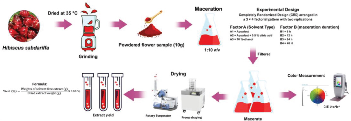
Fig. 1. Flow diagram of the process of anthocyanin extraction from H. sabdariffa flowers and extract yield.
Experimental design
The experiment was arranged in a completely randomized design using a two-factor factorial scheme (3 × 4) with two replications. Factor A represents the solvent type, consisting of distilled water, distilled water + 0.5% citric acid, and 70% ethanol. Factor B corresponded to the maceration duration, which was 6, 12, 24, and 48 h.
Measurement
Color measurement
The color characteristics of the anthocyanin extract from H. sabdariffa flowers were determined using a modified method described by Polat et al. (2022). Color measurements were performed using a Minolta CR-400 colorimeter based on the CIE Lab* color system (L*, a*, and b*). The color intensity of the anthocyanin extracts was recorded using a colorimeter, and the mean and standard deviation values were calculated for each sample.
The total color difference (ΔE) for each sample was subsequently calculated using the following equation:

Explanation:
ΔE=Total color change
L* =Lightness;
a* =Negative values indicate green, while positive values indicate red;
b* =Negative values indicate blue, while positive values indicate yellow;
p =Blank
bp =Sample extract
Extract yield
The extract yield (%) of the H. sabdariffa flower extract was calculated using the formula described by Gonfa et al. (2020)as follows:

Total anthocyanin content
The TAC of H. sabdariffa flower extracts was determined using the pH differential method as described by Lee et al. (2005). Absorbance measurements were performed using a SmartReader UV–Vis microplate absorbance reader (96-well plate, 15 V). The anthocyanin extract (10 mg) was dissolved in two different buffer solutions: potassium chloride buffer (0.025 M KCl, pH 1.0) and sodium acetate buffer (0.4 M CH3COONa, pH 4.5). The pH of each buffer solution was adjusted to 1.0 and verified to be 4.5.
After thorough mixing, the solutions were incubated in the dark at room temperature for 15–30 min. The absorbance of each sample was then measured at two wavelengths, 510 nm and 700 nm, using a microplate reader.
A=(A510-A700) pH 1.0 - (A510-A700) pH 4.5
The total anthocyanin content of the extract was calculated as cyanidin-3-glucoside monomer, based on the following formula (Lee et al., 2005):

Explanation:
A= Absorbance of A and B solutions;
MW=Molecular weight of cyanidin-3-glucoside (448.8)
DF= Dilution factor (0.004)
ɛ= Molar absorptivity of cyanidin-3-glucoside (26.900);
1= Cuvette path length (cm)
Profile of thin-layer chromatography
TLC analysis was conducted following the procedure described in Sampath and Vasanthi (2013)with minor modifications. The H. sabdariffa flower extract was first dissolved in 96% methanol to obtain a final concentration of 2 mg/ml. A small aliquot of the solution was carefully applied onto an aluminum-backed silica gel GF254 TLC plate (10 cm × 12 cm) at a position approximately 1 cm above the lower edge of the plate. After air-drying, the plate was placed in a chromatography chamber previously saturated with the mobile phase. The solvent system used for anthocyanin separation consisted of ethyl acetate, acetic acid, formic acid, and water in the ratio of 60:6:6:15 (v/v/v/v). Following chromatographic development, the plate was observed under ultraviolet light at wavelengths of 254 and 365 nm. The plate was then sprayed with a citroborate reagent to assist in the identification of compound groups. The appearance of yellow, blue, and green fluorescence spots indicated flavonoid compounds in the extract. The detected spots were marked, and their migration distances were recorded to determine the Rf values using the following equation:

Analysis of anthocyanin compounds using LC–MS/MS
The identification of anthocyanin compounds in H. sabdariffa flower extract was performed using liquid chromatography–tandem mass spectrometry (LC–MS/MS) at the Forensic Laboratory Center (Puslabfor), Bogor. The analysis was performed according to the method reported by Ismed et al. (2021)using an ACQUITY UPLC H-Class system (Waters, USA) coupled with a Xevo G2-S QTof mass spectrometer (Waters, USA). Chromatographic separation was achieved on a C18 column (1.8 μm, 2.1 × 100 mm) maintained at 50 °C. The mobile phase consisted of water containing 5 mM ammonium formate (solvent A) and acetonitrile containing 0.05% formic acid (solvent B). The flow rate was 23 min. Before analysis, the sample solution was filtered through a 0.2-μm membrane filter, and 5 μL of the filtrate was injected into the system. Mass spectrometric detection was performed in positive ESI mode (ESI+) over a mass range of 50–1200 m/z. The ion source temperature was set at 100 °C, and the desolvation temperature was maintained at 350 °C. The cone and desolvation gas flow rates were adjusted to 0 and 793 L/h, respectively. Collision energy was applied in the range of 4–60 eV.
Determination of total phenolic content
The TPC of H. sabdariffa extracts was determined using the Folin–Ciocalteu method described by Siddiqui et al. (2017)with slight modifications. The calibration curve was established using gallic acid as the reference standard at concentrations of 0, 20, 40, 60, 80, and 100 µg/ml, with each concentration prepared in duplicate. The extract samples were prepared at a concentration of 1000 µg/ml and analyzed in two independent replicates, with measurements performed at three different times. For the assay, 0.1 ml of the sample solution was mixed with 0.5 ml of 10% (v/v) Folin–Ciocalteu reagent and allowed to react for 3 min. Subsequently, 0.4 ml of 7.5% (w/v) Na2CO3 solution was added to the mixture. Then, the reaction mixture was incubated at room temperature for 60 min. After incubation, absorbance was measured at 765 nm using a UV–Vis spectrophotometer. All measurements were conducted in duplicate and repeated three times. The average absorbance values of the gallic acid standards were used to generate the calibration curve and the corresponding regression equation. The linearity of the calibration curve was evaluated based on the coefficient of determination (R²). The total phenolic content of the samples was calculated using the regression equation and expressed as mg GAE per gram of dry extract (mg GAE/g). The total phenolic content was calculated using the following equation:

where C = total phenolic content in mg/g, in GAE (gallic acid equivalent); C1 = concentration of gallic acid established from the calibration curve in mg/ml; V = volume of extract in ml; and m = the weight of the plant extract in g.
DPPH Radical Scavenging Activity of H. sabdariffa Extracts
The antioxidant capacity of the extracts was evaluated using the DPPH (1,1-diphenyl-2-picrylhydrazyl) radical scavenging assay following the method described by Baliyan et al. (2022). In brief, 2 ml of 0.5 mM DPPH solution was mixed with 0.6 ml of 100 mM Tris–HCl buffer (pH 7.4) in a test tube. To prepare the standard curve, 0.4 ml of ascorbic acid solution at various concentrations was added, and distilled water was used as the blank control. The reaction mixture was thoroughly homogenized and incubated for 20 min in the dark at room temperature to ensure complete reaction. The absorbance was then measured at 517 nm using a UV–Vis spectrophotometer. To determine the antioxidant activity, a calibration curve was constructed by plotting the absorbance against the concentration of ascorbic acid. For sample analysis, 0.4 ml of each extract sample was used in place of the ascorbic acid standard, and the same analytical procedure was followed. Subsequently, the antioxidant capacity of the samples was calculated based on the generated calibration curve.
Data analysis
Statistical analysis of the data on color parameters, extract yield, and total anthocyanin content was performed using analysis of variance based on a completely randomized design (CRD) arranged in a 3 × 4 factorial design. Differences among treatment means were determined using Duncan’s multiple range test (Steel and Torrie, 1995). In addition, anthocyanin profiles obtained from LC–MS/MS analysis were processed and visualized in the form of tables, chromatograms, and mass spectra using MassLynx software (Version 4.1) (Koley et al., 2020).
Ethical approval
Not needed for this study.
Results
Color measurement
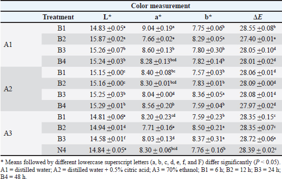
The results of the color analysis of H. sabdariffa flower extract are presented in Table 1. A significant interaction (P < 0.05) was observed between solvent type and maceration duration for the color parameters L*, a*, b*, and ΔE. The total color difference (ΔE) ranged from 27.40 to 28.72. The highest ΔE value was obtained in treatment A3B3 (28.72 + 0.06), while the lowest was recorded in treatment A1B2 (27.40 + 0.01). Although statistically significant differences were observed among treatments, the overall ΔE range remained relatively narrow.
Table 1. Effect of solvent type and maceration duration on H. sabdariffa flower extract color.
Extract yield
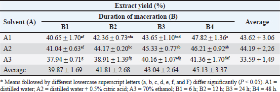
Table 2 presents the extract yield of H. sabdariffa flowers. Statistical analysis revealed a significant interaction (P < 0.05) between the solvent type and maceration duration on the yield of the extract. The extract yields obtained using the water-based solvents A1 and A2 ranged from 40.65% to 47.82% and from 41.04% to 46.21%, respectively, whereas the extract yields obtained using 70% ethanol (A3) ranged from 37.94% to 41.36%. Across maceration durations, the average extract yields were 39.87%, 41.81%, 43.04%, and 45.13% at 6, 12, 24, and 48 hours, respectively. The extract yields obtained using water at 48 hours and water supplemented with 0.5% citric acid at 24 and 48 hours ranged from 45.33% to 47.82%.
Table 2. Effect of solvent type and extraction time on H. sabdariffa flower extract yield (%).
Total anthocyanin content
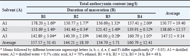
Table 3 presents the TAC of H. sabdariffa flower extract. The statistical analysis revealed a significant interaction (p < 0.05) between solvent type and maceration duration on the TAC values. The highest TAC was recorded in treatment A1B1 (178.20 + 1.69 mg/L). Variations in TAC values were observed across different solvent types and maceration durations, indicating that both extraction factors significantly influenced the recovery of anthocyanin from H. sabdariffa flowers.
Table 3. Effect of solvent type and maceration duration on the total anthocyanin content of H. sabdariffa flower extract (mg/l).
Profile thin-layer chromatography
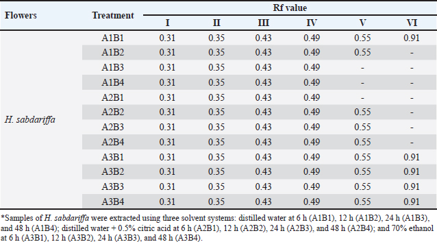
The chromatographic profiles of compounds extracted from H. sabdariffa flowers using different solvent systems and maceration durations are shown in Fig. 2(a–c), and the Rf values (Table 4). Observations were conducted under UV light at 254 nm (Fig. 2a) and 365 nm (Fig. 2b), followed by visualization at 365 nm (Fig. 2c). We observed faint spots under UV light at 254 nm, indicating relatively weak fluorescence of the separated compounds. In contrast, 365-nm observations revealed several bluish to purplish fluorescent bands, suggesting the presence of bioactive compounds in the extracts. Band intensity and distribution differences were observed among treatments. After visualization with the citroborate reagent, the chromatographic bands became clearer and more intense. The TLC analysis also revealed variations in the number of detected bands among treatments. When distilled water was used as the solvent, 6 bands were observed at maceration for 6 h, whereas 4 major bands were detected at 24 hours and 48 hours. The band at Rf 0.55 appeared consistently across several maceration durations in the extracts obtained using distilled water + 0.5% citric acid, while bands with higher Rf values (0.91) were not consistently detected. Extraction with 70% ethanol produced relatively consistent TLC patterns across maceration durations.
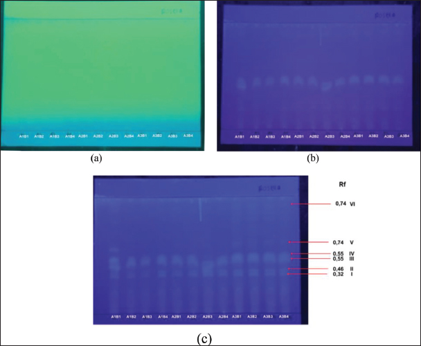
Fig. 2. TLC profiling of extracts of H. sabdariffa flowers obtained using different solvent systems and extraction durations. Samples included distilled water at 6 h (A1B1), 12 h (A1B2), 24 h (A1B3), and 48 h (A1B4); distilled water + 0.5% citric acid at 6 h (A2B1), 12 h (A2B2), 24 h (A2B3), and 48 h (A2B4); and 70% ethanol at 6 h (A3B1), 12 h (A3B2), 24 h (A3B3), and 48 h (A3B4). Chromatographic patterns are visualized under UV 254 nm (a), UV 365 nm (b), and UV 365 nm, and (c) 365-nm UV after visualization with citroborate reagent. The solvent system used for anthocyanin separation consisted of ethyl acetate, acetic acid, formic acid, and water in the ratio of 60:6:6:15 (v/v/v/v).
Table 4. Rf values of the TLC profile of H. sabdariffa flower extract.
Analysis of anthocyanin compounds using LC–MS/MS
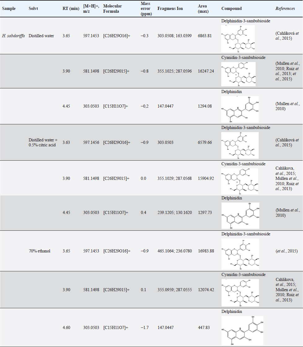
Based on the LC–MS/MS profiles, the H. sabdariffa flower extracts exhibited multiple anthocyanin peaks. The identified anthocyanin compounds and their corresponding LC–MS/MS characteristics are summarized in Table 5. LC–MS/MS analysis (Fig. 3) identified three anthocyanin compounds: delphinidin-3-sambubioside, cyanidin-3-sambubioside, and delphinidin. In extracts obtained using distilled water acidified with 0.5% citric acid (Fig. 4), the same three anthocyanins, namely, delphinidin-3-sambubioside, cyanidin-3-sambubioside, and delphinidin, were consistently detected. Similarly, the extraction with 70% ethanol (Fig. 5) also detected the three compounds. In the distilled water extract, delphinidin-3-sambubioside was detected at a retention time of 3.65 min with an [M+H]+ ion at m/z 597.1453, producing fragment ions at m/z 303.0508 and 163.0399. Cyanidin-3-sambubioside was eluted at 3.90 min of RT (m/z 581.1498) with fragment ions at m/z 355.1025 and 287.0596. The addition of 0.5% citric acid slightly increased the intensity of cyanidin-3-sambubioside detection, with a peak area of 16,247 compared with 15,904 in the distilled water extract. Extraction with 70% ethanol produced the highest peak area for delphinidin-3-sambubioside (16,983.88), whereas all extracts contained relatively low levels of free delphinidin.
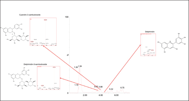
Fig. 3. LC chromatogram and MS/MS spectra of H. sabdariffa flower extract obtained using distilled water. Identified compounds: delphinidin-3-sambubioside, cyanidin-3-sambubioside, and delphinidin.
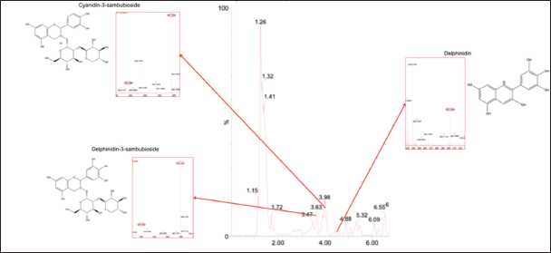
Fig. 4. LC chromatogram and MS/MS spectra of H. sabdariffa flower extract using distilled water + 0.5% citric acid. Identified compounds: delphinidin-3-sambubioside, cyanidin-3-sambubioside, and delphinidin.
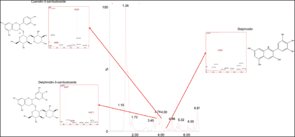
Fig. 5. LC chromatogram and MS/MS spectra of H. sabdariffa flower extract prepared using 70% ethanol. Identified compounds: delphinidin-3-sambubioside, cyanidin-3-sambubioside, and delphinidin.
Table 5. List of structure anthocyanins tentatively identified in H. sabdariffa flower extract using LC–MS/MS.
Total polyphenol content and comparative antioxidant activity of extracts based on the highest anthocyanin content
The highest total phenolic content (TPC) and DPPH radical scavenging activity of H. sabdariffa flower extract were obtained using distilled water with a maceration time of 6 h (Table 6). The total phenolic content of the extract was 19.37 + 0.51 mg GAE/ml. The extract also exhibited a DPPH radical scavenging activity of 0.308 + 0.008 mg AAE/ml, resulting in specific antioxidant activity of 0.016 mg/mg based on the total phenolic content.
Table 6. Total phenolic content of H. sabdariffa flower extract containing three Anthocyanins.

Discussion
Color measurement
Variations in L* values were associated with differences in anthocyanin stability, which are influenced by solvent polarity and pH. Acidified extraction systems are known to maintain anthocyanins in the form of flavylium cations, thereby improving pigment stability and color retention (Xue et al., 2024; Divya et al., 2025).
The a* parameter reflects redness intensity and is closely related to the abundance of anthocyanins in their stable flavylium form under acidic conditions, which gives H. sabdariffa extracts their characteristic red color (Nistor et al., 2022). Lower a* values may indicate anthocyanin degradation or structural transformation into less intensely colored forms, such as hemiketal and chalcone structures (Peñaloza et al., 2022). Higher b* values may reflect a shift toward yellowish tones due to anthocyanin hydration, copigmentation, or other phenolic compounds (Magalhães et al., 2024).
Despite the relatively small differences in ΔE and statistical differences, visual color variations among treatments were limited. Overall, solvent composition and extraction conditions played an important role in preserving anthocyanin stability and maintaining the color quality of H. sabdariffa extracts. Acidified aqueous systems are more effective in retaining redness intensity and producing more color-stable extracts for potential food and feed applications.
Extract yield
The higher extraction yield obtained using water-based solvents may be attributed to the polarity of the major bioactive compounds in H. sabdariffa, particularly anthocyanins and organic acids, which are more soluble in polar solvents. Aqueous extraction systems improve the recovery of these compounds compared with hydroalcoholic solvents (Izquierdo-Vega et al., 2020). In addition, acidified extraction may further enhance extraction efficiency by promoting cell wall disruption and stabilizing anthocyanins in their flavylium cation form, thereby increasing the release of soluble compounds into the solvent (Enaru et al., 2021).
The effect of maceration duration is associated with mass transfer during solvent penetration into plant tissues. Longer extraction times generally allow greater diffusion of secondary metabolites into the solvent until equilibrium is reached, resulting in improved extraction efficiency (Dougherty et al., 2019). Overall, the use of aqueous acidified solvents combined with appropriate maceration duration appears to be more effective in maximizing the extraction yield from H. sabdariffa.
Total anthocyanin content
The high anthocyanin content in H. sabdariffa extracts may be related to the naturally high concentration of organic acids, including citric, hydroxycitric, hibiscus, tartaric, malic, and ascorbic acids, in the flower tissues, which create an acidic environment favorable for anthocyanin stability (Da-Costa-Rocha et al., 2014). Under acidic conditions, anthocyanins predominantly exist in the flavylium cation form, which is more stable, highly soluble in water, and responsible for the characteristic red coloration of H. sabdariffa extracts (Khoo et al., 2017; Izquierdo-Vega et al., 2020).
The acidic matrix of H. sabdariffa petals may also facilitate anthocyanin diffusion from plant tissues into the solvent and improve pigment release by partially disrupting cell wall structures (Mattioli et al., 2020). These findings suggest that the interaction between plant matrix characteristics, solvent polarity, and extraction duration influences anthocyanin extraction efficiency. Overall, aqueous extraction combined with shorter maceration time appeared to be more effective in preserving anthocyanin stability and maximizing pigment recovery, which is consistent with previous findings on flower-based anthocyanin extraction (Tena and Asuero, 2022).
Profile thin-layer chromatography
The stronger fluorescent bands observed at 365 nm than at 254 nm that the extracted compounds exhibit greater fluorescence under longer UV wavelengths, which is characteristic of phenolic compounds such as anthocyanins and flavonoids. The increased band visibility after citroborate treatment may be related to the formation of borate complexes with hydroxyl groups in flavonoid and anthocyanin structures, thereby enhancing fluorescence intensity and facilitating compound detection (Riasari et al., 2025; Jannah et al., 2026).
The reduced number of bands in aqueous extracts with prolonged maceration may indicate partial degradation or lower stability of certain compounds, particularly anthocyanins, which are sensitive to extended extraction conditions (Enaru et al., 2021). The more consistent chromatographic profiles observed in acidified water and 70% ethanol extracts indicate that these solvent systems are more effective in maintaining compound stability and extracting phenolic compounds with different polarities.
The inconsistency of bands with higher Rf values indicates that less polar compounds were either less stable or less efficiently extracted under the tested conditions. Overall, solvent type and extraction duration played important roles in determining the diversity, stability, and persistence of bioactive compounds in H. sabdariffa extracts. Similar chromatographic patterns among treatments may also have relatively comparable chemical compositions and potentially similar biological activities (Zych and Pyka-Pająk, 2025).
Analysis of anthocyanin compounds using LC–MS/MS
The LC–MS/MS analysis showed that nonacylated sambubioside derivatives, particularly delphinidin-3-sambubioside and cyanidin-3-sambubioside, dominated the anthocyanin profile of H. sabdariffa flowers. These compounds are the major anthocyanins responsible for the characteristic red color and biological activity of roselle extracts (Mullen et al., 2010; Ruiz et al., 2013; Cahlíková et al., 2015).
The increased detection of cyanidin-3-sambubioside under acidified conditions suggests that anthocyanin stability is maintained in acidic environments by promoting the flavylium cation form, which is less susceptible to degradation during extraction and analysis (Ruiz et al., 2013; Cahlíková et al., 2015). Although ethanol extraction resulted in higher detection of delphinidin-3-sambubioside, the overall anthocyanin composition remained relatively similar across solvent systems, indicating that the dominant anthocyanins in H. sabdariffa are highly polar and can be effectively extracted using aqueous solvents. Overall, distilled water is an effective and environmentally friendly solvent for preserving nonacylated sambubioside anthocyanins in H. sabdariffa. These findings support the potential application of roselle anthocyanins in functional foods and feed additives targeting lipid metabolism. In particular, delphinidin-3-sambubioside has been reported to attenuate hyperlipidemia and oleic acid-induced steatosis in high-fat diet-induced obese rats and HepG2 cells, respectively (Long et al., 2021). In addition, cyanidin-3-sambubioside-rich extracts significantly reduce aortic cholesterol accumulation and improve HDL function in hyperlipidemic rat models, suggesting a protective effect against cardiovascular disease (Farrell et al., 2015).
Total polyphenol content and comparative antioxidant activity of extracts based on the highest anthocyanin content
The relatively high total phenolic content in H. sabdariffa extract indicates the presence of abundant phenolic constituents, including the major anthocyanins identified in this study, namely delphinidin-3-sambubioside, cyanidin-3-sambubioside, and delphinidin. These compounds are well recognized for their strong contribution to roselle extracts’ antioxidant capacity. The observed DPPH radical scavenging activity reflects the phenolic compounds’ strong hydrogen-donating ability, enabling effective free radical neutralization. The positive association between total phenolic content and antioxidant activity is consistent with previous findings, which reported that nonacylated sambubioside anthocyanins play a key role in the radical scavenging properties of H. sabdariffa extracts (Tena et al., 2020). The relatively higher phenolic retention in distilled water extracts indicates that aqueous systems are effective in preserving anthocyanin stability during extraction. Overall, aqueous extraction using distilled water provides an efficient and environmentally friendly approach for obtaining phenolic-rich H. sabdariffa extracts. These characteristics support their potential application as natural feed additives to enhance antioxidant status and support lipid metabolism in poultry.
Conclusion
It can be concluded that the extraction conditions influenced the change in anthocyanin color, extract yield, and total anthocyanin content. The 70% ethanol solvent with a maceration time of 24 h produced the greatest ΔE color change, whereas distilled water at 48 h and distilled water supplemented with 0.5% citric acid at 24 and 48 h resulted in greater anthocyanin extract yields. Distilled water with a maceration time of 6 h produced the highest total anthocyanin content. Anthocyanins extracted from H. sabdariffa flowers contained delphinidin-3-sambubioside and cyanidin-3-sambubioside, compounds known to inhibit HMG-CoA reductase activity in the synthesis of cholesterol. The extract also exhibited considerable total phenolic content, DPPH radical scavenging activity, and specific antioxidant activity, indicating its ability to scavenge free radicals. Therefore, anthocyanin extract from H. sabdariffa flowers may serve as a potential natural feed additive for reducing cholesterol levels in poultry.
Acknowledgments
The authors would like to sincerely acknowledge the laboratory staff and technicians for their valuable technical assistance. The authors also express their deepest gratitude to Shimane University, Japan, for the academic support and guidance provided during the PMDSU PKPI and PKP programs, particularly in facilitating the polyphenol and DPPH analyses of H. sabdariffa flower extract samples at the Laboratory of the Faculty of Life and Environmental Science, Shimane University.
Conflict of interest
The authors declare no conflicts of interest.
Funding
This research was funded by the PMDSU grant program from the Ministry of Research, Technology, and Higher Education of the Republic of Indonesia in 2024 (Master Contract No. 041/E5/PG.02.00.PL/2024 and Subsidiary Contract No. 134/UN16.19/PT.01.03/PL/2024).
Authors’ contributions
MJ and MEM were responsible for the conceptual framework and design of the study. FI, YR, MK, and AN provided technical support and supervision throughout the study. MJ, AH, VFS, and YM conducted the laboratory work, performed the data analysis, and prepared the initial draft of the manuscript. All authors have read and approved the final version of the manuscript.
Data availability
All data underlying the results of this study are included in the manuscript.
References
Adisakwattana, S., Pasukamonset, P. and Chusak, C. 2020. Clitoria ternatea beverages and antioxidant usage. In Pathology. Cambridge, MA: Academic Press, pp: 189–196; doi: 10.1016/B978-0-12-815972-9.00018-4
Amer, S.A., Al-Khalaifah, H.S., Gouda, A., Osman, A., Goda, N.I.A., Mohammed, H.A., Darwish, M.I.M., Hassan, A.M. and Mohamed, S.K.A. 2022. Potential Effects of Anthocyanin-Rich Roselle (Hibiscus sabdariffa L.) Extract on the Growth, Intestinal Histomorphology, Blood Biochemical Parameters, and the Immune Status of Broiler Chickens. Antioxidants 11(3), 544; doi: 10.3390/antiox11030544
Baliyan, S., Mukherjee, R., Priyadarshini, A., Vibhuti, A., Gupta, A., Pandey, R.P. and Chang, C.M. 2022. Determination of Antioxidants by DPPH Radical Scavenging Activity and Quantitative Phytochemical Analysis of Ficus religiosa. Molecules 27(4), 1326; doi: 10.3390/molecules27041326
Cahlíková, L., Ali, B.H., Havlíková, L., Ločárek, M., Siatka, T., Opletal, L. and Blunden, G. 2015. Anthocyanins of Hibiscus sabdiffera calyces from Sudan. Nat. Prod. Commun. 10(1), 77–79.
Chen, C., Yang, X., Liu, S., Zhang, M., Wang, C., Xia, X., Lou, Y. and Xu, H. 2021. The effect of lipid metabolism regulator anthocyanins from Aronia melanocarpa on 3T3-L1 preadipocytes and C57BL/6 mice via activating AMPK signaling and gut microbiota. Food Function 12(14), 6254–6270; doi: 10.1039/D1FO00907A
Da-Costa-Rocha, I., Bonnlaender, B., Sievers, H., Pischel, I. and Heinrich, M. 2014. Hibiscus sabdariffa L.–A phytochemical and pharmacological review. Food. Chem. 165, 424–443; doi: 10.1016/j.foodchem.2014.05.002
Divya., Joshi, S., Appukuttan, J., Chandrapala, J. and Majzoobi, M. 2025. Impact of Conventional and Advanced Techniques on Stability of Natural Food Colourants. Foods 14(18), 3187; doi: 10.3390/foods14183187
Dougherty, P.G., Sahni, A. and Pei, D. 2019. Understanding Cell Penetration of Cyclic Peptides. Chem. Rev. 119(17), 10241–10287; doi: 10.1021/acs.chemrev.9b00008
Enaru, B., Drețcanu, G., Pop, T.D., Stǎnilǎ, A. and Diaconeasa, Z. 2021. Anthocyanins: factors Affecting Their Stability and Degradation. Antioxidants. (Basel). 10(12), 1967; doi: 10.3390/antiox10121967
Falowo, A.B. 2022. Potential of medicinal plants as Hypocholesterolemic agents in chicken meat production. Sci. Lett. 10(1), 24–31; doi: 10.47262/SL/10.1.132021028
Farombi, E.O. and Ige, O.O. 2007. Hypolipidemic and antioxidant effects of ethanolic extract from dried calyx of Hibiscus sabdariffa in alloxan-induced diabetic rats. Fundam. & Clin. Pharmacol. 21(6), 601–609; doi: 10.1111/j.1472-8206.2007.00525.x
Farrell, N., Norris, G., Lee, S.G., Chun, O.K. and Blesso, C.N. 2015. Anthocyanin-rich black elderberry extract improves markers of HDL function and reduces aortic cholesterol in hyperlipidemic mice. Food. & Function. 6(4), 1278–1287; doi: 10.1039/c4fo01036a
Giampietro-Ganeco, A., Boiago, M.M., Mello, J.L.M., Souza, R.A., Ferrari, F.B., Souza, P.A. and Borba, H. 2020. Lipid assessment, cholesterol and fatty acid profile of meat from broilers raised in four different rearing systems. Anais da Academia Brasileira de Ciencias 92(suppl 1), e20190649; doi: 10.1590/0001-3765202020190649
Godyla-Jabłoński, M., Raczkowska, E., Jodkowska, A., Kucharska, A.Z., Sozański, T. and Bronkowska, M. 2024. Effects of Anthocyanins on Components of Metabolic Syndrome-A Review. Nutrients 16(8), 1103; doi: 10.3390/nu16081103
Gonfa, T., Teketle, S. and Kiros, T. 2020. Effect of extraction solvent on qualitative and quantitative analysis of major phyto-constituents and in-vitro antioxidant activity evaluation of Cadaba rotundifolia Forssk leaf extracts. Cogent Food & Agriculture 6(1), 1853867; doi: 10.1080/23311932.2020.1853867
Hanifi, L., Chaher-Bazizi, N., Bellik, Y., Karou, L., Kaddour, T., Saidene, N., Kilani-Atmani, D., Atmani, D. and Bachir-Bey, M. 2025. Optimization of Monomeric Anthocyanin Extraction Conditions From a New Source (Crataegus azarolus L. Fruits) Sequential Methodology Combined With a Central Composite Design, and Evaluation of Their Antioxidant Properties. J. Food Sci. 90(5), e70300; doi: 10.1111/1750-3841.70300
Hina Farheen., Ambrish Kumar., Devesh Kumar., Shivam Parmar., Deeksha Pathak., Preetika Sharma. and Saroj Singh Chahar*. 2025. Anthocyanins: natural Pigments with Multifaceted Health Benefits for Humans. J. Sci. Innov. Nature Earth 5(3), 1–3; doi: 10.59436/jsiane.407.2583-2093
Ismed, F., Desti, W. N., Arifa, N., Rustini, R., & Putra, D. P. 2021. TLC-Bioautographic and LC-MS/MS Detection of Antimicrobial Compounds from Four Semipolar Extracts of Cladonia Species. https://doi.org/10.2991/ahsr.k.211105.008
Izquierdo-Vega, J., Arteaga-Badillo, D., Sánchez-Gutiérrez, M., Morales-González, J., Vargas-Mendoza, N., Gómez-Aldapa, C., Castro-Rosas, J., Delgado-Olivares, L., Madrigal-Bujaidar, E. and Madrigal-Santillán, E. 2020. Organic Acids from Roselle (Hibiscus sabdariffa L.)-A Brief Review of Its Pharmacological Effects. Biomedicines 8(5), 100; doi: 10.3390/biomedicines8050100
Jannah, M., Harahap, A., Sardi, V.F., Rizal, Y., Ismed, F. and Mahata, M.E. 2026. Anthocyanin Identification in Miana Leaf (Coleus scutellarioides L. Benth) Extracts Using Different Solvents and Maceration Times for Application as a Poultry Feed Additive. Trends. Sci. 23(7), 12496; doi: 10.48048/tis.2026.12496
Khoo, H.E., Azlan, A., Tang, S.T. and Lim, S.M. 2017. Anthocyanidins and anthocyanins: colored pigments as food, pharmaceutical ingredients, and the potential health benefits. Food Nutr. Res. 61(1), 1361779; doi: 10.1080/16546628.2017.1361779
Koley, T.K., Khan, Z., Oulkar, D., Singh, B.K., Maurya, A., Singh, B. and Banerjee, K. 2020. High resolution LC-MS characterization of phenolic compounds and the evaluation of antioxidant properties of a tropical purple radish genotype. Arabian J. Chem. 13(1), 1355–1366; doi: 10.1016/j.arabjc.2017.11.007
Krein, D.D.C., Emer, C.D., Dettmer, A. and Piccin, J.S. 2024. Anthocyanin: a review of the technologies for obtaining the compound. Ciência. E. Natura. 46, e84237; doi: 10.5902/2179460X84237
Lee, J., Durst, R.W., Wrolstad, R.E., Eisele, T., Giusti, M.M., Hach, J., Hofsommer, H., Koswig, S., Krueger, D.A., Kupina;, S., Martin, S.K., Martinsen, B.K., Miller, T.C., Paquette, F., Ryabkova, A., Skrede, G., Trenn, U. and Wightman, J.D. 2005. Determination of Total Monomeric Anthocyanin Pigment Content of Fruit Juices, Beverages, Natural Colorants, and Wines by the pH Differential Method: collaborative Study. J. AOAC. Int. 88(5), 1269–1278; doi: 10.1093/jaoac/88.5.1269
Long, Q., Chen, H., Yang, W., Yang, L. and Zhang, L. 2021. Delphinidin-3-sambubioside from Hibiscus sabdariffa. L attenuates hyperlipidemia in high fat diet-induced obese rats and oleic acid-induced steatosis in HepG2 cells. Bioengineered 12(1), 3837–3849; doi: 10.1080/21655979.2021.1950259
Magalhães, D., Gonçalves, R., Rodrigues, C.V., Rocha, H.R., Pintado, M. and Coelho, M.C. 2024. Natural Pigments Recovery from Food By-Products: health Benefits towards the Food Industry. Foods 13(14), 2276; doi: 10.3390/foods13142276
Mattioli, R., Francioso, A., Mosca, L. and Silva, P. 2020. Anthocyanins: a Comprehensive Review of Their Chemical Properties and Health Effects on Cardiovascular and Neurodegenerative Diseases. Molecules 25(17), 3809; doi: 10.3390/molecules25173809
Mullen, W., Larcombe, S., Arnold, K., Welchman, H. and Crozier, A. 2010. Use of accurate mass full scan mass spectrometry for the analysis of anthocyanins in berries and berry-fed tissues. J. Agric. Food Chem. 58(7), 3910–3915; doi: 10.1021/jf902267v
Nistor, M., Pop, R., Daescu, A., Pintea, A., Socaciu, C. and Rugina, D. 2022. Anthocyanins as Key Phytochemicals Acting for the Prevention of Metabolic Diseases: an Overview. Molecules 27(13), 4254; doi: 10.3390/molecules27134254
Nugroho, B.A., 2020. Indonesia’s broilers business facing oversupply difficulties. IOP Conf. Ser. Earth Environ. Sci. 478, 012010. Bristol, United Kingdom: IOP Publishing; doi: 10.1088/1755-1315/478/1/012010
Peñaloza, S., Delesma, C., Muñiz, J. and López-Ortiz, A. 2022. The anthocyanin’s role on the food metabolic pathways, color and drying processes: an experimental and theoretical approach. Food Bioscience 47, 101700; doi: 10.1016/j.fbbio.2022.0101700
Polat, S., Guclu, G., Kelebek, H., Keskin, M. and Selli, S. 2022. Comparative elucidation of colour, volatile and phenolic profiles of black carrot (Daucus carota L.) pomace and powders prepared by five different drying methods. Food Chem. 369, 130941; doi: 10.1016/j.foodchem.2021.130941
Riasari, H., Fitriansyah, S.N., Hasanah, S.U., Aulia, M., Zavella, K. and Padilah, N. 2025. Quantification of Phenolics, Flavonoids, and In Vitro Antioxidant Activity in Rosella and Breadfruit Leaf Extracts. Pharmacognosy. J. 17(6), 17; doi: 10.5530/pj.2025.17.86
Ruiz, A., Hermosín-Gutiérrez, I., Vergara, C., von Baer, D., Zapata, M., Hitschfeld, A., Obando, L. and Mardones, C. 2013. Anthocyanin profiles in South Patagonian wild berries by HPLC-DAD-ESI-MS/MS. Food Res. Int. 51(2), 706–713; doi: 10.1016/j.foodres.2013.01.043
Sampath, M. and Vasanthi, M. 2013. Isolation and structural elucidation of flavonoids from Polyalthia longifolia (Sonn.) Thwaites and evaluation of antibacterial, antioxidant and anticancer potential. Int. J. Pharm. Pharm. Sci. 5(1), 336–341.
Siddiqui, N., Rauf, A., Latif, A. and Mahmood, Z. 2017. Spectrophotometric determination of the total phenolic content, spectral and fluorescence study of the herbal Unani drug Gul-e-Zoofa ( Nepeta bracteata Benth). J. Taibah. Univ. Med. Sci. 12(4), 360–363; doi: 10.1016/j.jtumed.2016.11.006
Singh, S., Nwabor, O.F., Syukri, D.M. and Voravuthikunchai, S.P. 2021. Chitosan-poly(vinyl alcohol) intelligent films fortified with anthocyanins isolated from Clitoria ternatea and Carissa carandas for monitoring beverage freshness. Int. J. Biol. Macromolecules 182, 1015–1025; doi: 10.1016/j.ijbiomac.2021.04.027
Steel, R.G.D. and Torrie, J.H. 1995. Prinsip dan Prosedur Statistika (4th ed).Indonesia: Penerbit Gramedia Pustaka Utama.
Tena, N. and Asuero, A.G. 2022. Up-To-Date Analysis of the Extraction Methods for Anthocyanins: principles of the Techniques, Optimization, Technical Progress, and Industrial Application. Antioxidants (Basel). 11(2), 286; doi: 10.3390/antiox11020286
Tena, N., Martín, J. and Asuero, A.G. 2020. State of the Art of Anthocyanins: antioxidant Activity, Sources, Bioavailability, and Therapeutic Effect in Human Health. Antioxidants (Basel). 9(5), 451; doi: 10.3390/antiox9050451
Xu, Y., Ke, H., Li, Y., Xie, L., Su, H., Xie, J., Mo, J. and Chen, W. 2021. Malvidin-3-O-Glucoside from Blueberry Ameliorates Nonalcoholic Fatty Liver Disease by Regulating Transcription Factor EB-Mediated Lysosomal Function and Activating the Nrf2/ARE Signaling Pathway. J. Agric. Food Chem. 69(16), 4663–4673; doi: 10.1021/acs.jafc.0c06695
Xue, H., Zhao, J., Wang, Y., Shi, Z., Xie, K., Liao, X. and Tan, J. 2024. Factors affecting the stability of anthocyanins and strategies for improving their stability: a review. Food Chem. X. 24, 101883; doi: 10.1016/j.fochx.2024.101883
Zhang, Y., Ge, H., Yu, Y., Gao, H., Fan, X., Li, Q. and Zhou, Z. 2024. Dietary salidroside supplementation improves meat quality and antioxidant capacity and regulates lipid metabolism in broilers. Food Chem. X. 22, 101406; doi: 10.1016/j.fochx.2024.101406
Zych, M. and Pyka-Pająk, A. 2025. TLC in the Analysis of Plant Material. Processes 13(11), 3497; doi: 10.3390/pr13113497








