





| Research Article | ||
Open Vet. J.. 2026; 16(7): 4397-4409 Open Veterinary Journal, (2026), Vol. 16(7): 4397-4409 Research Article In vivo two-dimensional ultrasonographic characterization of rabbit kidneysAvche Dineva1, Kamelia Stamatova-Yovcheva1*, Rosen Dimitrov1, Anton Russenov2, Diyana Vladova1 and Betina Boneva-Marutsova31Department of Veterinary Anatomy, Histology and Embryology, Faculty of Veterinary Medicine, Trakia University, Stara Zagora, Bulgaria 2Department of Internal Medicine, Faculty of Veterinary Medicine, Trakia University, Stara Zagora, Bulgaria 3Department of Veterinary Microbiology, Infectious and Parasitic Diseases, Faculty of Veterinary Medicine, Trakia University, Stara Zagora, Bulgaria *Corresponding Author: Kamelia Stamatova-Yovcheva. Department of Veterinary Anatomy, Histology and Embryology, Faculty of Veterinary Medicine, Trakia University, Stara Zagora, Bulgaria. Email: kameliastamatovayovcheva [at] gmail.com Submitted: 06/03/2026 Revised: 01/06/2026 Accepted: 09/06/2026 Published: 11/07/2026 © 2025 Open Veterinary Journal
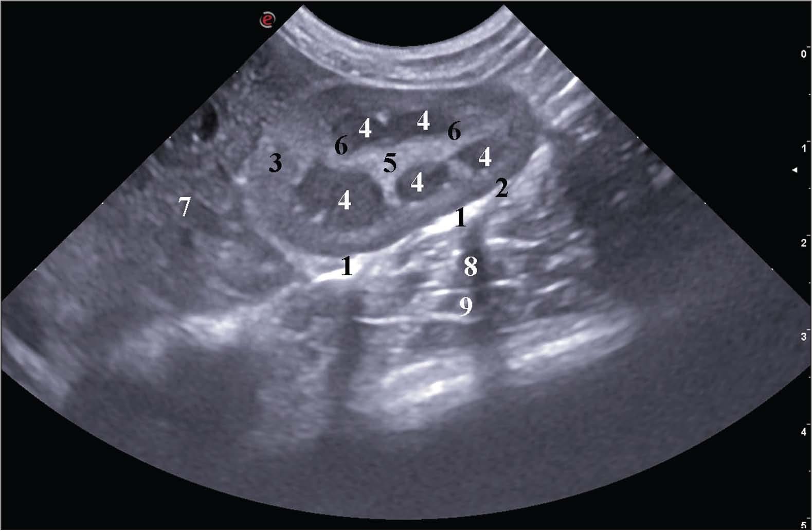
AbstractBackground: Ultrasonography (US) is an amply sufficient, definite, and precise method for obtaining objective anatomical data on rabbit kidneys. Aim: The aim of the present study was to investigate in vivo the specific ultrasonographic anatomical features of kidneys in New Zealand White rabbits. Methods: Ten clinically healthy, sexually mature New Zealand White rabbits, weighing 2.8–3.5 kg, were investigated. The experiment was separated into two parts because the animals were divided into two groups—five males and five females. The male rabbits were studied with a diagnostic ultrasound system (model Vetus 80) equipped with a microconvex multifrequency transducer with a working frequency of 8–10 MHz and a radius of 20 mm, suited for Doppler US. Female animals were investigated with the US device Еsaote MyLabX8Vet and a microconvex multifrequency transducer with working frequency 5 MHz. Results: The in vivo longitudinal US anatomical scanning of the right kidney showed the anatomical contact of the organ with the caudate process of the liver, descending part of the duodenum, and the ascending colon. Within the renal medulla, five to eight hypoechoic renal pyramids with indistinct ends, terminating in the sharply delineated hyperechoic renal papilla, were observed. The longitudinal US imaging of the left kidney provided data about the organ’s multipyramidal features. The renal pyramids were five to eight, of hypoechoic profile, and terminated in the hyperechoic renal papilla and the adjacent renal crest. The color Doppler imaging of the right kidney identified two arterial branches—a dorsal primary and a ventral primary branch. The color Doppler mode of the left renal artery and its dorsal primary branch depicted a flow direction travelling away from the transducer of dark blue color. The interlobar arteries were visible in the vicinity of the renal pyramids. Conclusion: The data from the present investigation suggest a new aspect for the anatomy of the rabbit kidneys. These organs are multipyramidal organs, composed of renal pyramids, whose ends fuse in one renal papilla. The color Doppler provides the right and the left renal artery and their division into dorsal and ventral primary branches. Keywords: Doppler ultrasonography, Morphometry, Renal anatomy, Ultrasonography. IntroductionIn small domestic mammals, ultrasonography (US) is a precise technique for the visualization of abdominal parenchymal organs. The obtained results in B-mode and in real time are interpreted according to the grayscale variations of the images. The similarity between US and surgical findings is of utmost importance in determining the indications for surgery. An advantage of the method is that it allows sedation or general anesthesia (Pennick and D’Anjou, 2008; Schreurs et al., 2008; Webster and N, 2008; Agthe, 2011a; Garcia and Froes, 2012). Ultrasonographically, the kidneys of small mammals have a moderately hypoechoic, homogeneous cortex. In contrast to human kidneys, the medullary area is nearly anechoic. The corticomedullary differentiation is distinct (Albury, 2015). The imaging planes for US visualization of the right and left kidneys in small mammals do not always match the transverse, sagittal, and dorsal anatomical planes. In these cases, the organs are imaged obliquely to achieve detailed visualization (Mauragis and Berry, 2016). Ultrasound imaging is sufficiently definite and precise for obtaining objective anatomical data in horses, large ruminants, and carnivores. The right bovine kidney is visualized in the right fossa paralumbalis via a microconvex 3.5–5.0 MHz transducer using a percutaneous transabdominal approach. The left kidney is evaluated transrectally with a 6.0–8.0 MHz linear transducer. The renal parenchyma is more hypoechoic than the renal sinus. In large dog breeds, the kidneys are depicted most clearly with a 5-MHz transducer, and in small dog breeds and cats, the optimum frequency is from 7 tо 10 MHz (Kealy and Mcallister, 2000; Mannion, 2006; Webster, 2008). According to Seiler et al. (2022)objective macrometric US data for the right and left kidneys of dogs and cats are obtained in the sagittal plane. The dimensions of the renal papillae and the renal pelvis are achieved in the transverse plane. According to Konde (1985); Nyland et al. (1995) and Nyland and Mattoon (2002) the US visualization of the right kidney in cats is more difficult than that of the left kidney due to the right kidney’s intrathoracic position. The right and left kidneys are examined in the transverse and longitudinal planes. The cortex renis has a normoechogenic homogeneous fine-grained echo structure, while it is hypoechoic compared to the liver and spleen. The liver and the spleen are US anatomical markers and acoustic windows for the US examination of the right and left kidneys. The medulla renis is more hypoechoic than the renal cortex. For optimum visualization of the kidneys, a linear 8-MHz transducer is most often used. In carnivores, there are three US imaging planes for visualization of kidneys—two along the long axis of studied organs—dorsal and sagittal, and one along the short axis, transverse (Webster, 2008; Huynh and Berry, 2017). The anatomical US examination of the urinary system of the New Zealand White rabbit has been performed in dorsally positioned animals. The real-time scan was conducted with an 8-MHz linear transducer. The anatomical US dimensions in clinically healthy rabbits varied between the right and left kidneys from 27.80 tо 35.70 mm (length) and from 16.90 to 22.40 mm (width) (Moarabi et al., 2011). Barone (2020)describes the rabbit kidney as similar to that of small ruminants and dogs. Hilus renalis is considerably narrowed and located in the immediate vicinity of extremitas cranialis. The kidney has 6 to 8 pyramids with poorly distinct margins. The crista renalis is well differentiated. Extremitas cranialis of the right kidney reaches the 13th rib, whereas extremitas caudalis reaches L3. The left kidney extends between L3–L4, and extremitas caudalis reaches L5. The kidneys of the New Zealand White rabbit are easily accessible for US anatomy examination in dorsal and lateral recumbency of animals. The US features of the right and left kidneys are similar to those described in the dog and cat. The cortex renis is a hyperechoic finding compared to the medulla renis and has a fine-grained echostructure. On the other hand, the cortex of the left kidney is more hypoechoic than the spleen, while that of the right kidney is hypoechoic to isoechoic compared to the liver parenchyma. The sinus renalis is hyperechoic compared to the cortex renis and medulla renis. A specific feature of the medulla renis is the presence of anechoic and round-shaped renal pyramids. The US anatomical dimensions of the two kidneys are almost identical (Pennick and D’Anjou, 2008; Moarabi et al., 2011; Penninck and D’Anjou, 2015). According to Banzato et al. (2014)the US examination is a highly informative method for the precise visualization of the abdominal organs in the rabbit, including both kidneys. With the positioning of the animals in dorsal recumbency and scanning with a linear 8.5-MHz transducer, real anatomy imaging results are achieved for the kidneys. In both sexes, the sizes of the right and left kidneys were approximately equal. The same relationship was observed with respect to the renal pelvis height. The values of the US parameters were directly proportional to body weight. On the transverse scan image, a sharp demarcation between the cortex renis and the medulla renis was demonstrated. Such differentiation was not observed on longitudinal and parasagittal images. The sinus renalis, pelvis renalis, and crista renalis were visualized in the transverse and longitudinal imaging planes. The pelvis renalis height was measured in the transverse plane as the distance between the sinus renalis and crista renalis. (Banzato et al., 2014). A US anatomy study of rabbit kidneys was conducted by Dimitrov (2012). In the longitudinal and transverse planes, the US anatomy images of both kidneys depicted them as oval-shaped findings. Their margins were clearly distinct from the adjacent organs. The capsula adiposa was hyperechoic with irregular margins. The capsula fibrosa was more hyperechoic than the cortex renis. The right kidney’s acoustic density was inferior to that of the liver. The pelvis renalis had hyperechoic septa. The medulla renis was hypoechoic in relation to the renal cortex. The pelvis renalis was more hypoechoic than the peripelvic adipose tissue (Dimitrov, 2012). Contrast-enhanced US scanning is an accurate technique for presentation of both rabbit kidneys. The precision of obtained results is similar to that of computed tomography and magnetic resonance imaging of the organs in terms of objective evaluation of renal hemodynamics and perfusion (Ryu et al., 2016). The US anatomy imaging of the right and left kidneys of men is performed in ventral recumbency. When the examined individual is in dorsal recumbency, the liver serves as an acoustic window for visualization of the right kidney. The right and left lateral transabdominal approaches are appropriate alternatives for visualization of the two kidneys. The kidney is divided into two zones—a hypoechoic peripheral and a hyperechoic central. The right kidney is hypoechoic in relation to the liver (Tomov and Naumov, 1992; Block, 2004; Chakarski et al., 1996). US combined with color Doppler scanning is a suitable method for quantitative analysis of intrarenal anatomical features in cats. Three distribution patterns of renal interlobar arteries’ point of origin are known. Ultrasonographically, the kidneys of cats are scanned in the dorsal, transverse, and sagittal planes. They are observed as oval findings with a smooth hyperechoic fibrous capsule. The cortex of the right kidney demonstrates a uniform echogenicity and is isoechoic to hypoechoic in relation to the liver parenchyma. The cortex of the left kidney is more hypoechoic than the spleen parenchyma. The renal recesses are hyperechoic and linear, crossing the medulla by forming a hyperechoic band corresponding to the C-shaped profile of crista renalis. The length, width, and height of the right kidney exceed the respective dimensions of the left one (Hoskins and McDicken, 1997; Maher et al., 2024). The positive correlation between kidney length and volume and body weight is not of clinical interest due to the wide range of the standard deviation (SD). Kidney length is of higher morphological and clinical value due to its lower variability with body size. The increased volume of the organs could hardly be determined in the absence of changes in echogenicity and echotexture (Heng, 2025). Perirenal adipose collections are often a prerequisite for increased echogenicity of cortex renis (Yeager and Anderson, 1989). Cortex renis is a homogeneous, peripherally located band surrounding the medulla. It is hypoechoic compared to the spleen and hypoechoic to isoechoic compared to the liver parenchyma. In some instances, the higher echogenicity of the renal cortex in relation to the liver may be assumed as a normal finding. The medulla renis is hypoechoic in relation to the renal cortex—a marker of corticomedullary differentiation (Ivančić and Mai, 2008). At the renal hilus level, the vessels are found along the short axis of the kidney. The identification of arteries and veins is done by color Doppler scanning. They are differentiated from ureters, which lack colored blood flow. The length of the kidney is determined by the highest value obtained in the three orthogonal planes. This parameter is influenced by the breed, the gender, and the body weight. In the cat, the kidney length varied between 30 and 45 mm. The width is between 22 and 28 mm, and the height is from 19 tо 25 mm. The renal pelvis width is 1.00–3.00 mm in cats and 2.00 mm in dogs (Boysen and Chalhoub, 2004; Agthe, 2011b; Huynh and Berry, 2017). The left kidney of the dog is located caudal to the spleen. Most often, it is ventral to the cranial lumbar segment of the vertebral column, but in some cases it can present with a partially subcostal localization (in dogs with a deep chest). The kidneys are examined along their long and short axes. For visualisation of the right kidney, the patient is positioned in left lateral recumbency. Access to the right kidney is less easy compared to that of the left, due to its subcostal position. It is observed in the XI–XII intercostal space. In the cat, the right and left kidneys are presented at similar levels (Mannion, 2006; Agthe, 2011b; Huynh and Berry, 2017). B-mode US in the three planes defines three components in the kidney of carnivores and rats: a centrally located sinus renalis with the highest echogenicity, an almost anechoic and homogeneous medulla renis, and a cortex renis of fine-grained echotexture. The renal pelvis and ureters are not visualized in clinically healthy patients. The relationship between the echogenicity of the medulla and the cortex is described as corticomedullary differentiation. Reduced corticomedullary differentiation is a marker of kidney diseases (Mannion, 2006; Agthe, 2011b; Courtiade and A, 2011; Huynh and Berry, 2017; Varshney and Chaudhary, 2021). The left kidney of the marmoset is hyperechoic compared to the spleen. The right kidney is hyperechoic or isoechoic in relation to the liver. The corticomedullary differentiation is poorly defined and not always established, with absence of a corticomedullary rim. The length of both kidneys in female individuals is higher than that measured in males. The right kidney in both sexes is shorter than the left one. No relationship between the length of the kidneys and body mass is found. The increasing use of the rabbit as an anatomical research model, its popularity as a pet, and the frequent specific renal pathologies in this experimental animal species are the incentives for performing the present study. Considering that US is a precise, rapid, and gentle imaging technique for obtaining reliable US anatomical and macrometric data, a US study algorithm was applied for morphological basis creation. Materials and MethodsTen clinically healthy, sexually mature New Zealand White rabbits, weighing 2.8–3.5 kg, were investigated. The studied cohort was divided into two groups—five males and five females. Prior to the studies, 2–5 ml urine samples were obtained through catheterization in sterile vials for microbiological testing. To eliminate the potential effects on renal blood flow, the color Doppler study was conducted without anesthesia. An anesthetic protocol was used for the anatomical US study. The rabbits were anesthetized with 15 mg/kg Zoletil® 50 IM (tiletamine hydrochloride 125 mg and zolazepam hydrochloride 125 mg in 5 ml of solution) (Virbac, Carros-Cedex, France). The experiment was separated into two parts because the study was designed as an exploratory descriptive ultrasonographic investigation. The male rabbits were studied with an ultrasound device diagnostic ultrasound system (model Vetus 80 Shenzhen Mindray Animal Medical Technology Company Ltd., China) with a microconvex multifrequency model GE 8C transducer with working frequency 8–10 MHz and radius 20 mm, suited for Doppler US. The animals were studied in the University Veterinary Hospital, Stara Zagora, Bulgaria. Female animals were investigated with the US device Еsaote MyLabX8Vet (Italy) and a multiconvex multifrequency transducer (mC 3–11, with working frequency 5 MHz). The animals were studied in Veterinary Clinic Karagiozov Vet, Plovdiv, Bulgaria. The abdominal hair was clipped from the xiphoid process to the pelvic brim using an animal clipper device (Moser Animalline: ARCO, Type 1854, Wahl GmbH, Germany). Contact gel (TRANSSOUND cristal version, EF Medica Srl, Italy) was applied to achieve better contact between the transducer and the skin. The results were interpreted in line with NAV (2017). The animals from both parts of the experiment were positioned in supine recumbency (Dimitrov, 2012). The percutaneous transabdominal right-side and left-side US scanning was conducted. L2 was used as an anatomical landmark. Morphometric measurements were obtained for the right and left kidneys. The measurements were height, width, length, thickness of the cortex, and renal pelvis width. All of them were presented in mm. Statistical analysisStatistical analysis was performed using IBM SPSS Statistics version 29.0 (IBM Corp., Armonk, NY). Continuous variables were initially assessed for distribution normality using the Shapiro–Wilk test. Due to the limited sample size and the non-normal distribution of several variables, non-parametric statistical methods were applied throughout the analysis. Morphometric measurements are presented as mean ± SD. Paired comparisons between the right and left kidneys were evaluated using the Wilcoxon signed-rank test. Exploratory comparisons between male and female rabbits were performed using the Mann–Whitney U test. The statistical unit in the present study was the individual rabbit (n=10), not the individual kidney measurement. Accordingly, bilateral renal measurements were treated as paired observations within the same animal during statistical analysis. The level of statistical significance was set at p < 0.05. Effect size interpretation for non-parametric comparisons was based on rank-biserial correlation coefficients. The reproducibility of ultrasonographic measurements was evaluated using the intraclass correlation coefficient (ICC). Twenty ultrasonographic images with clearly visualized renal contours and optimal image resolution were selected for reliability assessment. Intraobserver reproducibility was assessed by repeated measurements performed by the same investigator after a 1-week interval under blinded conditions without access to the initial measurements. ICC analysis was performed using a two-way mixed-effects model with absolute agreement definition for single measurements. Reliability was interpreted according to established thresholds as follows: poor (<0.50), moderate (0.50–0.75), good (0.75–0.90), and excellent (>0.90). Confidence intervals (95% CI) were calculated for all ICC estimates. A. renalis and v. renalis were continuously scanned on a flat-panel and high-resolution wide-angle matrix. The blood flow direction in relation to the transducer was determined through the red and blue color grading. The optimal angle correction was 40°, the PRF was 4 kHz, the scale was 60 cm/s, the gate was 1–2 mm, and the gain was 65%. The frequency was 9 MHz. In order to validate the findings of the 2D ultrasonographic examination, native rabbit kidney specimens obtained from a certified slaughterhouse for lagomorph processing were included in the study. Ethical approvalThe study has been conducted by permission No. 377, issued by the Ethics Committee of the Ministry of Agriculture and Food, Bulgarian Animal Safety Agency, with opinion No. 293 of 29.02.2024, Sofia, Bulgaria, in compliance with the provisions of the Animal Protection Act in Bulgaria (promulgated in State Gazette No. 13/8 February 2008) and the European Convention for the Protection of Vertebrate Animals Used for Experimental and Other Scientific Purposes (ETS No. 123, OJ L 222, 24 August 1999, p. 31–37). All animal experiments were in accordance with the ARRIVE guidelines and complied with the U.K. Animals (Scientific Procedures) Act, 1986 and associated guidelines, EU Directive 2010/63/EU for animal experiments. ResultsThe in vivo US scan of the right kidney in the longitudinal plane demonstrated the anatomical contact of the organ with the proc. caudatus of the liver, the duodenal pars descendens, and the ascending colon. The right kidney was isoechoic compared to the liver, and according to the grayscale pattern, its echogenicity was fine-grained and similar to that of the proc. caudatus. The capsula adiposa and capsula fibrosa were more hyperechoic findings than the cortex renis. Five to eight hypoechoic renal pyramids were found in the medulla renis, with indistinct edges that terminated into the sharply demarcated hyperechoic papilla renalis. The crista renalis was a hyperechoic linear finding (Fig. 1).
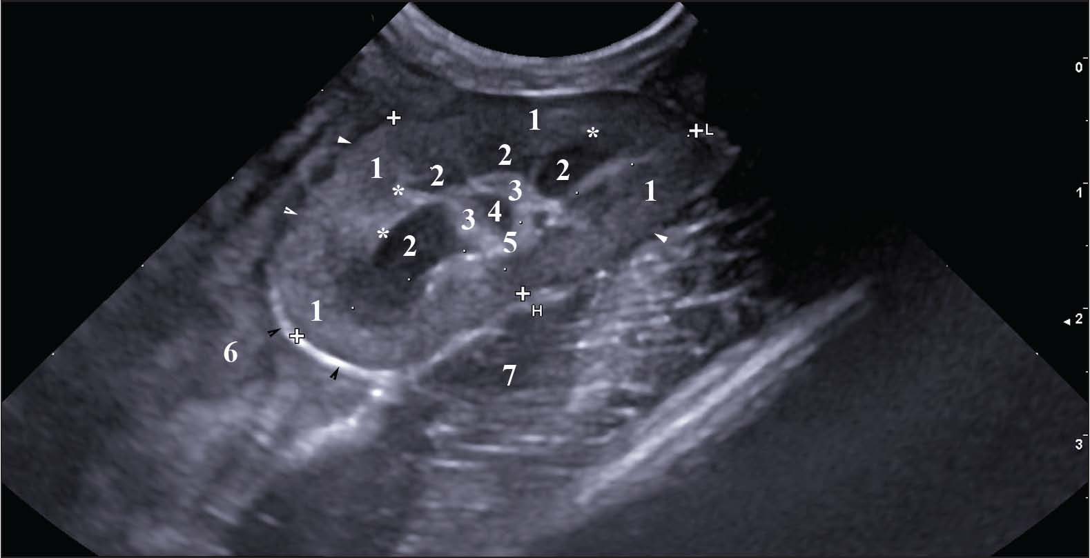
Fig. 1. In vivo longitudinal 2D US anatomical image of the right rabbit kidney. (1) capsula adiposa; (2) capsula fibrosa; (3) cortex renis; (4) pyramis renalis; (5) papilla renalis; (6) crista renalis; (7) proc. caudatus; (8) pars descendens of the duodenum; (9) colon ascendens (5-MHz microconvex probe). The in vivo examination of the right kidney in the oblique plane revealed a multipyramidal kidney structure with clearly delineated hyperechoic corticomedullary differentiation. On the oblique image, the sinus renalis appeared as a hyperechoic structure relative to the pelvis renalis (Fig. 2).
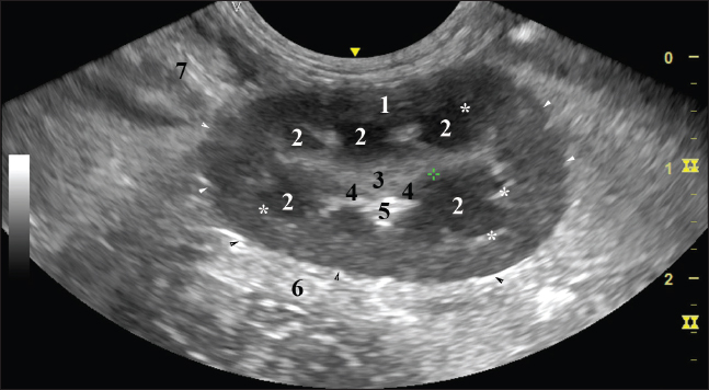
Fig. 2. In vivo oblique 2D US anatomical image of the right rabbit kidney. (1) cortex renis; (2) pyramis renalis; (3) papilla renalis; (4) pelvis renalis; (5) sinus renalis; (6) proc. caudatus; (7) pars descendens of the duodenum (8-MHz microconvex probe). The left kidney was multipyramidal. The renal pyramids terminated in the hyperechoic papilla renalis and the adjacent crista renalis. The organ’s corticomedullary differentiation was seen as a distinct hyperechoic linear structure compared to the relatively hypoechoic cortex renis. The latter had a fine-grained echotexture and was more hypoechoic than the spleen (Fig. 3).
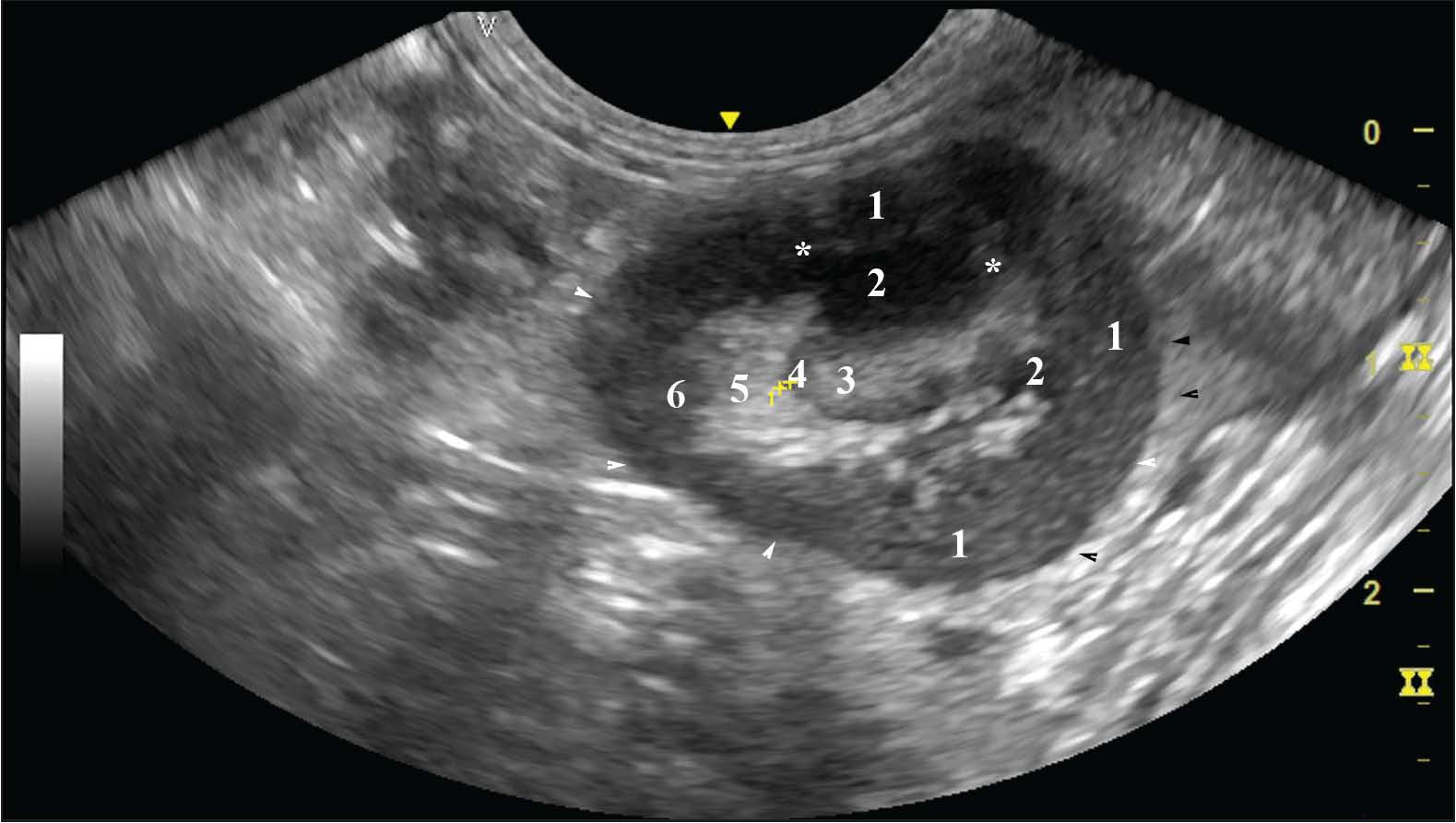
Fig. 3. In vivo longitudinal 2D US anatomical image of the left rabbit kidney. (1) cortex renis; (2) pyramis renalis; (3) papilla renalis; (4) pelvis renalis; (5) sinus renalis; (6) pars ascendens of the duodenum; (7) spleen. (*) corticomedullary differentiation (white arrowhead) capsula fibrosa; (black arrowhead) capsula adiposа (8-MHz microconvex probe). The hyperechoic renal papilla was observed in the transverse plane. The renal pelvis was visualised as a linear arc-shaped hypoechoic finding relative to the hyperechoic renal sinus. The renal hilus was a hypoechoic structure close to the medial margin of the organ. The corticomedullary differentiation was seen as a fine, relatively hyperechoic finding compared to the renal cortex (Fig. 4).
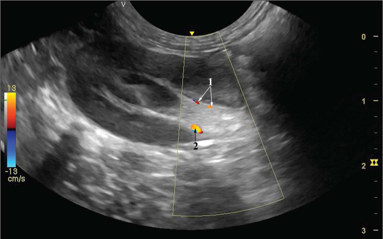
Fig. 4. In vivo transverse 2D US anatomical image of the left rabbit kidney. (1) cortex renis; (2) pyramis renalis; (3) papilla renalis; (4) pelvis renalis; (5) sinus renalis; (6) hilus renalis; (*) corticomedullary differentiation; (white arrowhead) capsula fibrosa; (black arrowhead) capsula adiposa (8-MHz microconvex probe). The color Doppler imaging of the right kidney’s a. renalis revealed two branches of the artery —a primary dorsal and a primary ventral branch. The blood flow pattern was from bright yellow to dark red. The light yellow blood flow hue determined its turbulent nature and high velocity, whereas the dark red hue defined the blood flow’s parabolic character and low velocity. The renal blood vessels were observed in depth as short segments and had a straight course (Fig. 5).
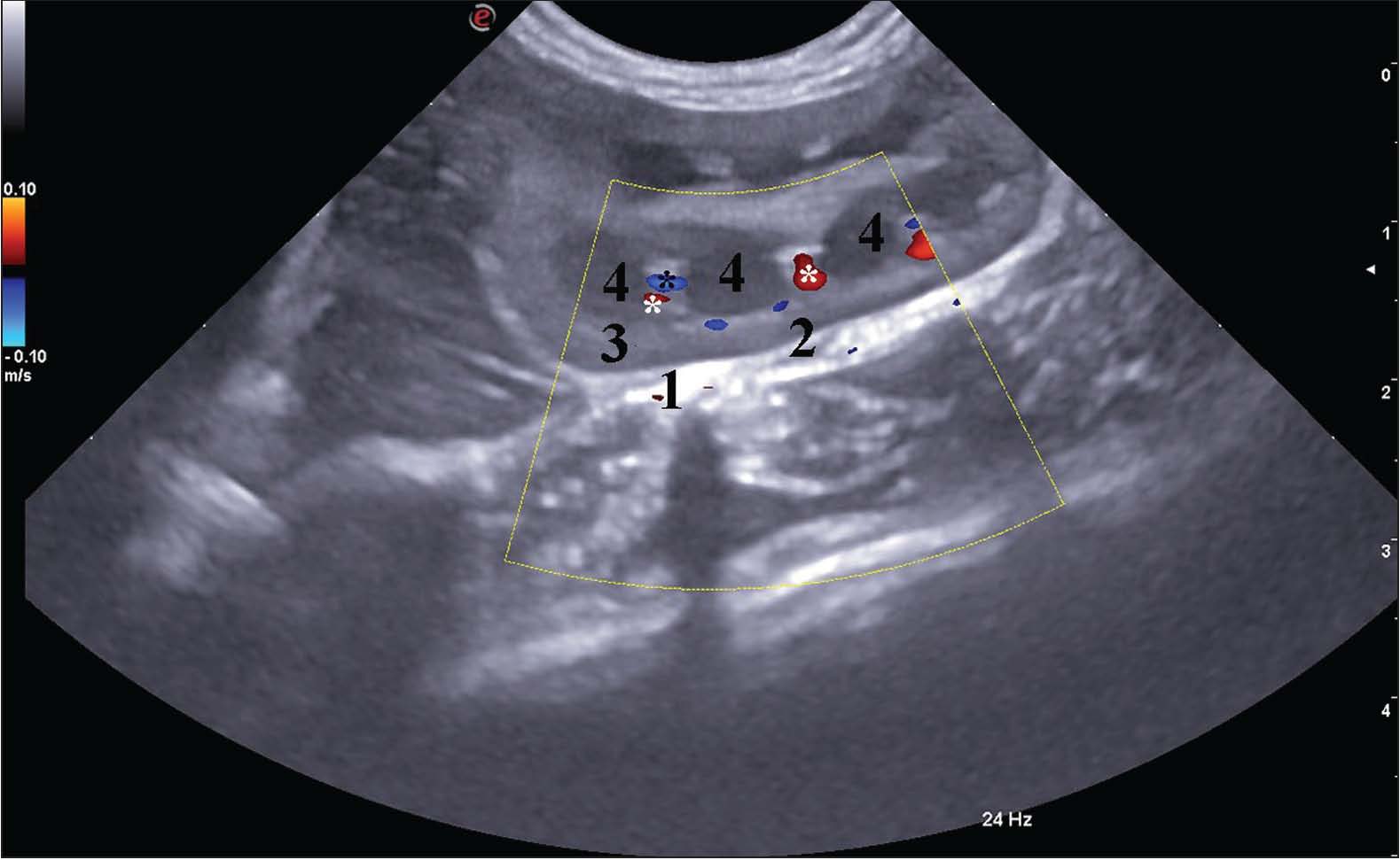
Fig. 5. Color Doppler US image of the right rabbit kidney (color and pulse Doppler transducer, 8 MHz). (1) dorsal primary branch; (2) ventral primary branch. The color Doppler scan displayed the blood flow in the right kidney’s interlobar arteries in dark red. This feature was indicative of low flow velocity in the direction toward the transducer. The interlobar veins demonstrated dark blue color, a marker of the parabolic nature of the blood flow in a direction moving away from the transducer (Fig. 6).
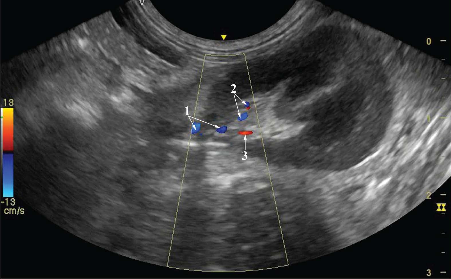
Fig. 6. Color Doppler US image of the right rabbit kidney (longitudinal section, color and pulse Doppler transducer, 5 MHz). (1) capsula adiposa; (2) capsula fibrosa; (3) cortex renis; (4) papilla renalis; (white star) interlobar arteries; (black star) interlobar veins. The color Doppler scan of the blood flow in the left renal artery and its dorsal primary branch was directed away from the transducer and imaged in dark blue due to its parabolic nature. The primary ventral branch appeared in red hues, which centrally were in the medium red color range and peripherally were dark red. The direction of blood flow was toward the transducer (Fig. 7).
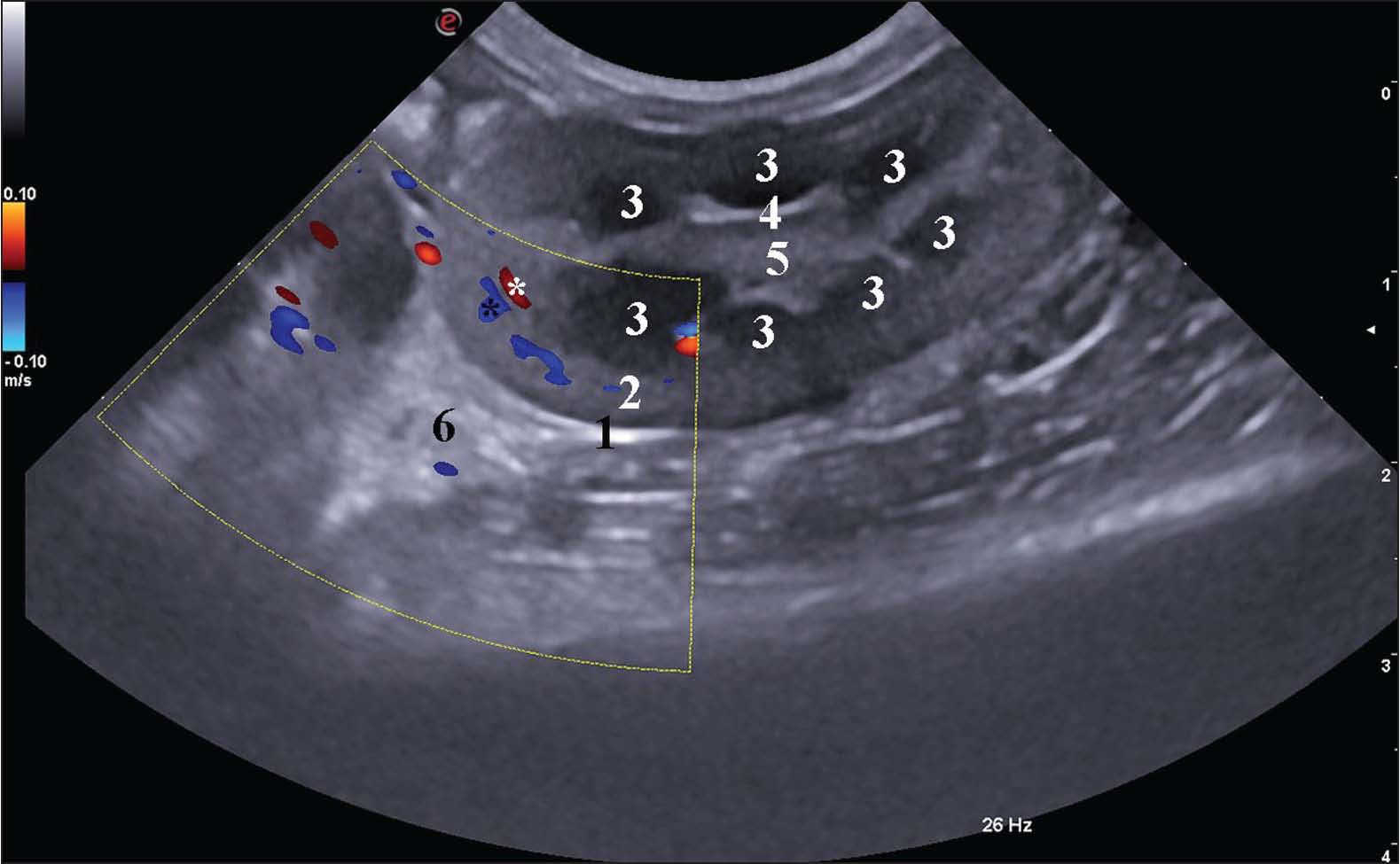
Fig. 7. Color Doppler US image of the left rabbit kidney (color and pulse Doppler transducer, 8 MHz). (1) a. renalis; (2) dorsal primary branch; (3) ventral primary branch. The interlobar arteries in the left kidney were of dark red hues (the direction of blood flow was toward the transducer). Conversely, the interlobar veins were imaged in dark blue (the blood flow was moving away from the transducer). They were visible in the vicinity of the renal pyramids (Fig. 8).
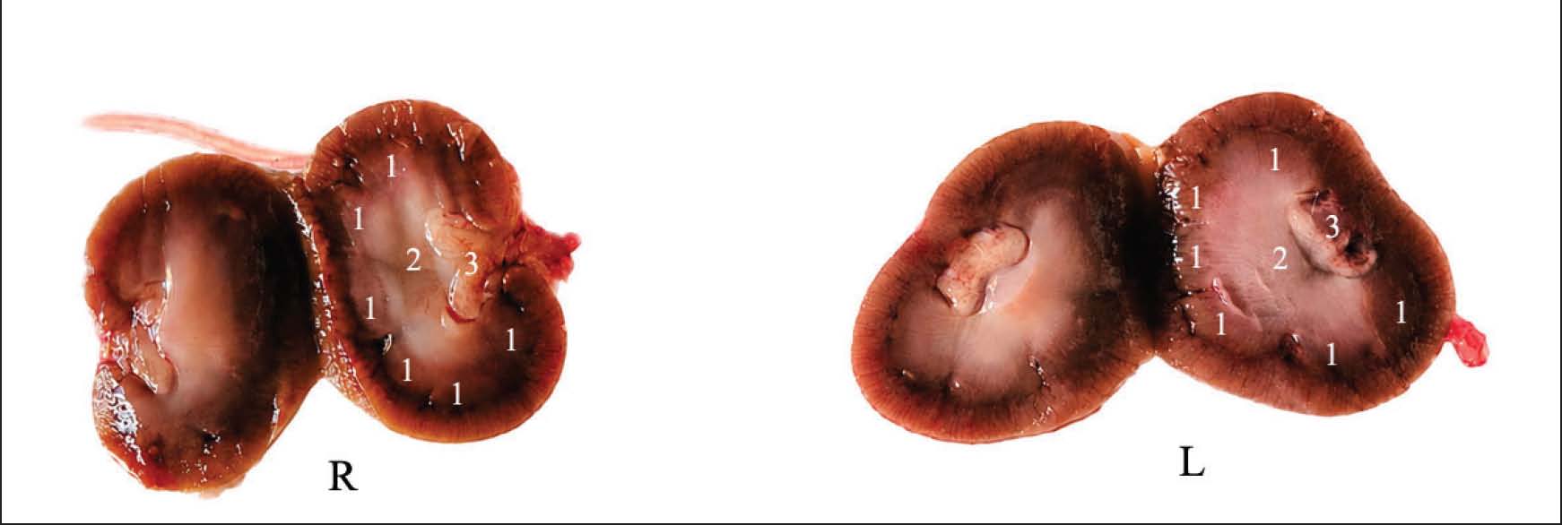
Fig. 8. Color Doppler US image of the left rabbit kidney (color and pulse Doppler transducer, 5 MHz). (1) capsula fibrosa; (2) cortex renis; (3) pyramis renalis; (4) crista renalis; (5) pelvis renalis; (6) spleen; (white star) interlobar arteries; (black star) interlobar veins. The obtained 2D US results were confirmed by anatomical gold-standard images (Fig. 9).
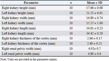
Fig. 9. Right (R) and left (L) kidneys in the rabbit. (1) pyramis renalis; (2) papilla renalis; (3) pelvis renalis. Morphometric ultrasonographic measurements of the kidneys are summarized in Table 1. Paired statistical analysis demonstrated significant lateral asymmetry for kidney height and width. The left kidney height was significantly greater compared to the right kidney (Wilcoxon signed-rank test, p=0.002), whereas the right kidney width was significantly greater compared to the left kidney (Wilcoxon signed-rank test, p=0.0098) (Table 2). Table 1. Descriptive statistics of kidney morphometric parameters (mean ± SD).
Table 2. Paired comparison between right and left kidneys (Wilcoxon signed-rank test).
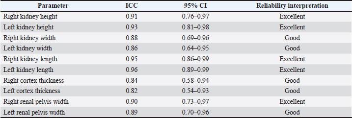
No statistically significant differences were identified between the right and left kidneys regarding kidney length (p=0.846), cortical thickness (p=0.481), or renal pelvis width (p=0.942) (Table 3). Table 3. Intraobserver reproducibility of ultrasonographic morphometric measurements assessed by ICC.
DiscussionThe findings were defined according to the grayscale variations in B-mode and real-time scanning modes. The method has a number of advantages over conventional radiography, which provides a less detailed image despite the large field of view. The Doppler component of the present study was intended primarily for descriptive visualization of renal vascular architecture rather than quantitative hemodynamic assessment (Pennick and D’Anjou, 2008; Schreurs et al., 2008; Webster and N, 2008; Agthe, 2011a; Garcia and Froes, 2012). The obtained results related to the hypoechoic renal cortex, its fine-grained echogenicity, the anechoic characteristics of the renal pyramids, as well as the distinct hyperechoic corticomedullary differentiation correspond to the reported literature data (Albury, 2015). Our data were obtained in longitudinal, oblique, and transverse planes. Therefore, the scanning planes did not match the classical anatomical planes. The applied study algorithm was in accordance with the aim to achieve a detailed anatomical visualization of the kidneys (Mauragis and Berry, 2016). The planes selected by us complement the data established by some authors (Seiler et al., 2022) for macrometric values of canine and feline kidneys in the applied scanning planes. The transducer frequency (from 5 to 8 MHz), as well as the hyperechoic characteristics of the sinus renalis compared to the renal parenchyma, support some research findings. It should be noted that the transducer frequency of 5 MHz is sufficient for investigation of small animals, opposite to the belief that it applies only to large animals (Kealy and Mcallister, 2000; Mannion, 2006; Webster, 2008). The applied examination algorithm corresponds to the established approach for examination of these organs in the cat (Konde, 1985; Nyland et al., 1995; Nyland and Mattoon, 2002). The paired organs were easily accessible, which contradicts the established fact about the relatively more difficult visualization of the right feline kidney (Konde, 1985; Nyland et al., 1995; Nyland and Mattoon, 2002). The obtained images of the right and left kidneys through the liver and spleen as acoustic windows are in line with the US examination of both organs in the cat (Konde, 1985; Nyland et al., 1995; Nyland and Mattoon, 2002). The examination of the kidneys in the longitudinal and transverse planes, the US features of the cortex renis and the hypoechogenicity of the medulla renis compared to the renal cortex complement the data reported for the cat (Konde, 1985; Nyland et al., 1995; Nyland and Mattoon, 2002). A limitation of the present study is the use of different ultrasound systems and transducer frequencies between male and female rabbits due to the two-stage experimental design. Therefore, sex-related comparisons should be interpreted cautiously, and the study was not intended as a standardized comparison between imaging platforms or frequencies. (Moarabi et al., 2011). The obtained US anatomical characteristics of the right and left kidneys were in rabbits in dorsal and lateral recumbency. Considering the US features of the right and left kidney in the rabbit, the lower echogenicity of the left kidney’s cortex compared to the spleen, and the lower echogenicity of the right kidney’s cortex in relation to the liver parenchyma, support the similarity between the kidneys of rabbits and those of dogs and cats reported by several researchers (Pennick and D’Anjou, 2008; Moarabi et al., 2011; Penninck and D’Anjou, 2015). An important specific feature observed by us was the presence of anechoic round-shaped renal pyramids, which evidenced the multipyramidal structure of the organs. Paired morphometric analysis demonstrated significant lateral asymmetry between the kidneys, particularly regarding renal height and width. The left kidney exhibited significantly greater height values, whereas the right kidney demonstrated greater width measurements. On the contrary, kidney length, cortical thickness, and renal pelvis width did not differ significantly between the paired organs (Banzato et al., 2014). Our results show clear visualization of the sinus renalis and pelvis renalis, which adds to the information about the rabbit kidney (Banzato et al., 2014). The presented US anatomy images of both rabbit kidneys in longitudinal and transverse planes correspond to the data for these organs in the rabbit (Dimitrov, 2012). Our results differ from those described by Dimitrov (2012)regarding the presence of septa in the renal pelvis. Our US anatomy imaging data are detailed and reflect the real-time anatomical features of the rabbit kidney. The results obtained may be used as an anatomical model in the diagnosis of diseases affecting the renal parenchyma in humans and the diagnosis of common kidney diseases in rabbits (e.g., hydronephrosis or renal failure) (Ryu et al., 2016). Considering the size of the studied animals and the similar US anatomical imaging of the studied organs to that described in humans, it may be argued that the features of rabbit kidneys may obtained ultrasonographic anatomical data may contribute to the future experimental use of rabbits as imaging models in nephrological research settings. This hypothesis supports the opinion of some authors on the application of the rabbit as a model in human medicine (Wang et al., 2007). The right/left lateral transabdominal access is an appropriate alternative for the visualization of both kidneys. Therefore, the algorithm chosen by us for examining the kidneys in the rabbit corresponds to the research approach in humans (Tomov and Naumov, 1992; Block, 2004; Chakarski et al., 1996). The hyperechoic US characteristics of the cortex renis, the relatively lower cortical echogenicity compared to that of the liver parenchyma and the spleen, add information to the existing findings for these organs in the cat (Yeager and Anderson, 1989; Boysen and Chalhoub, 2004; Ivančić and Mai, 2008; Agthe, 2011b; Huynh and Berry, 2017). The hypoechoic features of the right and left kidneys in relation to the liver and spleen, the clearly defined corticomedullary differentiation, and the lack of sex-associated differences in US parameters are not in line with the findings reported for the US features of the kidneys in the marmoset. The presented ultrasonographic findings support the applicability of two-dimensional US combined with color Doppler imaging as a useful and informative method for visualization of rabbit kidneys and their vascular architecture under clinical conditions (Boysen and Chalhoub, 2004; Agthe, 2011b; Huynh and Berry, 2017). The morphometric analysis demonstrated selective lateral asymmetry between the paired kidneys, primarily affecting renal height and width, whereas renal length and renal pelvis dimensions remained comparable. Similar side-associated morphometric variability has been described in feline ultrasonographic studies (Maher et al., 2024). On the contrary, Banzato et al. (2014)reported approximately equal renal dimensions in healthy rabbits, suggesting that the observed asymmetry in the present study may reflect biological variability, methodological differences, or differences in imaging protocols. The absence of significant differences in renal length between paired kidneys corresponds to previous observations that renal length represents one of the most stable ultrasonographic morphometric parameters with relatively low biological variability (Heng, 2025). Furthermore, the good to excellent intraobserver reproducibility of the ultrasonographic measurements supports the methodological consistency of the applied imaging protocol and the reliability of the obtained morphometric data. ConclusionThe kidneys demonstrated a multipyramidal ultrasonographic appearance. They are composed of five to eight hypoechoic renal pyramids found in the medulla renis, with indistinct edges that terminated into the sharply demarcated hyperechoic papilla renalis. The study provides descriptive ultrasonographic reference data regarding rabbit kidney morphology and vascular visualization in clinically healthy New Zealand White rabbits. AcknowledgmentsThe authors would like to express their sincere gratitude to the Faculty of Veterinary Medicine, Trakia University, Stara Zagora, Bulgaria, for providing the facilities, equipment, and institutional support necessary for conducting this study. Conflict of interestThe authors declare that there is no conflict of interest. FundingThis research received no specific grant. Authors' contributionsConceptualization: Avche Dineva and Kamelia Stamatova-Yovcheva; Validation: Rosen Dimitrov; Methodology: Anton Russenov and Betina Boneva-Marutsova. Data availabilityData are available from the authors upon reasonable request and with permission from the corresponding author, Kamelia Stamatova-Yovcheva. ReferencesAgthe, P. 2011a. Small animal abdominal ultrasonography: how to get started: Part 1. Companion Anim. doi: 10.1111/j.2044-3862.2011.00084.x Agthe, P. 2011b. Small animal abdominal ultrasonography: how to get started: Part 2. Companion Anim. doi: 10.1111/j.2044-3862.2011.00096.x Albury, K. 2015. An overview of small animal veterinary sonography. JDMS 31(3), 160–167. Banzato, T., Bellini, L., Contiero, B., Martin, A., Balikçi, S. and Zotti, A. 2014. Abdominal anatomic features and reference values determined by use of ultrasonography in healthy common rats (Rattus norvegicus). Am. J. Vet. Res. 75(1), 67–76. Barone, R. 2020. Chapitre, I. In Anatomie comparée des mam mifères domestiques. Splanchnologie II. Troisième edition. Ed., Barone, R. Paris, France: Vigot, pp: 3–80. Block, B. 2004. Color atlas of ultrasound anatomy: kidneys. Stuttgart, New York: Thieme, pp: 180–201. Boysen, S. and Chalhoub, S. 2004. Veterinary point of care ultrasound (POCUS): abdomen, pleural space, lung, heart and vascular systems. Calgary, Canada: Faculty of Veterinary Medicine, University of Calgary. Available via https://vet.ucalgary.ca/vcds/podcasts Chakarski, V., Mincheva, E., Lazarova, I., Bueva, A., Popov, D., Oien, R. and Katen, J. 1996. Atlas of abdominal echography, 1st ed. Sofia, Bulgaria: Medicine and Physical Education. Courtiade, A. 2011. Échographie abdominale chez le rat: images normales et mesures de référence sur une population de rats fisher. Thèse, Faculté de Médecine de Créteil, Créteil, France. Dimitrov, R. 2012. Ultrasound features of kidneys in the rabbit (Oryctolagus cuniculus). Vet. World 5(5), 274–278. Garcia, D.A.A. and Froes, T.R. 2012. Errors in abdominal ultrasonography in dogs and cats. J. Small Anim. Pract. doi: 10.1111/j.1748-5827.2012.01249.x Heng, H. 2025. Small animal abdominal ultrasound: what is the significance of abnormal findings. Available via https://cabidigitallibrary.org Hoskins, P.R. and Mcdicken, W.N. 1997. Colour ultrasound imaging of blood flow and tissue motion. Br. J. Radiol. 70(837), 878–890. Huynh, E. and Berry, C.R. 2017. Ultrasonography of the urinary tract: kidneys and ureters. Today’s Vet. Pract. 7, 31–44. Ivančić, M. and Mai, W. 2008. Qualitative and quantitative comparison of renal vs. hepatic ultrasonographic intensity in healthy dogs. Vet. Radiol. Ultrasound 49(4), 368–373. Kealy, K. and Mcallister, H. 2000. Soft tissues. In Diagnostic radiology and ultrasonography of the dog and cat, Konde, L.J. 1985. Sonography of the kidney. Vet. Clinics North Amer. Small Anim. Pract. 15(6), 1149–1158. Maher, M.A., Palacio, L., Henao, J.C. and Elsharkawy, S.H. 2024. Gross morphological and ultrasonographic dimensions of normal feline kidney with reference to resistive index. Sci. Rep. 14, 25662; doi: 10.1038/s41598-024-75341-0 Mannion, P. 2006. Diagnostic ultrasound in small animal practice: kidneys. Oxford, UK: Blackwell Publishing, pp: 109–144. Mauragis, D. and Berry, C.R. 2016. Small animal abdominal ultrasonography liver & gallbladder: part 2. Today’s Vet. Pract. 6, 79–86. Moarabi, A., Mosallanejad, B., Ghadiri, A.R. and Borujeni, M.P. 2011. Ultrasonographic evaluation of the urinary system in New Zealand White rabbit and Tolai hare. Vet. Res. Forum 2(2), 113–120. Nyland, G.T. and Mattoon, J.S. 2002. Small animal diagnostic ultrasound. Philadelphia, PA: W.B. Saunders. Nyland, G.T., Mattoon, J.S. and Wisner, E.R. 1995. Ultrasonography of the urinary tract and adrenal glands. In Veterinary diagnostic ultrasound. Eds., Nyland, T.G. and Mattoon, J.S. Philadelphia, PA: W.B. Saunders, pp: 95–125. Pennick, M.D. and D'Anjou. 2008. Atlas of small animal ultrasonography. Ames, IA: Blackwell Publishing. Penninck, D. and D'Anjou, M. 2015. Gastrointestinal tract. In Atlas of small animal ultrasonography. Eds., Penninck, D. and D’Anjou, M.A., Ryu, J.A., Kim, B., Kim, S.K., Seo, J.W. and Kim, J.S. 2016. Detection of parenchymal abnormalities in experimentally induced acute pyelonephritis in rabbits using contrast-enhanced ultrasonography, CT, and MRI. J. Korean Soc. Radiol. 75(6), 487–494. Schreurs, E., Vermote, K., Barberet, V., Daminet, S., Rudorf, H. and Saunders, J.H. 2008. Ultrasonographic anatomy of abdominal lymph nodes in the normal cat. Vet. Radiol. Ultrasound. 49(1), 68–72. Seiler, G.S., Cohen, E.G., D’Anjou, M.A., French, J., Gaschen, L., Knapp, S., Salwei, R.M. and Saunders, H.M. 2022. Abdominal ultrasound study guidelines. Vet. Radiol. Ultrasound 63(6), 661–674; doi: 10.1111/vru.13151 Tomov, I. and Naumov, N. 1992. Liver. In Ultrasound diagnostics of internal diseases. Sofia, Bulgaria: Medicine and Physical Education, pp: 41–90. Varshney, J.P. and Chaudhary, P.S. 2021. Ultrasound of the urinary tract. In Ultrasound in veterinary medicine: fundamentals and applications. Eds., Varshney, J.P. and Chaudhary, P.S. New Delhi, India: Nipa Genx, pp: 113–130. Wang, Y.X.J., Betton, G., Floettmann, E., Fantham, E. and Ridgwell, G. 2007. Imaging kidney in conscious rats with high-frequency ultrasound and detection of two cases of unilateral congenital hydronephrosis. Ultrasound. Med. Biol. 33(3), 483–486. Webster, N. 2008. Abdominal ultrasonography: practice makes perfect sense. Veterinary Times. Available via https://www.vettimes.co.uk Yeager, A.E. and Anderson, W.I. 1989. Study of association between histologic features and echogenicity of architecturally normal cat kidneys. Am. J. Vet. Res. 50(6), 860–863. | ||
| How to Cite this Article |
| Pubmed Style Dineva A, Stamatova-yovcheva K, Dimitrov R, Russenov A, Vladova D, Boneva-marutsova B. In vivo two-dimensional ultrasonographic characterization of rabbit kidneys. Open Vet. J.. 2026; 16(7): 4397-4409. doi:10.5455/OVJ.2026.v16.i7.26 Web Style Dineva A, Stamatova-yovcheva K, Dimitrov R, Russenov A, Vladova D, Boneva-marutsova B. In vivo two-dimensional ultrasonographic characterization of rabbit kidneys. https://www.openveterinaryjournal.com/?mno=312862 [Access: July 10, 2026]. doi:10.5455/OVJ.2026.v16.i7.26 AMA (American Medical Association) Style Dineva A, Stamatova-yovcheva K, Dimitrov R, Russenov A, Vladova D, Boneva-marutsova B. In vivo two-dimensional ultrasonographic characterization of rabbit kidneys. Open Vet. J.. 2026; 16(7): 4397-4409. doi:10.5455/OVJ.2026.v16.i7.26 Vancouver/ICMJE Style Dineva A, Stamatova-yovcheva K, Dimitrov R, Russenov A, Vladova D, Boneva-marutsova B. In vivo two-dimensional ultrasonographic characterization of rabbit kidneys. Open Vet. J.. (2026), [cited July 10, 2026]; 16(7): 4397-4409. doi:10.5455/OVJ.2026.v16.i7.26 Harvard Style Dineva, A., Stamatova-yovcheva, . K., Dimitrov, . R., Russenov, . A., Vladova, . D. & Boneva-marutsova, . B. (2026) In vivo two-dimensional ultrasonographic characterization of rabbit kidneys. Open Vet. J., 16 (7), 4397-4409. doi:10.5455/OVJ.2026.v16.i7.26 Turabian Style Dineva, Avche, Kamelia Stamatova-yovcheva, Rosen Dimitrov, Anton Russenov, Diyana Vladova, and Betina Boneva-marutsova. 2026. In vivo two-dimensional ultrasonographic characterization of rabbit kidneys. Open Veterinary Journal, 16 (7), 4397-4409. doi:10.5455/OVJ.2026.v16.i7.26 Chicago Style Dineva, Avche, Kamelia Stamatova-yovcheva, Rosen Dimitrov, Anton Russenov, Diyana Vladova, and Betina Boneva-marutsova. "In vivo two-dimensional ultrasonographic characterization of rabbit kidneys." Open Veterinary Journal 16 (2026), 4397-4409. doi:10.5455/OVJ.2026.v16.i7.26 MLA (The Modern Language Association) Style Dineva, Avche, Kamelia Stamatova-yovcheva, Rosen Dimitrov, Anton Russenov, Diyana Vladova, and Betina Boneva-marutsova. "In vivo two-dimensional ultrasonographic characterization of rabbit kidneys." Open Veterinary Journal 16.7 (2026), 4397-4409. Print. doi:10.5455/OVJ.2026.v16.i7.26 APA (American Psychological Association) Style Dineva, A., Stamatova-yovcheva, . K., Dimitrov, . R., Russenov, . A., Vladova, . D. & Boneva-marutsova, . B. (2026) In vivo two-dimensional ultrasonographic characterization of rabbit kidneys. Open Veterinary Journal, 16 (7), 4397-4409. doi:10.5455/OVJ.2026.v16.i7.26 |


