





| Case Report | ||
Open Vet. J.. 2026; 16(7): 4974-4981
Open Veterinary Journal, (2026), Vol. 16(7): 4974-4981 Case Report Exploratory single-case feasibility report of ventral bladder wall-preserving subtotal cystectomy for trigonal transitional cell carcinoma in a catToshikazu Sakai, Masaki Fukutomi, Shushi Yamamoto, Yuji Hamamoto, Eunryel Nam, Mitsuhiro Isaka and Shidow Torisu*Department of Small Animal Clinical Sciences, School of Veterinary Medicine, Rakuno Gakuen University, Ebetsu, Japan *Corresponding Author: Shidow Torisu. Department of Small Animal Clinical Sciences, School of Veterinary Medicine, Rakuno Gakuen University, Ebetsu, Japan. Email: s-torisu [at] rakuno.ac.jp Submitted: 09/03/2026 Revised: 27/05/2026 Accepted: 12/06/2026 Published: 27/07/2026 © 2025 Open Veterinary Journal
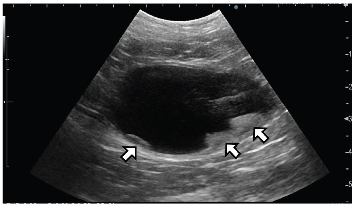
AbstractBackground: The surgical excision of transitional cell carcinoma (TCC) of the bladder, the second most common urinary tract tumor in cats, improves survival. When TCC involves the trigone, resection is particularly challenging because the primary blood supply to the bladder, the caudal vesical artery, runs along its dorsal aspect, and removal of this region can lead to bladder necrosis. Therefore, a total cystectomy is generally required. Urinary diversion following total cystectomy is technically demanding in cats owing to the extremely narrow ureters and urethra, and successful ureterourethral anastomosis has not yet been reported. Case Description: An 11-year-old neutered male domestic shorthaired cat was diagnosed with trigonal TCC. Subtotal cystectomy, including the trigone, was performed while preserving the ventral bladder wall, allowing for bilateral ureterovesical reimplantation without ureterourethral anastomosis. Postoperatively, right ureteral dehiscence developed, requiring revision surgery and right nephrectomy. Five months later, pyelonephritis developed, and tumor recurrence was confirmed 185 days postoperatively. Conclusion: Preservation of the ventral bladder wall during subtotal cystectomy demonstrated the technical possibility of ureteral reimplantation without ureterourethral anastomosis in a cat with trigonal TCC. However, this procedure was associated with major postoperative complications, including ureteral dehiscence, pyelonephritis, persistent urinary incontinence, and tumor recurrence. The reconstructive concept itself, which involves preservation of residual urothelium, may carry risks of local recurrence and secondary urinary tract obstruction. Therefore, the clinical applicability of this reconstructive strategy remains uncertain and should be interpreted cautiously. Future studies should accumulate additional cases and long-term follow-up data to determine the safety, reproducibility, and oncological suitability of this technique in feline TCC. Keywords: Bladder trigone, Cat, Subtotal cystectomy, Transitional cell carcinoma, Ureterovesical anastomosis. IntroductionDespite urinary tract tumors being relatively uncommon in cats, transitional cell carcinoma (TCC) of the bladder remains the second most common urinary tumor, preceded only by renal lymphoma (Wimberly and Lewis, 1979; Schwarz et al., 1985). In dogs, bladder TCCs most frequently occur in the trigone region, whereas in cats, tumors often arise outside the trigone region, with trigonal involvement accounting for approximately one-fourth of all reported cases (Griffin et al., 2020; Hildebrandt et al., 2023). Partial cystectomy prolongs survival in feline TCC (Griffin et al., 2020). On the contrary, the surgical resection of trigonal tumors is challenging because resection of this region requires transection of the caudal vesical artery, which courses along the dorsal surface of the bladder and provides the primary blood supply. Compromise of this vessel may result in bladder necrosis (Liptak et al., 2004); therefore, total cystectomy is typically indicated for complete removal of trigonal tumors (Boston and Singh, 2014). However, total cystectomy requires urinary reconstruction, such as ureterourethral anastomosis (Bacon et al., 2016; Skinner et al., 2020). In cats, the ureters and urethra are extremely narrow (approximately 0.4 and 0.9–2.0 mm in diameter, respectively) (Kochin et al., 1993; Wang et al., 1999; Mehl et al., 2005), making such anastomosis technically demanding. Although ureterourethral anastomosis has been described in dogs, to the best of our knowledge, no successful reconstructions of this type have been reported in cats (Bacon et al., 2016; Skinner et al., 2020). To date, only one previous report has described ureterocutaneous anastomosis following total cystectomy in cats. However, ureteral stenosis occurred approximately 1 year postoperatively (Maeta et al., 2022). As the skin epithelium tends to undergo keratinization and contraction, anastomosis between mucosal tissues reduces the risk of stenosis. Herein, we describe a case of trigonal TCC in a cat managed via subtotal cystectomy, including the trigone, with preservation of the ventral bladder wall, allowing for ureteral reimplantation without requiring ureterourethral anastomosis. Case DetailsHistory and clinical findingsAn 11-year-old neutered male domestic shorthair cat was referred for the evaluation of recurrent lower urinary tract disease. Thirty-seven months earlier, the cat had undergone partial cystectomy of the bladder apex for TCC, achieving complete margin-free excision. Sixteen months later, local recurrence was identified, and a second partial cystectomy was performed, achieving complete excision with tumor-free margins. At presentation, the cat presented with hematuria and dysuria. Abdominal ultrasonography and computed tomography revealed a bladder mass measuring approximately 1.5 cm at the trigone, and multiple additional masses in the central region of the bladder (Fig. 1). No evidence of metastasis was observed. A catheter-guided aspiration biopsy confirmed TCC. Based on these findings, subtotal cystectomy, including the trigone, was planned, and the procedure was performed with informed consent from the owner.
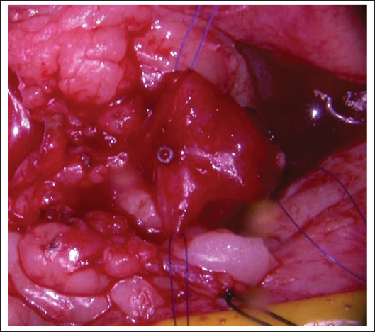
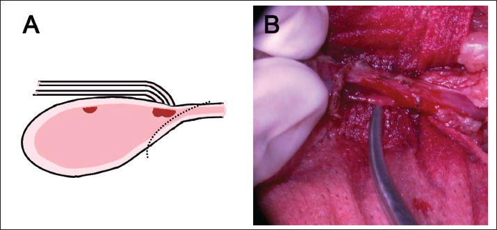
Fig. 1. Abdominal ultrasonographic image. Longitudinal view of the abdomen showing a tumor in the urinary bladder (arrow). Multiple lesions are present, including those in the trigone region of the bladder. Anesthesia and surgical procedureSurgery was performed under premedication and general anesthesia. A 4-Fr urethral catheter was placed before surgery, and a caudal midline celiotomy was performed with the cat in dorsal recumbency. After confirming the absence of tumor invasion into the ureters by palpation, each ureter was ligated and transected at a sufficient distance from the trigone. The caudal and cranial vesical arteries and the vesical mesentery were dissected to mobilize the bladder. Following transection of these vessels, diffuse discoloration of much of the bladder wall was observed, suggesting reduced perfusion, whereas the ventral bladder wall adjacent to the urethra retained relatively normal coloration. Therefore, the extent of bladder wall preservation was determined intraoperatively primarily based on a gross visual assessment of tissue coloration, and areas showing evident discoloration were excluded from the preserved segment. Although the preservation of a larger bladder segment would have increased the available mucosal surface area for ureteral anastomosis, the incision line was determined by prioritizing retention of tissue judged to have relatively preserved perfusion. Consequently, subtotal cystectomy including the trigone was performed while preserving the ventral bladder wall, and an oblique incision was made from the dorsal aspect of the urethra toward the ventral bladder wall (Fig. 2). The dorsal aspect of the urethral stump was further incised by approximately 5 mm to increase the anastomotic surface area. The preserved ventral bladder wall was retracted and stabilized with stay sutures to improve surgical exposure (Fig. 3).
Fig. 3. Preserved bladder wall. After the subtotal cystectomy, the remaining ventral bladder wall was spread using supporting sutures. The supporting sutures placed at the tip of the remaining bladder wall were reflected caudally so that the mucosal surface faced ventrally. The tip of the urethral catheter is visible within the bladder wall.
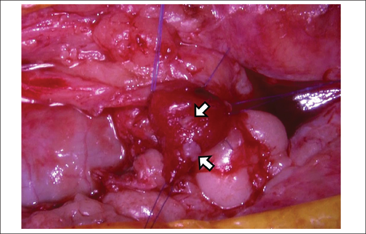
Fig. 2. (A) Schematic illustration of the bladder incision. Subtotal cystectomy was performed while preserving the ventral bladder wall. (B) Intraoperative photograph. The bladder was rotated 90°, and the incision was made with scissors from the dorsal aspect of the urethra toward the ventral aspect of the bladder. Although the bladder wall appeared pale and poorly perfused, the preserved ventral wall remained well perfused, and active bleeding was observed. Consequently, approximately 10 mm of the bladder was preserved. Openings were created in the ventral bladder wall, through which the left and right ureters were passed. Ureterovesical anastomosis was performed circumferentially using simple interrupted sutures with 9–0 absorbable material, approximating the ureteral and bladder mucosa (Fig. 4).
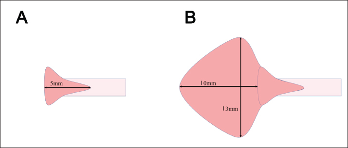
Fig. 4. Bladder wall with ureteral anastomosis. Openings were created in the expanded ventral bladder wall, through which the left and right ureters were passed and anastomosed circumferentially to the bladder mucosa using simple interrupted sutures with 9–0 absorbable material. The ureters are indicated by arrows. A 29-G catheter was used as a ureteral stent. On the left side, the stent was successfully placed from the renal pelvis to the tip of the penis. However, right-sided placement was unsuccessful because of the extremely small ureteral diameter and resistance encountered during catheter advancement. Minor ureteral injury occurred during the attempt and was repaired via resuturing. To avoid further iatrogenic damage, only the left ureter was stented, not the right ureter. This asymmetry resulted in cranial traction on the left ureter, causing mild caudal kinking of the right ureter at the anastomotic site. The ventral bladder wall was closed using a simple continuous suture pattern. Postoperative courseThe cat initially recovered uneventfully. On postoperative day 3, right ureteral dehiscence occurred, resulting in a uroabdomen. After several revision surgeries, a right nephrectomy was performed. The left ureter remained functional, and urine was continuously discharged from the penile orifice. The cat was managed with diapers that maintained an acceptable quality of life, although occasional fecal contamination was observed within the diapers. Five months after surgery, pyelonephritis caused by Klebsiella spp. was diagnosed and successfully treated with antibiotics. On postoperative day 185, TCC recurrence was identified at the ureteral anastomosis site, necessitating total lower urinary tract resection. Histopathological examination confirmed the presence of multifocal noncontiguous TCC lesions. At the time of writing, the cat was alive 8 months after subtotal cystectomy and 51 months after the initial partial cystectomy. Ethical approvalEthical approval was not required for this case report according to institutional policy, as the procedure was performed for therapeutic purposes within the scope of routine clinical practice. Written informed consent was obtained from the owner for the procedure and for publication of this case report and accompanying images. DiscussionThe present surgical approach was designed to enable the resection of a tumor involving the bladder trigone while facilitating ureteral reimplantation. In cats with TCC, various treatment modalities, including partial cystectomy, nonsteroidal anti-inflammatory drugs, chemotherapy, and radiation therapy, have been employed either alone or in combination. However, a standardized treatment protocol remains lacking (Griffin et al., 2020). Partial cystectomy combined with nonsteroidal anti-inflammatory drug therapy has been associated with prolonged survival in cats with TCC (Griffin et al., 2020). However, surgical resection of trigonal tumors remains challenging because trigone excision often requires transection of the caudal vesical artery, the primary blood supply to the bladder, leading to a risk of bladder necrosis. In dogs, a surgical technique that preserves the caudal vesical artery while resecting trigonal tumors has been described, allowing removal of the trigonal region without affecting the dorsal bladder vessels and nerves (Saulnier-Troff et al., 2008). However, this approach is not feasible in cats because of anatomical limitations and small bladder capacity, or in cases where the tumor has invaded the serosa. Consequently, complete resection of trigonal tumors may require total cystectomy. However, in cats, the extremely small diameter of the ureters and urethra makes urinary reconstruction following total cystectomy, particularly ureterourethral anastomosis, technically demanding (Wang et al., 1999; Adin and Scansen, 2011). Although ureterourethral anastomosis has been reported in dogs, it is associated with complications such as uroabdomen, ureteral obstruction due to anastomotic stricture, and pyelonephritis (Bacon et al., 2016; Skinner et al., 2020). Given these risks, similar procedures are expected to be even more challenging in cats. Preserving the ventral bladder wall offers several advantages. The presence of a preserved bladder wall enabled the anastomosis to be performed in the abdominal cavity rather than deep in the pelvis, thereby improving surgical accessibility and visualization. Furthermore, the cranial positioning of the anastomotic site helped reduce traction and tension on the ureters. In addition, the preservation of the ventral bladder wall increases the mucosal area available for ureteral anastomosis, facilitating easier, more reliable suturing. If the bladder is completely resected without preservation of the bladder wall, then even a 5-mm dorsal incision in the urethra provides a limited mucosal surface. Assuming an opening rate of approximately 75%, the expanded urethral mucosal area derived from a urethral diameter of 2.0 mm is estimated to be approximately 24 mm². On the contrary, when a portion of the ventral bladder wall is preserved as an elliptical segment (major axis, 13 mm; minor axis, 10 mm), the combined mucosal surface area of the urethra and preserved bladder is approximately 125 mm² (Fig. 5). These findings indicate that preservation of a portion of the ventral bladder wall markedly increases the mucosal surface area available for ureteral anastomosis.
Fig. 5. (a) Schematic illustration of urethral incision alone. A 5-mm dorsal incision was made in the urethra (inner diameter, 2.0 mm), and with an estimated 75% opening, the expanded mucosal area was approximately 24 mm². (b) Schematic illustration of partial bladder preservation combined with urethral incision. When the bladder was preserved as an elliptical segment (major axis, 13 mm; minor axis, 10 mm), the total expanded mucosal area was approximately 125 mm². Owing to the limited urethral mucosal surface, ureteral anastomosis is not only technically challenging but may also make subsequent urethral closure difficult, which is necessary to prevent urine leakage. Furthermore, within such a restricted space, ureteral anastomotic sites may become closely approximated or come into contact after urethral reconstruction, potentially leading to adhesion formation or mutual compression and consequently compromising luminal patency. Conversely, preserving a larger portion of the bladder may be advantageous for suturing from a technical standpoint; however, the risk of impaired blood supply may increase as the extent of preservation becomes greater. The bladder is primarily supplied by the caudal vesical artery, with additional contributions from the cranial vesical artery; however, in the procedure reported herein, both of these vessels were transected. Following transection of these arteries, the bladder wall became discolored, and only a limited portion that was contiguous with the urethra maintained normal coloration. The extent of bladder preservation was therefore determined intraoperatively based on a visual assessment of the bladder wall color, and only well-perfused tissue was preserved. As the remaining bladder was connected only to the urethra, its blood supply was presumed to depend largely on retrograde perfusion from the urethra. As this retrograde perfusion would be expected to reach the caudal portion of the bladder first and diminish toward the cranial region, the preservation of a larger cranial portion may increase the risk of inadequate perfusion. Accordingly, as the extent of bladder preservation increases, particular attention should be paid to the potential risk of compromised perfusion, especially in the cranial region. Taken together, this technique may provide a more favorable substrate considering the mucosal surface area for reconstruction than total cystectomy. Nevertheless, postoperative complications were observed in the present case, including ureteral anastomotic dehiscence, ascending urinary tract infection, and tumor recurrence. The ureteral dehiscence observed in the present case was most likely associated with unilateral ureteral stenting. As only the left ureter was stented, asymmetric mechanical forces may have developed between the ureters. As the ureters tend to align in a straight configuration, this asymmetry likely resulted in torsion of the bladder remnant and caudal traction on the unstented right ureter, ultimately leading to dehiscence at the anastomotic site. Bilateral ureteral stenting had initially been planned to reduce the risk of postoperative urinary stasis caused by transient inflammation, edema, or functional impairment of the ureters during the perioperative period. However, stent placement on the right side was ultimately abandoned because of the extremely small ureteral diameter and concerns regarding potential iatrogenic injury during attempted insertion. Therefore, asymmetric tension associated with unilateral stenting was considered to be a major contributing factor to this complication. To mitigate this risk, either bilateral stenting or omission of stents on both sides may help eliminate asymmetric tension between the ureters. However, unilateral stenting alone may not fully explain the occurrence of dehiscence. Additional factors inherent to the procedure may also have contributed, including the extremely small diameter of the feline ureters and potentially compromised vascular perfusion of the preserved bladder wall. In the present case, bladder wall viability was assessed intraoperatively primarily based on the gross coloration. However, subtle perfusion deficits not detectable in visual inspection alone may have affected anastomotic healing. Ascending urinary tract infection is a potential complication associated with ureteral reimplantation. Under normal conditions, the ureters traverse the bladder wall obliquely, providing an anti-reflux mechanism. However, this physiological barrier may be compromised following ureteral reimplantation, allowing vesicoureteral reflux during micturition (Stefanovlč et al., 1991). Chronic reflux could also increase the risk of ascending infection. In addition, intraoperative ureteral catheter placement may have further contributed to infection risk in this case (Kehinde et al., 2002). Furthermore, as urinary continence could not be maintained after surgery, long-term diaper management was required, potentially leading to an environment favorable for ascending bacterial contamination. In the present case, pyelonephritis developed following right nephrectomy, leaving only a single functional kidney. Therefore, postoperative urinary tract infection should be regarded as a potentially serious complication in this reconstructive approach. Although the infection in the present case was successfully managed with antimicrobial therapy, whether such infections represent an inherent limitation of the procedure itself warrants careful consideration in future cases. Local tumor recurrence was observed 185 days postoperatively, which is comparable to the reported median recurrence time (205 days) following partial cystectomy in cats (Griffin et al., 2020). Recurrence after partial cystectomy has been reported in 63.6% of cases (Griffin et al., 2020). These findings support that feline TCC may exhibit a “field cancerization” effect or intraluminal tumor cell dissemination, leading to multifocal recurrence throughout the lower urinary tract (Azémar et al., 2011; Fulkerson and Knapp, 2015). Although the preservation of the ventral bladder wall enabled ureteral reimplantation, the remaining urothelium may also have served as a substrate for tumor regrowth or implantation. In addition, as ureteral anastomosis was performed directly onto the preserved urothelial tissue, local recurrence at the anastomotic site may result in secondary ureteral obstruction. Therefore, this reconstructive strategy can involve a risk of tumor recurrence and a subsequent risk of urinary tract obstruction. Despite these oncological limitations, trigonal tumors may eventually cause life-threatening urinary obstruction if left untreated or if medical management fails. Griffin et al. (2020)described severe postoperative complications, including septic peritonitis and uroabdomen, following attempts at surgical treatment of trigonal tumors in cats. Therefore, even temporary maintenance of urinary flow and delay of obstruction represent a potential clinical benefit in selected cases, and recurrence may not occur in all cases. The present cat had already undergone two previous partial cystectomies over a prolonged clinical course, potentially resulting in prolonged exposure of the lower urinary tract to intraluminal tumor cell dissemination. This extensive prior disease history may also have contributed to recurrence in the present case. In a previous report describing total cystectomy with cutaneous ureterostomy in a cat, ureteral obstruction secondary to subcutaneous nodules developed approximately 14 months postoperatively (Maeta et al., 2022). Although the present case was not followed long enough to evaluate long-term anastomotic patency because of tumor recurrence, one conceptual advantage of the current technique is the use of mucosa-to-mucosa anastomosis, which may theoretically reduce the risk of stenosis compared with cutaneous diversion techniques. However, further accumulation of cases and long-term follow-up will be required to determine whether this theoretical advantage translates into durable clinical benefit. This report has several limitations. First, it is based on a single case, and the generalizability of the findings is inherently limited. Second, several significant postoperative complications occurred, including ureteral dehiscence, ascending urinary tract infection, and tumor recurrence, which may influence the overall evaluation of this technique. Finally, the follow-up period was relatively short, precluding assessment of long-term outcomes. Accordingly, the clinical applicability of this approach should be interpreted with caution and verified in future studies. ConclusionAlthough this report describes only a single case, preservation of the ventral bladder wall during subtotal cystectomy demonstrated the technical possibility of ureteral reimplantation without ureterourethral anastomosis in a cat with trigonal TCC. However, this experience should be regarded solely as an experimental proof-of-concept observation rather than evidence of clinical feasibility or therapeutic efficacy. Given the exploratory nature of the procedure, the occurrence of major postoperative complications, and the limited follow-up period, no definitive conclusions regarding the safety, reproducibility, or clinical applicability of this technique can be drawn from this single case. Further evaluation through accumulation of additional cases, ideally including multicenter studies with standardized long-term follow-up, will be necessary to determine whether this approach can represent a clinically applicable surgical option for feline TCC. AcknowledgmentsNone. Conflict of interestThe authors declare that there is no conflict of interest. FundingThis research received no specific grant. Authors' contributionsT.S. managed the case as the primary clinician and performed the surgery. M.F. collected the clinical data. Y.S. performed the imaging evaluation. H.Y., E.N., and I.M. were responsible for perioperative management. S.T. conducted the literature review and supervised the study. T.S. drafted the manuscript, and all authors critically revised and approved the final version. Data availabilityThe data supporting the findings of this study are available from the corresponding author upon reasonable request. ReferencesAdin, C.A. and Scansen, B.A. 2011. Complications of upper urinary tract surgery in companion animals. Vet. Clin. North Am. Small Anim. Pract. 41, 869–888. Azémar, M.D., Comperat, E., Richard, F., Cussenot, O. and Rouprêt, M. 2011. Bladder recurrence after surgery for upper urinary tract urothelial cell carcinoma: frequency, risk factors, and surveillance. Urol. Oncol. 29, 130–136. Bacon, N., Souza, C.H. and Franz, S. 2016. Total cysto-prostatectomy: technique description and results in two dogs. Can. Vet. J. 57, 491–496. Boston, S. and Singh, A. 2014. Total cystectomy for treatment of transitional cell carcinoma of the urethra and bladder trigone in a dog. Vet. Surg. 43, 294–300. Fulkerson, C.M. and Knapp, D.W. 2015. Management of transitional cell carcinoma of the urinary bladder in dogs: a review. Vet. J. 205, 217–225. Griffin, M.A., Culp, W.T.N., Giuffrida, M.A., Ellis, P., Tuohy, J., Perry, J.A., Gedney, A., Lux, C.N., Milovancev, M., Wallace, M.L., Hash, J., Mathews, K., Liptak, J.M., Selmic, L.E., Singh, A., Palm, C.A., Balsa, I.M., Mayhew, P.D., Steffey, M.A., Rebhun, R.B., Burton, J.H. and Kent, M.S. 2020. Lower urinary tract transitional cell carcinoma in cats: clinical findings, treatments, and outcomes in 118 cases. J. Vet. Intern. Med. 34, 274–282. Hildebrandt, I., Culp, W.T.N. and Griffin, M.A. 2023. A systematic review of canine cystectomy: indications, techniques, and outcomes. Animals (Basel) 13, 2896. Kehinde, E.O., Rotimi, V.O., Al-Awadi, K.A., Abdul-Halim, H., Boland, F., Al-Hunayan, A. and Pazhoor, A. 2002. Factors predisposing to urinary tract infection after J ureteral stent insertion. J. Urol. 167, 1334–1337. Kochin, E.J., Gregory, C.R., Wisner, E., Cain, G. and Gourley, I.M. 1993. Evaluation of a method of ureteroneocystostomy in cats. J. Am. Vet. Med. Assoc. 202, 257–260. Liptak, J., Dernell, W. and Withrow, S. 2004. Haemangiosarcoma of the urinary bladder in a dog. Aust. Vet. J. 82, 215–217. Maeta, N., Kutara, K., Saeki, K. and Hoshi, F. 2022. Modified Toyoda technique for total cystectomy and cutaneous ureterostomy in a cat. Vet. Surg. 51, 1280–1286. Mehl, M.L., Kyles, A.E., Pollard, R., Jackson, J., Kass, P.H., Griffey, S.M. and Gregory, C.R. 2005. Comparison of 3 techniques for ureteroneocystostomy in cats. Vet. Surg. 34, 114–119. Saulnier‐Troff, F.G., Busoni, V. and Hamaide, A. 2008. A technique for resection of invasive tumors involving the trigone area of the bladder in dogs: preliminary results in two dogs. Vet. Surg. 37, 427–437. Schwarz, P.D., Greene, R.W. and Patnaik, A.K. 1985. Urinary bladder tumors in the cat: a review of 27 cases. J. Am. Anim. Hosp. Assoc. 21, 237–245. Skinner, O.T., Boston, S.E. and Maxwell, P.L. 2020. Interventions and experience after complicated total cystectomy in a dog with transitional cell carcinoma. Vet. Surg. 49, 811–817. Stefanovlč, K.B., Bukurov, N.S. and Marinkovlč, J.M. 1991. Non-antireflux versus antireflux ureteroneocystostomy in adults. Br. J. Urol. 67, 263–266. Wang, B., Bhadra, N. and Grill, W.M. 1999. Functional anatomy of the male feline urethra: morphological and physiological correlations. J. Urol. 161, 654–659. Wimberly, H.C. and Lewis, R.M. 1979. Transitional cell carcinoma in the domestic cat. Vet. Pathol. 16, 223–228. | ||
| How to Cite this Article |
| Pubmed Style Sakai T, Fukutomi M, Yamamoto S, Hamamoto Y, Nam E, Isaka M, Torisu S. Exploratory single-case feasibility report of ventral bladder wall-preserving subtotal cystectomy for trigonal transitional cell carcinoma in a cat. Open Vet. J.. 2026; 16(7): 4974-4981. doi:10.5455/OVJ.2026.v16.i7.75 Web Style Sakai T, Fukutomi M, Yamamoto S, Hamamoto Y, Nam E, Isaka M, Torisu S. Exploratory single-case feasibility report of ventral bladder wall-preserving subtotal cystectomy for trigonal transitional cell carcinoma in a cat. https://www.openveterinaryjournal.com/?mno=313189 [Access: July 27, 2026]. doi:10.5455/OVJ.2026.v16.i7.75 AMA (American Medical Association) Style Sakai T, Fukutomi M, Yamamoto S, Hamamoto Y, Nam E, Isaka M, Torisu S. Exploratory single-case feasibility report of ventral bladder wall-preserving subtotal cystectomy for trigonal transitional cell carcinoma in a cat. Open Vet. J.. 2026; 16(7): 4974-4981. doi:10.5455/OVJ.2026.v16.i7.75 Vancouver/ICMJE Style Sakai T, Fukutomi M, Yamamoto S, Hamamoto Y, Nam E, Isaka M, Torisu S. Exploratory single-case feasibility report of ventral bladder wall-preserving subtotal cystectomy for trigonal transitional cell carcinoma in a cat. Open Vet. J.. (2026), [cited July 27, 2026]; 16(7): 4974-4981. doi:10.5455/OVJ.2026.v16.i7.75 Harvard Style Sakai, T., Fukutomi, . M., Yamamoto, . S., Hamamoto, . Y., Nam, . E., Isaka, . M. & Torisu, . S. (2026) Exploratory single-case feasibility report of ventral bladder wall-preserving subtotal cystectomy for trigonal transitional cell carcinoma in a cat. Open Vet. J., 16 (7), 4974-4981. doi:10.5455/OVJ.2026.v16.i7.75 Turabian Style Sakai, Toshikazu, Masaki Fukutomi, Shushi Yamamoto, Yuji Hamamoto, Eunryel Nam, Mitsuhiro Isaka, and Shidow Torisu. 2026. Exploratory single-case feasibility report of ventral bladder wall-preserving subtotal cystectomy for trigonal transitional cell carcinoma in a cat. Open Veterinary Journal, 16 (7), 4974-4981. doi:10.5455/OVJ.2026.v16.i7.75 Chicago Style Sakai, Toshikazu, Masaki Fukutomi, Shushi Yamamoto, Yuji Hamamoto, Eunryel Nam, Mitsuhiro Isaka, and Shidow Torisu. "Exploratory single-case feasibility report of ventral bladder wall-preserving subtotal cystectomy for trigonal transitional cell carcinoma in a cat." Open Veterinary Journal 16 (2026), 4974-4981. doi:10.5455/OVJ.2026.v16.i7.75 MLA (The Modern Language Association) Style Sakai, Toshikazu, Masaki Fukutomi, Shushi Yamamoto, Yuji Hamamoto, Eunryel Nam, Mitsuhiro Isaka, and Shidow Torisu. "Exploratory single-case feasibility report of ventral bladder wall-preserving subtotal cystectomy for trigonal transitional cell carcinoma in a cat." Open Veterinary Journal 16.7 (2026), 4974-4981. Print. doi:10.5455/OVJ.2026.v16.i7.75 APA (American Psychological Association) Style Sakai, T., Fukutomi, . M., Yamamoto, . S., Hamamoto, . Y., Nam, . E., Isaka, . M. & Torisu, . S. (2026) Exploratory single-case feasibility report of ventral bladder wall-preserving subtotal cystectomy for trigonal transitional cell carcinoma in a cat. Open Veterinary Journal, 16 (7), 4974-4981. doi:10.5455/OVJ.2026.v16.i7.75 |


