





| Research Article | ||
Open Vet. J.. 2026; 16(7): 4549-4557
Open Veterinary Journal, (2026), Vol. 16(7): 4549-4557 Research Article Hematological and biochemical safety of camel milk administration in ovalbumin-induced allergic Wistar ratsYoonis Dayr Mohamed1*, Rebecca Waihenya1,2, Kenneth Ogila2 and Raphael W. Lihana31Department of Molecular Biology and Biotechnology, Pan African University Institute for Basic Sciences Technology and Innovation, Nairobi, Kenya 2Department of Zoology, School of Biological Sciences, Jomo Kenyatta University of Agriculture and Technology, Nairobi, Kenya 3Kenya Medical Research Institute Center for Virus Research, Nairobi, Kenya *Corresponding Author: Yoonis Dayr Mohamed. Department of Molecular Biology and Biotechnology, Pan African University Institute for Basic Sciences Technology and Innovation, Nairobi, Kenya. Email: yoonisdayr [at] gmail.com Submitted: 13/03/2026 Revised: 22/05/2026 Accepted: 02/06/2026 Published: 17/07/2026 © 2025 Open Veterinary Journal
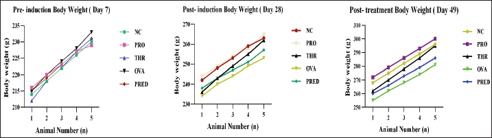
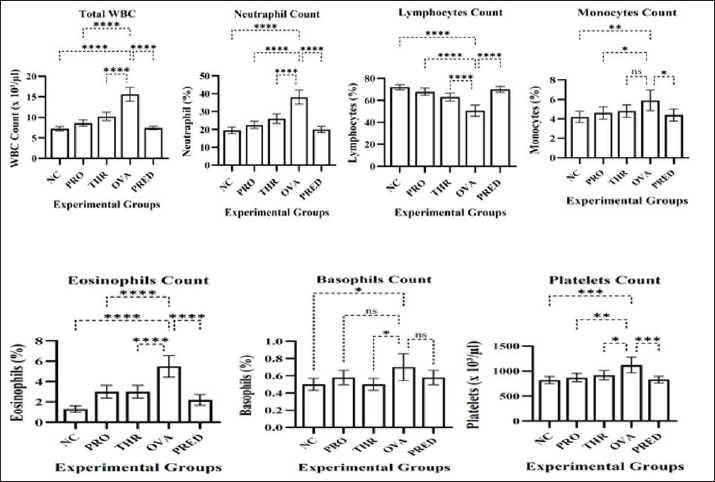
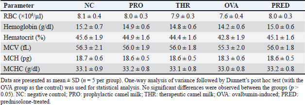
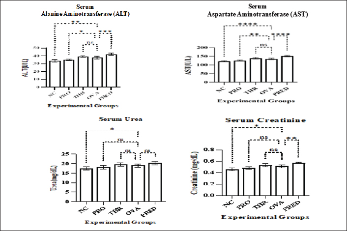
ABSTRACTBackground: Camel milk has been widely investigated for its therapeutic potential, particularly its immunomodulatory and anti-inflammatory properties; however, information regarding its systemic safety under allergic inflammatory conditions remains limited. Aim: To evaluate the systemic safety of camel milk administration in an ovalbumin (OVA)-induced allergic Wistar rat model. Methods: A total of 25 male Wistar rats (8–10 weeks old) were randomly allocated into five groups (n=5): negative control (NC), prophylactic camel milk (PRO), ovalbumin-induced allergic control (OVA), therapeutic camel milk (THR), and prednisolone-treated reference group (PRED). Allergic inflammation was induced through intraperitoneal sensitization and OVA challenge. Camel milk (10 ml/kg/day) was orally administered under PRO and THR regimens, while the PRED group received prednisolone (5 mg/kg/day) as the pharmacological reference treatment. Body weight progression was monitored throughout the experimental period, whereas hematological and serum biochemical parameters, including alanine aminotransferase (ALT), aspartate aminotransferase (AST), urea, and creatinine levels, were assessed at the endpoint. Results: All groups demonstrated progressive physiological weight gain without treatment-related growth suppression. The OVA group exhibited leukocytosis, neutrophilia, and eosinophilia, confirming systemic inflammatory activation, whereas camel milk-treated groups showed moderate leukocyte responses with eosinophil levels approaching physiological ranges. Red blood cell indices, platelet counts, and liver and renal function markers remained within the reference range. Conclusion: Oral camel milk administration is systemically well tolerated in OVA-induced allergic Wistar rats and supports its physiological safety as a complementary dietary intervention under allergic conditions. Keywords: Allergy, Camel milk, Ovalbumin model, Serum biochemistry, Wistar rats. IntroductionCamel milk has attracted increasing scientific interest as a functional dietary product with reported immunomodulatory, anti-inflammatory, and therapeutic effects (Alhassani, 2024). It has been investigated in various experimental models of immune-mediated and inflammatory disorders, where it has been shown to influence immune responses and attenuate inflammatory processes (Arab et al., 2014; He et al., 2022) . These findings have stimulated growing attention toward its potential application as a natural complementary intervention in allergic and inflammatory conditions by targeting key immune regulatory pathways (He et al., 2022). Allergic diseases are characterized by dysregulated immune responses, including elevated immunoglobulin E (IgE) production, eosinophilic infiltration, and a T helper 2 (Th2)-skewed cytokine profile (North, 2021). The ovalbumin (OVA)-induced allergic rat model is widely used to replicate key immunopathological features of allergic inflammation in humans (Thakur et al., 2019). Within such an inflammatory milieu, the administration of any dietary or therapeutic intervention warrants careful evaluation of systemic safety to ensure that it does not compromise physiological stability under immune stress. Accordingly, assessing whether camel milk administration exerts any unintended systemic effects in this model is essential. Although camel milk is traditionally consumed as a dietary product and is generally regarded as safe (Alhassani, 2024), its repeated administration at controlled doses in experimental settings constitutes a defined biological intervention that warrants formal safety evaluation. Although numerous studies have highlighted its immunomodulatory and anti-inflammatory properties (Behrouz et al., 2022), comparatively limited attention has been directed toward its systemic safety profile under conditions of allergic inflammatory stress (He et al., 2022; Rakhmatulina et al., 2025a, b). Previous studies have mainly evaluated the physiological or toxicological effects of camel milk, sometimes along with camel urine, in healthy Wistar rats under noninflammatory conditions (Abdulfatai et al., 2024). However, the safety profile of camel milk during allergic inflammatory responses remains to be explored. Therefore, this study aimed to systematically evaluate the systemic safety of camel milk administration in an OVA-induced allergic Wistar rat model by assessing body weight progression, hematological parameters, and biochemical markers of hepatic and renal function under both prophylactic (PRO) and therapeutic (THR) regimens. Materials and MethodsAnimals and their housingTwenty-five male Wistar rats (age 8–10 weeks; weight 200–250 g) were obtained from the Small Animal Facility for Research and Innovation at Jomo Kenyatta University of Agriculture and Technology, Nairobi, Kenya. Animals were housed in standard polypropylene cages (five rats per cage) under controlled environmental conditions (22°C ± 2°C; 50%–60% relative humidity; 12-hour light/dark cycle). Standard laboratory chow and clean drinking water were provided ad libitum throughout the study. Animals were monitored throughout the experimental period for general health status and morbidity, and a 7-day acclimatization period was allowed before the initiation of experimental procedures. Monitoring of body weightBody weight was recorded using a calibrated digital balance at pre-induction (Day 7), post-induction (Day 28), and at the study endpoint (Day 49). Measurements were performed throughout the experimental period to monitor growth progression and identify potential systemic or treatment-related adverse effects. Experimental groupsAnimals were randomly assigned to five groups (n=5 per group) as follows:
Experimental timelineFigure 1 shows a schematic representation of the experimental design. Day 0 represents the initiation of the acclimatization period. Animals were acclimatized from Days 0 to 7. Sensitization with OVA was performed on Days 7 and 14, followed by the challenge phase on Days 21, 24, and 27. Treatment was administered from Days 28 to 49. All animals were euthanized at the endpoint of the study (Day 49) for sample collection and analysis. Allergic inflammation inductionAn experimental allergic model was established by intraperitoneal administration of (OVA EndoFit™, InvivoGen, Toulouse, France; Cat. No. vac-pova; Lot No. 5821-46-02; endotoxin level <1 EU/mg) in combination with aluminum hydroxide as an adjuvant (Ahn et al., 2023). For sensitization, rats received intraperitoneal injections of OVA (75 µg) mixed with aluminum hydroxide (alum; 50 µl) and suspended in phosphate-buffered saline (PBS; 200 µl) on Days 7 and 14. The challenge phase was initiated on Day 21 and consisted of three intraperitoneal injections of OVA (50 µg) suspended in PBS (200 µl). These injections were administered at 3-day intervals on Days 21, 24, and 27 to induce acute allergic inflammation. Administration of camel milk and Prednisolone Camel milk was administered orally by gavage at a dose of 10 ml/kg body weight per day (Arab et al., 2021). For prophylactic (PRO) evaluation, animals in the PRO group received camel milk daily from days 7 to 28 before allergic induction, followed by OVA sensitization and challenge (Days 28–48) to evaluate its preventive effect against allergic induction. In the THR group, animals were first sensitized and challenged with OVA to induce allergic inflammation. Following induction, camel milk treatment was initiated and continued for 21 consecutive days (Days 28–49). The PRED group underwent OVA sensitization and challenge and subsequently received oral prednisolone at a dose of 5 mg/kg/day as a standard reference therapy for 21 consecutive days (Days 28–49) (Nashmi et al., 2024). At the end of the experimental period (Day 49), within 24 h after completion of the treatment phase or corresponding time point for control groups, all animals were euthanized under anesthesia, and blood samples were collected for hematological and biochemical analyses. Hematological and biochemical analysis of the samplesAnimals were anesthetized at the study endpoint (Day 49), and approximately 3–5 ml of whole blood was collected via cardiac puncture before euthanasia. Blood samples intended for hematological analysis were collected in Ethylenediaminetetraacetic acid-coated tubes for complete blood count (CBC) evaluation. Samples for biochemical analysis were collected in plain tubes, allowed to clot at room temperature to obtain serum. CBC parameters, including total leukocyte count (×10³/µl), differential leukocyte counts (neutrophils, lymphocytes, monocytes, eosinophils, and basophils; %), red blood cell count (RBC; ×106/µl), hemoglobin (g/dl), hematocrit (%), mean corpuscular volume (MCV; fl), mean corpuscular hemoglobin (MCH; pg), mean corpuscular hemoglobin concentration (MCHC; g/dl), and platelet count (×10³/µl), were analyzed using an automated hematology analyzer. A Mindray BC-series 3-part automated hematology analyzer (Mindray Bio-Medical Electronics Co., Ltd., Shenzhen, China) was used according to the manufacturer’s instructions. Serum samples were analyzed for liver and kidney function markers, including alanine aminotransferase (ALT), aspartate aminotransferase (AST), urea, and creatinine, using commercially available diagnostic kits following standard enzymatic colorimetric methods. Statistical analysisData are expressed as mean ± standard deviation. Differences among groups were analyzed using one-way analysis of variance followed by Dunnett’s post hoc test for comparisons against the appropriate control group. For body weight, the treatment groups were compared with the NC on Day 7 (pre-induction) and Day 28 (post-induction) and with the OVA-induced group on Day 49 (post-treatment). Hematological and biochemical parameters assessed on Day 49 were compared with those of the OVA-induced group. A p-value <0.05 was considered statistically significant. All statistical analyses and graphical representations were performed using GraphPad Prism (GraphPad Software, USA). Ethical approvalThe Institutional Scientific and Ethical Review Committee (ISERC) of Jomo Kenyatta University of Agriculture and Technology (JKUAT), Kenya, approved the use of experimental animals (Approval No. JKU/ISERC/02317/1345). All experimental procedures were conducted in accordance with the institutional guidelines for the care and use of laboratory animals. ResultsFigure 2 illustrates the body weight progression across the experimental period (n=5 per group). On Day 7 (pre-induction), no significant differences were observed among the experimental groups (p > 0.05), confirming comparable baseline body weights. Similarly, on Day 28 (post-induction), body weights remained statistically comparable across the groups (p > 0.05), indicating that allergic induction did not significantly affect overall growth at this stage. Variations in mean body weight were observed among groups at the endpoint (Day 49, post-treatment). The PRO group had the highest mean body weight (286 ± 10.5 g), followed by the NC group (282 ± 10.4 g). The THR (278 ± 12.5 g) and PRED (273 ± 11.3 g) groups showed slightly lower values, whereas the OVA group demonstrated the lowest mean body weight (268 ± 11.7 g). However, one-way ANOVA followed by Dunnett’s post hoc test (OVA group as control) indicated that these differences were not statistically significant (p > 0.05). All experimental groups exhibited a steady increase in body weight throughout the study period, reflecting normal physiological growth and the absence of treatment-related adverse effects. Hematological parametersLeukocyte and platelet parametersLeukocyte and platelet parameters are illustrated in Figure 3A–G. One-way ANOVA revealed significant differences among groups for total white blood cell (WBC) count, neutrophils, lymphocytes, eosinophils, monocytes, and platelets (p < 0.05), whereas no significant variation was observed in basophils (p > 0.05). The total WBC count was significantly elevated in the OVA group (15.6 ± 1.5 × 10³/µl) compared with the negative control (7.2 ± 0.6 × 10³/µl). Dunnett’s post hoc test (OVA as control) demonstrated that all other groups (NC, PRO, THR, and PRED) had significantly lower WBC values (p < 0.05). The PRO (8.6 ± 0.8 ×10³/µl) and THR (10.2 ± 1.0 ×10³/µl) group remained lower than OVA, whereas the PRED group (7.4 ± 0.4 ×10³/µl) was comparable to NC. Neutrophil (38.0% ± 3.5%) and eosinophil (5.5% ± 0.9%) percentages were significantly increased in the OVA group, whereas the percentage of lymphocytes was significantly reduced (50.5% ± 4.5%) compared with the control group. The treatment groups (PRO, THR, and PRED) showed significantly lower neutrophil and eosinophil levels and higher lymphocyte percentages than the OVA group (p < 0.05), indicating attenuation of inflammatory responses. ANOVA revealed significant variation in monocyte levels among groups; however, post hoc comparisons indicated only modest differences between groups. In contrast, basophil percentages remained statistically comparable across all groups (p > 0.05). The platelet counts were also significantly elevated in the OVA group (1,120 ± 150 ×10³/µl) compared with the negative control (820 ± 95 × 10³/µl). Dunnett’s post hoc test confirmed that the treatment groups exhibited significantly lower platelet counts than the OVA group (p < 0.05). Overall, OVA induction triggered a pronounced inflammatory response, whereas camel milk administration mitigated leukocyte alterations without inducing hematological toxicity. Erythrocytic parametersThe erythrocytic parameters are presented in Table 1. One-way ANOVA revealed no statistically significant differences among the experimental groups in red blood cell (RBC) count, hemoglobin concentration, hematocrit, mean corpuscular volume (MCV), mean corpuscular hemoglobin (MCH), or mean corpuscular hemoglobin concentration (MCHC) (p > 0.05). The RBC counts remained comparable across the groups, ranging from 7.6 ± 0.4 ×10⁶/µl in the OVA group to 8.1 ± 0.4 ×10⁶/µl in the normal control group. Similarly, hemoglobin concentrations showed minimal variation, ranging from 14.2 ± 0.6 g/dl (OVA) to 15.2 ± 0.7 g/dl (NC). Hematocrit values followed a similar pattern, ranging from 42.8% ± 1.9% in the OVA group to 45.6% ± 1.9% in the normal control group. Erythrocyte indices, including MCV, MCH, and MCHC, remained stable and within physiological ranges across all experimental groups, indicating that neither camel milk nor PRED administration adversely affected erythropoiesis. Overall, these findings demonstrate that allergic induction or treatment did not affect erythrocytic parameters, confirming the absence of hematological toxicity and preservation of red cell integrity. Serum biochemical parametersThe serum biochemical parameters are presented in Figure 4A–D. All measured liver and renal function markers remained within the physiological reference ranges across the experimental groups. For hepatic enzymes, ALT values ranged from 33.67 ± 1.53 U/l (NC) to 42.33 ± 1.53 U/l (PRED), while AST values ranged from 121.00 ± 3.00 U/l (NC) to 151.00 ± 3.61 U/l (PRED). One-way ANOVA followed by Dunnett’s post hoc test (OVA as control) showed that the NC and PRO groups had significantly lower ALT levels than the OVA group (p < 0.01 and p < 0.05, respectively), whereas the PRED group had significantly higher ALT levels (p < 0.01). A similar trend was observed for AST, where the NC and PRO groups had significantly lower AST values than OVA (p < 0.001 and p < 0.01, respectively), while the PRED group had significantly higher AST values (p < 0.001). The THR group did not differ significantly from the OVA group in terms of either ALT or AST. The renal function markers also showed significant variation among the groups. Urea concentrations ranged from 17.50 ± 0.70 mg/dl (NC) to 20.27 ± 0.86 mg/dl (PRED), whereas creatinine levels ranged from 0.463 ± 0.015 mg/dl (NC) to 0.570 ± 0.010 mg/dl (PRED). Dunnett’s post hoc analysis indicated that the NC group had significantly lower urea levels than the OVA group (p < 0.05), whereas the PRED group had significantly higher values (p < 0.05). For creatinine, the NC and PRO groups were significantly lower than the OVA group (p < 0.001 and p < 0.05, respectively), while the PRED group exhibited significantly higher levels (p < 0.001). The THR group did not differ significantly from the OVA group for either renal parameter. Overall, although statistically significant differences were observed among groups, all biochemical parameters remained within physiological limits, indicating preserved hepatic and renal function and absence of treatment-related toxicity. DiscussionThis study evaluated body weight progression, hematological indices, erythrocytic parameters, and serum biochemical markers in an OVA-induced allergic Wistar rat model to assess the systemic safety and immunomodulatory effects of camel milk administration under conditions of immune activation. The OVA-induced model employed is a well-established IgE-mediated type I hypersensitivity model characterized by Th2-driven immune responses, including leukocytosis, eosinophilia, and inflammatory activation (Casaro et al., 2019). In contrast to previous studies that predominantly investigated the broad therapeutic effects of camel milk across diverse experimental models, including both allergic and non-allergic conditions (Rakhmatulina et al., 2025a; Mihic et al., 2016), the present study advances current knowledge by systematically evaluating its hematological and biochemical safety within an active IgE-mediated allergic inflammatory context under both prophylactic (PRO) and therapeutic (THR), thereby providing novel insight into its tolerability under conditions of immune system dysregulation. Body weight progression remained within the expected physiological range throughout the experimental period, with baseline (~220–224 g) and endpoint (~268–286 g) values falling within the normal growth range for male Wistar rats (approximately 200–300 g) (Cossio-Bolaños et al., 2013). The absence of significant intergroup differences indicates that neither allergic induction nor camel milk administration adversely affected general health or metabolic stability. These findings agree with previous reports demonstrating that supplementation with camel milk does not impair growth or physiological development (Swelum et al., 2021). The marked leukocytosis observed in the OVA group (15.6 ×10³/µl) compared with the normal control (7.2 ×10³/µl) exceeds the typical physiological range for Wistar rats (approximately 4–9 ×10³/µl) (Patel et al., 2024), indicating pathological inflammatory activation. Similarly, eosinophil levels (5.5%) were substantially higher than the normal reference range (~0.7%–1.3%) (Patel et al., 2024), confirming a pronounced Th2-mediated allergic response. These findings are consistent with previous studies using OVA-induced allergic models, thereby validating the successful establishment of allergic inflammation in the present study (Casaro et al., 2019). Importantly, the PRO and THR groups exhibited marked reductions in neutrophil and eosinophil percentages compared with the OVA group, which showed elevated levels consistent with an active allergic inflammatory response (~35%–40% neutrophils and ~5%–6% eosinophils). Conversely, the camel milk–treated groups demonstrated lower values approaching physiological ranges. Conversely, lymphocyte percentages, which were reduced in the OVA group (~50%–55%), were restored in the PRO and THR groups (~65%–70%), indicating immune balance recovery. Platelet counts were also elevated in the OVA group (~1,100–1,200 ×10³/µl) and reduced in the treated groups, further supporting the attenuation of inflammatory activation. These findings demonstrate that camel milk not only avoids systemic toxicity but also actively modulates the dysregulated immune response (Behrouz et al., 2022). The observed immunomodulatory effects may be attributed to the presence of bioactive components, including lactoferrin, immunoglobulins, antioxidant vitamins, and bioactive peptides, in camel milk (Singh et al., 2024). These components exert anti-inflammatory effects through suppression of pro-inflammatory cytokines, reduction of oxidative stress, and regulation of immune cell activity (Swelum et al., 2021). Lactoferrin, in particular, is known to inhibit key inflammatory signaling pathways and promote immune homeostasis, which may explain the observed reduction in leukocytosis, neutrophilia, and eosinophilia in the treated groups (Mahala et al., 2022). Notably, erythrocytic parameters remained stable across all experimental groups, with RBC counts (7.6–8.1 ×10⁶/µl), hemoglobin (14.2–15.2 g/dl), and hematocrit (42.8%–45.6%) falling within normal physiological ranges for Wistar rats (Patel et al., 2024). The absence of significant differences indicates that neither allergic induction nor treatment affected erythropoiesis or oxygen-carrying capacity, confirming the preservation of red cell integrity and the absence of hematological toxicity. Similarly, serum biochemical parameters, including ALT (33–42 U/l), AST (121–151 U/l), urea (17.5–20.3 mg/dl), and creatinine (0.46–0.57 mg/dl), remained within physiological reference ranges reported for Wistar rats (Patel et al., 2024). These findings are consistent with previous studies demonstrating that camel milk does not induce hepatotoxic or nephrotoxic effects (Shakeel et al., 2022). Notably, the PRED-treated group exhibited relatively higher biochemical values than the other groups. These elevations are likely attributable to the known pharmacological effects of glucocorticoids on hepatic enzyme activity and protein metabolism rather than indicating organ toxicity (Pacheva et al., 2025). In comparison, camel milk-treated groups maintained biochemical stability without elevation, indicating a more favorable safety profile. Despite these strengths, several limitations should be acknowledged. The biochemical panel used to assess hepatic and renal safety was limited to ALT, AST, urea, and creatinine levels; although these are standard indicators of organ function (Askari et al., 2016), the inclusion of additional biomarkers such as alkaline phosphatase, bilirubin, albumin, and total protein would provide a more comprehensive evaluation. In addition, liver and kidney tissue samples were not histopathologically examined, which limits direct structural assessment of organ integrity (Abdulfatai et al., 2024). However, the primary objective of this study was to evaluate hematological and biochemical safety within an acute IgE-mediated allergic model rather than to conduct a full toxicological analysis. Future studies incorporating histopathology, expanded biochemical profiling, and targeted molecular markers are recommended to further validate these findings and elucidate the underlying mechanisms. ConclusionIn conclusion, ovalbumin induced an acute allergic inflammatory response, and camel milk administration under both PRO and THR regimens effectively mitigated these immune alterations without compromising hematological or biochemical integrity. These findings demonstrate that camel milk is well tolerated and exerts immunomodulatory effects under conditions of allergic immune activation. Collectively, camel milk administration maintained normal body weight progression while preserving hematological and biochemical parameters in the OVA-induced allergic model. Although the OVA group exhibited comparatively lower weight gain, body weight remained within physiological limits, suggesting that allergic inflammation did not result in severe systemic compromise. In contrast, the camel milk-treated groups maintained steady growth without evidence of growth suppression, indicating good tolerability under inflammatory conditions. Hematological alterations, including leukocytosis and eosinophilia, confirmed systemic inflammatory activation in the OVA group. Importantly, camel milk administration did not induce hematological toxicity but instead attenuated OVA-induced alterations in leukocyte profiles, indicating an immunomodulatory effect. Similarly, liver and renal biochemical markers remained within physiological ranges, confirming the absence of hepatotoxic or nephrotoxic effects following oral administration of 10 ml/kg/day. Overall, these findings demonstrate that camel milk is a safe and immunomodulatory dietary intervention in an experimental model of allergic inflammation. Nevertheless, future studies incorporating histopathological evaluation of vital organs, larger sample sizes, and long-term investigations are warranted to further validate its systemic safety and elucidate the underlying mechanisms of action. AcknowledgmentThe authors acknowledge the Pan-African University Institute for Basic Sciences, Technology and Innovation (PAUSTI), Kenya, for providing the laboratory facilities required for this study. The authors would like to thank the Small Animal Facility for Research and Innovation (SAFARI) at Jomo Kenyatta University of Agriculture and Technology (JKUAT), Nairobi, Kenya, for providing animal housing and technical assistance. Conflict of interestThe authors declare no conflict of interest regarding the publication of this study. FundingThe African Union provided financial support for this research through the Pan-African University Institute for Basic Sciences, Technology and Innovation (PAUSTI), Kenya. Authors’ contributionsYoonis Dayr Mohamed conceptualized the study, designed the experimental framework, performed the laboratory experiments, analyzed the data, and prepared the article. Rebecca Waihenya supervised the project, contributed to the study design, and critically reviewed the article. Kenneth Ogila supervised the project and contributed academic guidance during the research process. Raphael W. Lihana supervised the project, provided technical guidance, and critically reviewed the manuscript. All authors have reviewed and approved the final version of the article. Data availabilityAll data were presented in the study. ReferencesAbdulfatai, A., Hakeemat, O.M., Jimoh, A.G., Aishat, O., Afisu, B. and Akeem, A.O. 2024. A periodical of the Faculty of Natural and Applied Sciences, UMYU, Katsina assessment of physiological/toxicological effects of camel milk and urine on Wistar rats : a study on weight gain, hematological, biochemical, and histopathological changes. J. Am. Acad. Sci. 3(3), 31–37. Ahn, S.Y., Lee, J., Lee, D.H., Ho, T.L., Le, C.T.T. and Ko, E.J. 2023. Chronic allergic asthma induces T-cell exhaustion and impairs virus clearance in mice. Respiratory. Res. 24(160), 1–12; doi:10.1186/s12931-023-02448-9 Alhassani, W.E. 2024. Camel milk: nutritional composition, therapeutic properties, and benefits for human health. Open Vet. J. 14(12), 3164–3180; doi:10.5455/OVJ.2024.v14.i12.2 Arab, H.H., Ashour, A.M., Alqarni, A.M., Arafa, E.S.A. and Kabel, A.M. 2021. Camel milk mitigates cyclosporine-induced renal damage in rats: targeting p38/erk/jnk mapks, nf-κb, and matrix metalloproteinases. Biology 10(5) 442; doi: 10.3390/biology10050442 Arab, H.H., Salama, S.A., Eid, A.H., Omar, H.A., Arafa, E.S. and Maghrabi, I.A. 2014. Camel’s milk ameliorates TNBS-induced colitis in rats via downregulation of inflammatory cytokines and oxidative stress. Food. Chem. Toxicol. Int. J. Published. For. Br. Ind. Biol. Res. Assoc. 69, 294–302; doi:10.1016/j.fct.2014.04.032 Askari, H., Seifi, B. and Kadkhodaee, M. 2016. Evaluation of renal-hepatic functional indices and blood pressure based on the progress of time in a rat model of chronic kidney disease. Nephro-Urol. Monthly. 8(3), e37840; doi:10.5812/numonthly.37840 Behrouz, S., Saadat, S., Memarzia, A., Sarir, H., Folkerts, G. and Boskabady, M.H. 2022. The antioxidant, anti-inflammatory and immunomodulatory effects of camel milk. Front. Immunol. 13, 855342; doi:10.3389/fimmu.2022.855342 Casaro, M., Souza, V.R., Oliveira, F.A. and Ferreira, C.M. 2019. OVA-induced allergic airway inflammation mouse model. Methods Mol. Biol. (Clifton. NJ). 1916, 297–301; doi:10.1007/978-1-4939-8994-2_28 Cossio-Bolaños, M., Gómez Campos, R., Vargas Vitoria, R., Hochmuller Fogaça, R.T. and De Arruda, M. 2013. Reference curves for assessing the physical growth of male Wistar rats. Nutricion Hospitalaria 28(6), 2151–2156; doi:10.3305/nh.2013.28.6.6659 He, J., Guo, K., Chen, Q., Wang, Y. and Jirimutu. 2022. Camel milk modulates the gut microbiota and has anti-inflammatory effects in a mouse model of colitis. J. Dairy Sci. 105(5), 3782–3793; doi:10.3168/jds.2021-21345 Mahala, N., Mittal, A. and Dubey, U.S. 2022. Medicinal potential of camel milk lactoferrin. In Current issues and advances in the dairy industry. Ed., Ibrahim, S.A. IntechOpen; doi:10.5772/intechopen.108316 Mihic, T., Rainkie, D., Wilby, K.J. and Pawluk, S.A. 2016. The therapeutic effects of camel milk: a systematic review of animal and human trials. Anim. Hum. Trials 21(4), NP110–26; doi:10.1177/2156587216658846 Nashmi, A.D., Hasan, J.K. and Ibrahim, M.A. 2024. Study of anti-inflammatory effect of dipyridamole by evaluation inflammatory cells and histopathology in rat: airway models. J. Anim. Health Prod. 12(2), 240–248; doi:10.17582/journal.jahp/2024/12.2.240.248 North, P.S. 2021. Immunological basis for the development of allergic diseases-prevalence, diagnosis and treatment strategies. IntechOpen; doi:10.5772/intechopen.95804 Pacheva, M., Brugger, D., Riond, B. and Kook, P.H. 2025. Influence of prednisolone treatment on serum bile acid concentrations in cats. Vet. Sci. 12(10), 12; doi:10.3390/vetsci12100933 Patel, S., Patel, S., Kotadiya, A., Patel, S., Shrimali, B., Joshi, N., Patel, T., Trivedi, H., Patel, J., Joharapurkar, A. and Jain, M. 2024. Age – related changes in hematological and biochemical profiles of Wistar rats. Lab. Anim. Res. 40(7), 1–12; doi:10.1186/s42826-024-00194-7 Rakhmatulina, A., Kenenbay, S., Abuova, A., Kizatova, M., Ibraikhan, A., Makhmudov, F., Mukashev, A., Aitbaeva, A., Abilkaiyr, Z., Ibadullayeva, G., Chomanov, U., Murzabulatov, A., Azimova, S., Kulpiisova, A., Bayantassova, S., Aralbayev, N., Imanbayeva, N., Dikhanbayeva, F., Burambayeva, N., Smagulova, N., Issimov, A. and White, P. 2025b. The effect of camel milk on house dust mite allergen induced asthma model in BALB/C mice. PLoS One 20(6), 327504; doi:10.1371/journal.pone.0327504 Shakeel, K., Rabail, R., Iahtisham-Ul-Haq., Sehar, S., Nawaz, A., Manzoor, M.F., Walayat, N., Socol, C.T., Maerescu, C.M. and Aadil, R.M. 2022. Camel milk protectiveness toward multiple liver disorders: a review. Front. Nutr. 9, 944842; doi:10.3389/fnut.2022.944842 Singh, S., Mann, S., Kalsi, R., Singh, S., Taneja, N.K., Oberoi, H.S., Chauhan, K. and Taneja, P. 2024. Exploring the therapeutic and nutritional potential of camel milk: challenges and prospects: a comprehensive review. Appl. Food. Res. 4(2), 100622; doi:10.1016/j.afres.2024.100622 Swelum, A.A., El-Saadony, M.T., Abdo, M., Ombarak, R.A., Hussein, E.O.S., Suliman, G., Alhimaidi, A.R., Ammari, A.A., Ba-Awadh, H., Taha, A.E., El-Tarabily, K.A. and Abd El-hack, M.E. 2021. Nutritional, antimicrobial and medicinal properties of Camel’s milk: a review. Saudi J. Biol. Sci. 28(5), 3126–3136; doi:10.1016/j.sjbs.2021.02.057 Thakur, V.R., Khuman, V., Beladiya, J.V., Chaudagar, K.K. and Mehta, A.A. 2019. An experimental model of asthma in rats using ovalbumin and lipopolysaccharide allergens. Heliyon 5(11), 2864; doi:10.1016/j.heliyon.2019.e02864
Fig. 1. The experimental timeline of the study. Animals underwent acclimatization (Days 0–7), sensitization with OVA (Days 7 and 14), the challenge phase (Days 21–27), and the treatment period (Days 28–49), followed by endpoint evaluation on Day 49.
Fig. 2. Progression of body weight across the experimental period: (A) Pre-induction body weight (Day 7), (B) post-induction body weight (Day 28), and (C) post-treatment body weight (Day 49). Data are presented as mean ± SD (n=5 per group). No significant differences were observed between the groups (p > 0.05). NC: negative control; PRO: prophylactic camel milk; THR: therapeutic camel milk; OVA: ovalbumin-induced; PRED: prednisolone-treated.
Fig. 3. Leukocyte and platelet parameters in the experimental groups. (A) Total WBC count, (B) neutrophil count, (C) lymphocyte count, (D) monocyte count, (E) eosinophil count, (F) basophil count, and (G) platelet count. Data are presented as mean ± SD (n=5 per group). One-way analysis of variance followed by Dunnett’s post hoc test (with the OVA group as the control) was used for statistical analysis. Significant differences are indicated as *p < 0.05, **p < 0.01, ***p < 0.001, and ****p < 0.0001; ns, not significant. NC: negative control; PRO: prophylactic camel milk; THR: therapeutic camel milk; OVA: ovalbumin-induced; PRED: prednisolone-treated. RBC, red blood cell count; MCV, mean corpuscular volume; MCH, mean corpuscular hemoglobin; MCHC, mean corpuscular hemoglobin concentration. Table 1. Erythrocytic parameters in experimental groups (Mean ± SD).
Fig. 4. Serum biochemical parameters in the experimental groups. Serum levels of ALT (A), AST (B), urea (C), and creatinine (D) are presented as mean ± SD (n=5 per group). One-way analysis of variance followed by Dunnett’s post hoc test (with the OVA group as the control) was used for statistical analysis. Significant differences are indicated as *p < 0.05, **p < 0.01, ***p < 0.001, and ****p < 0.0001; ns, not significant. NC: negative control; PRO: prophylactic camel milk; THR: therapeutic camel milk; OVA: ovalbumin-induced; and PRED: prednisolone-treated. | ||

| How to Cite this Article |
| Pubmed Style Mohamed YD, Waihenya R, Ogila K, Lihana RW. Hematological and biochemical safety of camel milk administration in ovalbumin-induced allergic Wistar rats. Open Vet. J.. 2026; 16(7): 4549-4557. doi:10.5455/OVJ.2026.v16.i7.38 Web Style Mohamed YD, Waihenya R, Ogila K, Lihana RW. Hematological and biochemical safety of camel milk administration in ovalbumin-induced allergic Wistar rats. https://www.openveterinaryjournal.com/?mno=313802 [Access: July 15, 2026]. doi:10.5455/OVJ.2026.v16.i7.38 AMA (American Medical Association) Style Mohamed YD, Waihenya R, Ogila K, Lihana RW. Hematological and biochemical safety of camel milk administration in ovalbumin-induced allergic Wistar rats. Open Vet. J.. 2026; 16(7): 4549-4557. doi:10.5455/OVJ.2026.v16.i7.38 Vancouver/ICMJE Style Mohamed YD, Waihenya R, Ogila K, Lihana RW. Hematological and biochemical safety of camel milk administration in ovalbumin-induced allergic Wistar rats. Open Vet. J.. (2026), [cited July 15, 2026]; 16(7): 4549-4557. doi:10.5455/OVJ.2026.v16.i7.38 Harvard Style Mohamed, Y. D., Waihenya, . R., Ogila, . K. & Lihana, . R. W. (2026) Hematological and biochemical safety of camel milk administration in ovalbumin-induced allergic Wistar rats. Open Vet. J., 16 (7), 4549-4557. doi:10.5455/OVJ.2026.v16.i7.38 Turabian Style Mohamed, Yoonis Dayr, Rebecca Waihenya, Kenneth Ogila, and Raphael W. Lihana. 2026. Hematological and biochemical safety of camel milk administration in ovalbumin-induced allergic Wistar rats. Open Veterinary Journal, 16 (7), 4549-4557. doi:10.5455/OVJ.2026.v16.i7.38 Chicago Style Mohamed, Yoonis Dayr, Rebecca Waihenya, Kenneth Ogila, and Raphael W. Lihana. "Hematological and biochemical safety of camel milk administration in ovalbumin-induced allergic Wistar rats." Open Veterinary Journal 16 (2026), 4549-4557. doi:10.5455/OVJ.2026.v16.i7.38 MLA (The Modern Language Association) Style Mohamed, Yoonis Dayr, Rebecca Waihenya, Kenneth Ogila, and Raphael W. Lihana. "Hematological and biochemical safety of camel milk administration in ovalbumin-induced allergic Wistar rats." Open Veterinary Journal 16.7 (2026), 4549-4557. Print. doi:10.5455/OVJ.2026.v16.i7.38 APA (American Psychological Association) Style Mohamed, Y. D., Waihenya, . R., Ogila, . K. & Lihana, . R. W. (2026) Hematological and biochemical safety of camel milk administration in ovalbumin-induced allergic Wistar rats. Open Veterinary Journal, 16 (7), 4549-4557. doi:10.5455/OVJ.2026.v16.i7.38 |


