





| Research Article | ||
Open Vet. J.. 2026; 16(7): 4358-4372 Open Veterinary Journal, (2026), Vol. 16(7): 4358-4372 Research Article Olive oil rich in hydroxytyrosol attenuates NaIO₃-induced acute retinal damage in miceWendong Chen, Xiujuan Zhu, Wenbin Ye* and Manchun SuSchool of Agriculture and Forestry Technology, Longnan Normal University, Longnan, China *Corresponding Author: Wenbin Ye. School of Agriculture and Forestry Technology, Longnan Normal University, Longnan, China. Email: lnszywb [at] 163.com Submitted: 16/03/2026 Revised: 06/06/2026 Accepted: 14/06/2026 Published: 11/07/2026 © 2025 Open Veterinary Journal
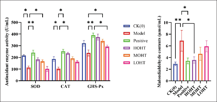
AbstractBackground: Hydroxytyrosol (HT) is a natural phenolic antioxidant found in olive oil and has been reported to exhibit antioxidative and anti-inflammatory bioactivities. However, its potential protective effect in chemically induced retinal injury remains insufficiently defined. Aim: This study investigated whether olive oil rich in hydroxytyrosol (OHT) could attenuate sodium iodate (NaIO₃)-induced acute retinal damage in mice. Methods: Retinal damage was induced by intraperitoneal injection of a 5% NaIO₃ solution at 25 mg/kg body weight. After model establishment, mice received OHT by gavage for 28 consecutive days at doses of 200, 100, or 50 mg/kg. These doses refer to the OHT preparation rather than pure HT. Serum catalase (CAT), superoxide dismutase (SOD), glutathione peroxidase (GSH-Px), malondialdehyde (MDA), interleukin-18 (IL-18), tumor necrosis factor-α (TNF-α), and transforming growth factor-β (TGF-β) were measured. Retinal morphology was assessed using paraffin-embedded eye sections, and retinal Serpin family F member 1 (SERPINF1) and Patatin-like phospholipase domain–containing 2 (PNPLA2) expression was examined by immunohistochemistry. Results: Compared with the model group, OHT treatment increased serum CAT, GSH-Px, and SOD activities, and TGF-β levels, while reducing serum MDA, IL-18, and TNF-α levels (p < 0.05 or p < 0.01). Histological observation showed that OHT, particularly at the high dose, partially preserved retinal structure after NaIO₃ exposure. Immunohistochemical analysis further showed higher retinal SERPINF1 and PNPLA2 immunopositive expression in OHT-treated mice than in model mice (p < 0.05 or p < 0.01). Conclusion: OHT attenuated NaIO₃-induced retinal damage in mice, possibly through improvement of oxidative stress and inflammatory responses. The concurrent changes in SERPINF1 and PNPLA2 expression suggest that these proteins may be associated with the retinal protective response, although direct mechanistic involvement requires further validation. Keywords: Hydroxytyrosol, Retinal injury, Oxidative stress, SERPINF1, PNPLA2. IntroductionRetinal degeneration is a major cause of irreversible visual impairment and is commonly associated with progressive damage to photoreceptors and the retinal pigment epithelium (RPE). Retinitis pigmentosa is one representative inherited retinal degenerative disease, typically beginning with night blindness and followed by progressive peripheral visual field loss and, in advanced cases, severe visual impairment or blindness. Its global prevalence has been estimated at approximately 1/3,000 to 1/8,000, while the reported prevalence in China is approximately 1/3,784 and may reach 1/1,000 in certain regions. Although current therapeutic strategies can delay disease progression or preserve residual visual function in selected patients, effective and broadly applicable interventions remain limited (Liu and Wu, 2021). Therefore, safe nutritional, or functional-food–derived compounds with antioxidant and anti-inflammatory activities may provide useful supportive strategies for retinal protection. However, their effects still require experimental validation in controlled retinal injury models. Hydroxytyrosol (HT; 3,4-dihydroxyphenylethanol) is an important phenolic compound derived from Olea europaea. It is widely present in olive fruits and branches and can also be generated through oleuropein hydrolysis (Li et al., 2023). HT has attracted attention because of its antioxidative, anti-inflammatory, anti-atherosclerotic, antibacterial, antithrombotic, antitumor, hemostatic, and endothelial-protective properties (Mosca et al., 2020). It has also been investigated as a natural food additive and as a potentially safe bioactive compound for cardioprotection, neuroprotection, and chemoprevention (Visioli, 2012). These biological activities suggest that HT-containing preparations may have protective potential in tissues vulnerable to oxidative stress, including the retina. Olive oil contains multiple natural nutrients, including polyphenols, squalene, triterpenoids, flavonoids, sterols, fat-soluble vitamins, and carotenoids (Ye et al., 2023a). It is widely regarded as a health-promoting vegetable oil because long-term consumption has been associated with improvements in lipid metabolism, blood glucose, blood pressure, platelet aggregation, and inflammatory status (Lu et al., 2023). Our previous studies showed that olive oil rich in hydroxytyrosol (OHT) exerted anti-aging and hypolipidemic effects in mouse models (Chen et al., 2023a; Ye et al., 2023b). Nevertheless, whether OHT can protect retinal tissue against chemically induced oxidative injury remains unclear. Sodium iodate (NaIO₃) is commonly used to induce acute retinal injury, particularly through selective damage to the RPE and subsequent secondary effects on photoreceptors. This model reproduces several pathological features relevant to retinal degeneration, such as oxidative stress, RPE disruption, and thinning of retinal layers, but it should not be considered a complete model of inherited retinitis pigmentosa. For this reason, the NaIO₃ model is suitable for evaluating retinal protective effects under acute oxidative injury conditions rather than for fully representing the clinical course of retinitis pigmentosa. In addition to histological injury, retinal damage may involve changes in proteins related to retinal homeostasis and function. Serpin family F member 1 (SERPINF1), also known as pigment epithelium–derived factor, is a multifunctional molecule with antiangiogenic and neurotrophic activities (Ren et al., 2005). Patatin-like phospholipase domain–containing 2 (PNPLA2) is a retinyl ester hydrolase expressed in the RPE and participates in the visual cycle by influencing retinoid metabolism and RPE65-related activity (Hara et al., 2023). Changes in SERPINF1 and PNPLA2 expression may therefore reflect retinal structural and functional responses after injury, although expression changes alone cannot establish a direct mechanistic pathway. Based on these considerations, the present study investigated the protective effects of OHT against NaIO₃-induced acute retinal damage in mice. We evaluated serum oxidative stress and inflammatory indicators, retinal histopathological changes, retinal layer thickness and cell number, and the immunohistochemical expression of SERPINF1 and PNPLA2. This study aimed to provide preclinical evidence for the retinal protective potential of OHT and to clarify whether changes in SERPINF1 and PNPLA2 expression changes are associated with this protective response. Materials and MethodsMaterials and reagentsSpecific pathogen-free-grade Kunming (KM) mice were obtained from the Lanzhou Institute of Veterinary Medicine, Chinese Academy of Agricultural Sciences (license No. SCXK [Gan] 2020-0002). NaIO₃; CAS No. 7681-55-2; purity ≥99%; Cat. No. S4007 was purchased from Sigma-Aldrich (St. Louis, MO, USA). OHT; HT content: 20 mg/100 g was obtained from the Key Laboratory of Chemistry of Northwest Specialty Plant Resources, Lanzhou Institute of Chemical Physics, Chinese Academy of Sciences. Andrographolide (CAS No. 5508-58-7; Cat. No. HY-N0191), used as an active comparator with reported retinal anti-inflammatory and antiangiogenic activity, was purchased from MedChemExpress (Monmouth Junction, NJ, USA). The 3,3′-diaminobenzidine (DAB) kit was purchased from Yeasen Biotechnology (Shanghai, China). Primary rabbit polyclonal antibodies against SERPINF1 and PNPLA2 were obtained from Mlbio (Shanghai, China). Immunohistochemistry SP hypersensitivity kits and endogenous biotin blocking kits were purchased from ACROBiosystems (Beijing, China). Isoflurane was obtained from Shanghai Hengfengqiang Animal Pharmaceutical Co., Ltd. (Shanghai, China). Zoletil, a tiletamine–zolazepam injectable anesthetic, was used for deep anesthesia before sample collection. Assay kits for catalase (CAT; Cat. No. BC0200), superoxide dismutase (SOD; Cat. No. BC0170), glutathione peroxidase (GSH-Px/GPX; Cat. No. BC1190 or BC1195, according to kit specifications), and malondialdehyde (MDA; Cat. No. BC0020) were purchased from Solarbio Science and Technology Co., Ltd. (Beijing, China). Enzyme-linked immunosorbent assay kits for interleukin-18 (IL-18), tumor necrosis factor-α (TNF-α), and transforming growth factor-β (TGF-β) were purchased from Inselisa Biotechnology Co., Ltd. (Huangshi, China). Instruments and equipmentThe instruments used in this study included an SM2010 R microtome (Asiapeptide Biotechnology Center, Nanjing, China), a DM500 light microscope (Asiapeptide Biotechnology Center), a benchtop centrifuge (Hunan Xiangyi Laboratory Instrument Development Co., Ltd., Hunan, China), a UV–Vis spectrophotometer (Shimadzu, Shanghai, China), an electronic analytical balance (Shimadzu), and an automatic microplate reader (Jingcheng Instrumentation Co., Ltd., Qingdao, China). Preparation and characterization of OHTFresh olive fruits were washed, pitted, crushed, passed through an 80-mesh sieve, and mixed with distilled water at a weight ratio of 1:6. The mixture was homogenized using a high-speed tissue homogenizer at 16,000 r/min for 10 minutes, with a 0.5-minute pause after every 2 minutes of homogenization. The olive fruit homogenate was stirred at 45°C until pulp formation. After physical pressing at room temperature, the pulp was centrifuged at 20,000 r/min, and the pomace and supernatant oil–water mixture were separated. The supernatant was filtered through a 400-mesh membrane, mixed with sodium sulfate at 10% of the filtrate weight, stirred thoroughly, and allowed to stand for 25 minutes. The HT-rich crude olive oil was then obtained by oil–water separation and allowed to stand for 24 hours to obtain HT-rich extra virgin olive oil. The HT content of the obtained OHT was determined by high-performance liquid chromatography. Separation was performed on a Sino Chrom ODS-BP column (4.6 × 250 mm, 5 μm), using 0.1% formic acid–water and acetonitrile (86:14, v/v) as the mobile phase. The flow rate was 1.0 ml/min, the column temperature was 30°C, the detection wavelength was 280 nm using a diode-array detector, and the injection volume was 20 μl. The final HT content of the OHT preparation was 20 mg/100 g. In comparison, the HT content of ordinary olive oil was approximately 2.5 mg/kg. Therefore, the OHT doses used in the animal experiment referred to the amount of OHT preparation administered, not to the amount of pure HT. Based on the measured HT content of 20 mg/100 g, the 50, 100, and 200 mg/kg OHT doses corresponded approximately to 0.01, 0.02, and 0.04 mg/kg HT, respectively. Animals, randomization, and experimental groupingSixty KM mice weighing 22–28 g, with equal numbers of males and females, were used in this study. The animals were acclimatized for 1 week under the same housing conditions before the experiment. Mice were housed in a specific pathogen-free animal facility under controlled temperature, humidity, and light–dark cycle conditions and were provided with standard chow and water ad libitum. After acclimatization, mice were randomly allocated into six groups using a random number method, with 10 mice in each group: blank control group (CK [0]), model group, andrographolide active comparator group, high-dose OHT group (HOHT; 200 mg/kg), medium-dose OHT group (MOHT; 100 mg/kg), and low-dose OHT group (LOHT; 50 mg/kg). The grouping information was concealed from the investigators responsible for histological observation, immunohistochemical image acquisition, and quantitative image analysis. Data analysis was performed after group coding was completed. Establishment of the NaIO₃-induced retinal injury model and drug administrationThe NaIO₃-induced model was used as an acute oxidative retinal injury model characterized primarily by RPE injury and secondary retinal structural damage. Except for the blank control group, mice received an intraperitoneal injection of a 5% NaIO₃ solution at 25 mg/kg body weight to induce retinal damage. The blank control group received an equal volume of normal saline. After model establishment, mice in the HOHT, MOHT, and LOHT groups were gavaged with OHT at doses of 200, 100, and 50 mg/kg, respectively, once daily for 28 consecutive days. These doses referred to the administered OHT preparation rather than pure HT. Mice in the active comparator group were gavaged with andrographolide at 50 mg/kg once daily for 28 days. Andrographolide was selected because previous studies reported its retinal anti-inflammatory and antiangiogenic effects in experimental retinal disease models, supporting its use as an active comparator rather than as a disease-specific standard positive control (Yu et al., 2015). Mice in the model group and blank control group received the same volume of normal saline by gavage. The gavage procedure was conducted with reference to GB 15193.1-2014 National Food Safety Standard: Procedures for Toxicological Assessment of Food. Measurement of serum CAT, SOD, GSH-Px, MDA, IL-18, TNF-α, and TGF-βAt the end of the 28-day intervention, mice were deeply anesthetized with Zoletil at 50–75 mg/kg by intramuscular injection. Blood was collected from the retro-orbital venous plexus using a blood collection tube inserted along the medial canthus at approximately 45° to the face. Blood samples were placed in yellow-capped coagulation tubes and kept at 4°C for 6 hours. The samples were then centrifuged, and the serum supernatant was collected for biochemical analysis. Serum CAT, SOD, GSH-Px, MDA, IL-18, TNF-α, and TGF-β levels were measured according to the manufacturers’ instructions for the corresponding assay kits. All measurements were performed using the same batch of kits for each indicator to reduce inter-assay variation. Paraffin sectioning and hematoxylin–eosin (H&E) stainingAfter deep anesthesia and blood collection, mice were euthanized, and bilateral eyeballs were collected. The eyeballs were fixed in 4% paraformaldehyde for 24 hours and rinsed under running water for 12 hours. After dehydration, clearing, and paraffin embedding, serial eye sections were prepared at a thickness of 7 μm using a microtome. Sections were spread, dried, and used for H&E staining or immunohistochemical staining. H&E-stained sections were mounted, observed under a light microscope, and photographed for assessment of retinal morphology. The remaining paraffin sections were stored for subsequent immunohistochemical analysis (Chen et al., 2024b). Immunohistochemical staining for SERPINF1 and PNPLA2Paraffin sections were deparaffinized in xylene, rehydrated through graded alcohols, and subjected to antigen retrieval using citrate buffer. Immunohistochemical staining was then performed according to the kit protocol. Briefly, sections were incubated with an endogenous peroxidase blocker for 15 minutes and non-immune goat serum for 15 minutes. The sections were then incubated with primary rabbit polyclonal antibodies against SERPINF1 or PNPLA2 at a dilution of 1:200 overnight at 4°C. After washing with phosphate-buffered saline three times for 3 minutes each, sections were incubated with biotin-labeled goat anti-rabbit immunoglobulin G diluted 1:500 for 20 minutes and then with streptavidin–biotin–peroxidase reagent for 20 minutes. Color development was performed with DAB for 5 minutes, followed by hematoxylin counterstaining. For negative control staining, the primary antibody was replaced with antibody diluent after incubation with non-immune goat serum, while the remaining procedures were kept unchanged. The negative control was used to assess nonspecific staining. After staining, sections were dehydrated, cleared, mounted, observed, and photographed under a light microscope (Chen et al., 2023c). Histological and image analysisH&E-stained retinal sections were used to assess retinal structural changes, retinal layer thickness, and retinal cell number. For each mouse, representative sections passing through comparable retinal regions were selected. Retinal thickness and layer-specific thickness were measured using calibrated image analysis software. Cell numbers in selected retinal layers were counted in comparable microscopic fields. The investigators conducting histological evaluation and image quantification were blinded to the experimental grouping. For immunohistochemical analysis, SERPINF1 and PNPLA2 staining was observed under a light microscope and photographed using a microimaging system. Positive staining was identified as brown or yellow-brown signals. Mean optical density was used to quantify SERPINF1 and PNPLA2 immunopositive expression in comparable retinal regions. For each animal, multiple non-overlapping microscopic fields were analyzed, and the mean value was used for statistical analysis to avoid pseudoreplication. Statistical analysisData were analyzed using GraphPad Prism 10. Quantitative data are expressed as mean ± standard deviation. Each experimental group included 10 mice unless otherwise specified in the figure legends. Before between-group comparison, the normality of data distribution was assessed using the Shapiro–Wilk test, and homogeneity of variance was assessed using Levene’s test or the Brown–Forsythe test, as appropriate. For normally distributed data with homogeneous variance, comparisons among multiple groups were performed using one-way analysis of variance (ANOVA), followed by the least significant difference post hoc test for pairwise comparisons. If variance homogeneity was not satisfied, Welch’s ANOVA followed by an appropriate post hoc test was used. A two-sided p value < 0.05 was considered statistically significant. Statistical significance was presented as p < 0.05 or p < 0.01. Ethical approvalAll animal procedures were approved by the Ethics Committee of Longnan Normal University (Approval No. 2025-0018; approval date: March 10, 2025). All procedures were conducted in accordance with institutional animal welfare requirements. Animal discomfort was minimized through acclimatization, appropriate anesthesia, and standardized handling. Mice were deeply anesthetized before blood collection and tissue harvesting, and euthanasia was performed before eyeball collection. ResultsEffects of OHT on serum SOD, CAT, GSH-Px, and MDA in mice with NaIO₃-induced retinal damageAs shown in Figure 1, NaIO₃ administration altered serum oxidative stress–related indicators. Compared with the blank control group, the model group showed significantly lower serum SOD and CAT activities (p < 0.05) and a significantly higher MDA level (p < 0.01), indicating that NaIO₃ induced systemic oxidative stress in mice. Compared with the model group, the andrographolide active comparator group showed higher serum SOD (p < 0.01), GSH-Px (p < 0.01), and CAT activities (p < 0.05), together with a lower MDA level (p < 0.05).
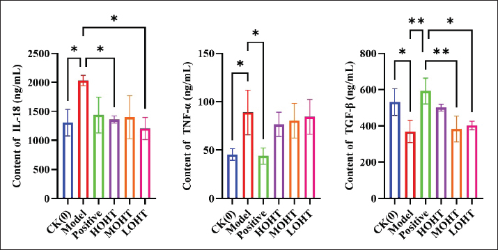
Fig. 1. Effects of OHT on serum CAT, SOD, GSH-Px, and MDA in mice with NaIO₃-induced retinal damage. CAT, catalase; SOD, superoxide dismutase; GSH-Px, glutathione peroxidase; MDA, malondialdehyde; OHT, olive oil rich in hydroxytyrosol; NaIO₃, sodium iodate. OHT treatment also improved several oxidative stress indicators. Compared with the model group, serum SOD and GSH-Px activities were significantly increased in the MOHT group (p < 0.05), CAT activity was significantly increased in the HOHT group (p < 0.05), and the serum MDA level was significantly decreased in the HOHT group (p < 0.05). The GSH-Px activity in the LOHT group was lower than that in the andrographolide active comparator group (p < 0.05). No significant differences were observed in the remaining comparisons with the active comparator group (p > 0.05). Effects of OHT on serum IL-18, TNF-α, and TGF-β in mice with NaIO₃-induced retinal damageAs shown in Figure 2, NaIO₃ exposure induced changes in serum inflammatory indicators. Compared with the blank control group, the model group had significantly higher serum IL-18 and TNF-α levels (p < 0.05) and a significantly lower TGF-β level (p < 0.05). Compared with the model group, the andrographolide active comparator group showed a lower TNF-α level (p < 0.05) and a higher TGF-β level (p < 0.01). In the OHT-treated groups, serum IL-18 levels were significantly lower in the HOHT and LOHT groups than in the model group (p < 0.05).
Fig. 2. Effects of OHT on serum IL-18, TNF-α, and TGF-β in mice with NaIO₃-induced retinal damage. IL-18, interleukin-18; TNF-α, tumor necrosis factor-α; TGF-β, transforming growth factor-β; OHT, olive oil rich in hydroxytyrosol; NaIO₃, sodium iodate. Compared with the andrographolide active comparator group, serum TGF-β levels were lower in the MOHT group (p < 0.01) and LOHT group (p < 0.05). No significant differences were observed in the remaining comparisons (p > 0.05). These results suggest that OHT partially attenuated the inflammatory response associated with NaIO₃-induced retinal injury. Effects of OHT on retinal histopathology in mice with NaIO₃-induced retinal damageRepresentative H&E-stained retinal sections are shown in Fig. 3. In the blank control group, the nine retinal layers were clearly identifiable, with regular cellular morphology, dark blue nuclear staining, and red cytoplasmic staining (Fig. 3I). In the model group, the retinal inner limiting membrane and RPE showed structural disruption. The photoreceptor cell layer was visibly atrophic, and the outer plexiform layer between the inner and outer nuclear layers appeared sparse (Fig. 3II).
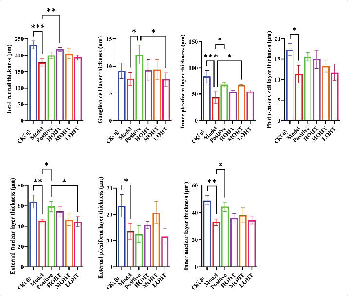
Fig. 3. H&E staining of mouse retinas after NaIO₃-induced retinal damage and OHT treatment (×400). I, CK (0) group; II, model group; III, andrographolide active comparator group; IV, HOHT group; V, MOHT group; VI, LOHT group. A, inner limiting membrane; B, nerve fiber layer; C, ganglion cell layer; D, inner plexiform layer; E, inner nuclear layer; F, outer plexiform layer; G, outer nuclear layer; H, photoreceptor cell layer; I, RPE. CK, blank control; HOHT, high-dose OHT; MOHT, medium-dose OHT; LOHT, low-dose OHT. In the andrographolide active comparator group, the retinal layers were relatively clear and better preserved than those in the model group (Fig. 3III). In the HOHT group, retinal cells showed relatively regular morphology and more uniform staining, suggesting partial preservation of retinal structure after NaIO₃ exposure (Fig. 3IV). In the MOHT group, the original retinal structure appeared blurred, with partial detachment of the RPE (Fig. 3V). In the LOHT group, atrophy of the outer plexiform layer and photoreceptor cell layer remained evident (Fig. 3VI). Overall, the histological findings indicated that HOHT showed a more apparent protective effect on retinal morphology than the lower OHT doses. Effects of OHT on retinal thickness and retinal cell number in mice with NaIO₃-induced retinal damageAs shown in Figure 4, NaIO₃ exposure significantly reduced retinal thickness. Compared with the blank control group, the model group showed significantly decreased total retinal thickness (p < 0.01), inner plexiform layer thickness (p < 0.01), outer nuclear layer thickness (p < 0.01), inner nuclear layer thickness (p < 0.01), photoreceptor cell layer thickness (p < 0.05), and outer plexiform layer thickness (p < 0.05).
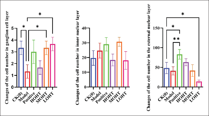
Fig. 4. Changes in retinal thickness in mice with NaIO₃-induced retinal damage after OHT treatment. OHT, olive oil rich in hydroxytyrosol; NaIO₃, sodium iodate. Compared with the model group, the andrographolide active comparator group showed significantly increased thickness of the ganglion cell layer, inner plexiform layer, outer nuclear layer, and inner nuclear layer (p < 0.05). In the OHT-treated groups, total retinal thickness was significantly increased in the HOHT group (p < 0.01), and inner plexiform layer thickness was significantly increased in the MOHT group (p < 0.05). Compared with the active comparator group, the ganglion cell layer and outer nuclear layer were significantly thinner in the LOHT group (p < 0.05). No significant differences were observed in the remaining comparisons (p > 0.05). As shown in Figure 5, the number of ganglion cell layer cells was significantly lower in the model group than in the blank control group (p < 0.05). The number of outer nuclear layer cells was significantly higher in the andrographolide active comparator group (p < 0.05) but significantly lower in the LOHT group (p < 0.05), compared with the blank control group. Compared with the model group, the active comparator group showed a significant increase in the number of outer nuclear layer cells (p < 0.01), while the MOHT and LOHT groups showed increased numbers of ganglion cell layer cells (p < 0.05). No significant differences were observed in the remaining comparisons (p > 0.05).
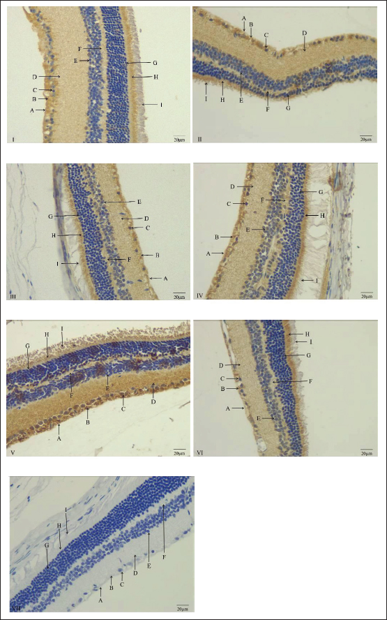
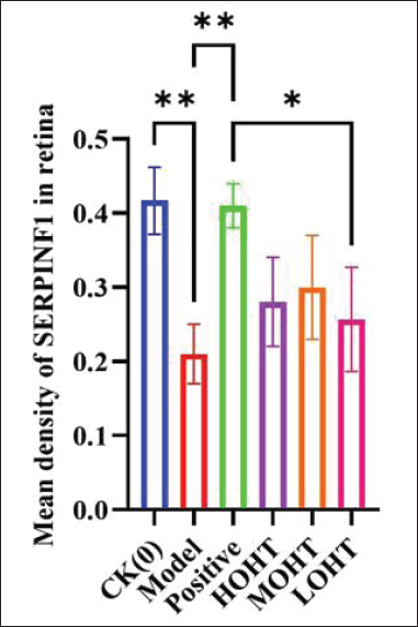
Fig. 5. Changes in retinal cell number in mice with NaIO₃-induced retinal damage after OHT treatment. OHT, olive oil rich in hydroxytyrosol; NaIO₃, sodium iodate. Effects of OHT on SERPINF1 expression in the retina of mice with NaIO₃-induced retinal damageSERPINF1 immunohistochemical staining results are shown in Figures 6 and 7. In the negative control sections, retinal cell bodies were stained light blue and nuclei were stained dark blue, with no obvious brown or yellow-brown positive staining, indicating no apparent nonspecific staining. In the experimental sections, SERPINF1-immunopositive signals appeared as tan, yellow-brown, or light yellow staining. SERPINF1-positive staining was mainly observed in the nerve fiber layer, ganglion cell layer, inner plexiform layer, outer plexiform layer, and photoreceptor cell layer.
Fig. 6. Immunohistochemical staining of SERPINF1 in mouse retinas after NaIO₃-induced retinal damage and OHT treatment (×400). I, CK (0) group; II, model group; III, andrographolide active comparator group; IV, HOHT group; V, MOHT group; VI, LOHT group; VII, negative control staining. A, inner limiting membrane; B, nerve fiber layer; C, ganglion cell layer; D, inner plexiform layer; E, inner nuclear layer; F, outer plexiform layer; G, outer nuclear layer; H, photoreceptor cell layer; I, RPE. SERPINF1, serpin family F member 1. In the blank control group, SERPINF1 immunopositive staining was relatively uniform and light yellow. In the model group, the nerve fiber layer and photoreceptor cell layer showed atrophic changes, and SERPINF1-positive staining was mainly distributed in the nerve fiber layer. Quantitative analysis showed that SERPINF1 expression was significantly lower in the model group than in the blank control group (p < 0.01). Compared with the model group, SERPINF1 expression was significantly higher in the andrographolide active comparator group (p < 0.01). In the HOHT group, SERPINF1-positive signals were mainly distributed in the nerve fiber layer and photoreceptor cell layer. In the MOHT group, stronger tan staining was observed, and SERPINF1-positive signals were mainly located in the nerve fiber layer, ganglion cell layer, and inner plexiform layer. In the LOHT group, positive staining was more evident in the photoreceptor cell layer. Compared with the andrographolide active comparator group, SERPINF1 expression was significantly lower in the LOHT group (p < 0.05). These findings indicate that NaIO₃ reduced retinal SERPINF1 immunopositive expression, while OHT treatment was associated with partial restoration of SERPINF1 expression. Effects of OHT on PNPLA2 expression in the retina of mice with NaIO₃-induced retinal damagePNPLA2 immunohistochemical staining results are shown in Figures 8 and 9. In the negative control sections, retinal cell bodies were stained light blue and nuclei were stained dark blue, with no obvious positive staining. In the experimental sections, PNPLA2-immunopositive signals appeared as tan, yellow-brown, or light yellow staining. PNPLA2-positive staining was distributed across several retinal layers, including the nerve fiber layer, ganglion cell layer, inner plexiform layer, outer plexiform layer, and photoreceptor cell layer.
Fig. 7. Mean optical density of SERPINF1 immunohistochemical staining in mouse retinas. SERPINF1, serpin family F
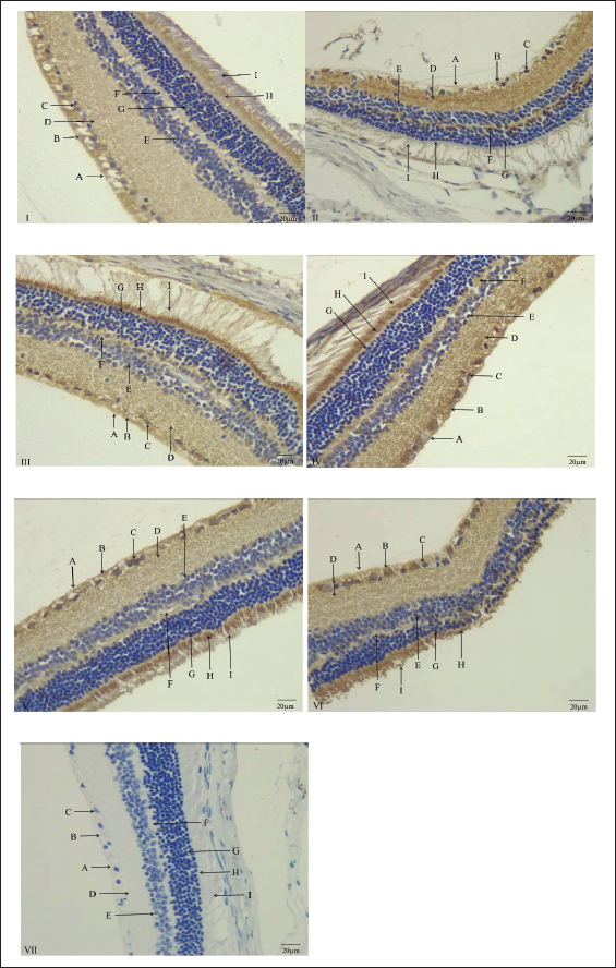
Fig. 8. Immunohistochemical staining of PNPLA2 in mouse retinas after NaIO₃-induced retinal damage and OHT treatment (×400). I, CK (0) group; II, model group; III, andrographolide active comparator group; IV, HOHT group; V, MOHT group; VI, LOHT group; VII, negative control staining. A, inner limiting membrane; B, nerve fiber layer; C, ganglion cell layer; D, inner plexiform layer; E, inner nuclear layer; F, outer plexiform layer; G, outer nuclear layer; H, photoreceptor cell layer; I, RPE. PNPLA2, patatin-like phospholipase domain–containing 2.
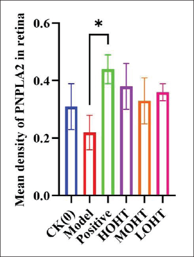
Fig. 9. Mean optical density of PNPLA2 immunohistochemical staining in mouse retinas. PNPLA2, patatin-like phospholipase domain–containing 2. In the blank control group, the photoreceptor cell layer showed relatively stronger brown staining. In the model group, the inner and outer plexiform layers showed yellow-brown staining, and irregular nuclear morphology was observed in the inner nuclear layer. In the andrographolide active comparator group, the nine retinal layers were clearly delineated, and PNPLA2-positive signals were relatively uniform and light yellow. Quantitative analysis showed that PNPLA2 expression was significantly higher in the active comparator group than in the model group (p < 0.05). In the HOHT, MOHT, and LOHT groups, PNPLA2-positive signals were mainly distributed in the nerve fiber layer and photoreceptor cell layer, with brown staining observed in these regions. These results indicate that OHT treatment was accompanied by changes in PNPLA2 immunopositive expression after NaIO₃-induced retinal injury. DiscussionThe present study evaluated the retinal protective effect of OHT in a mouse model of NaIO₃-induced retinal damage. The results showed that NaIO₃ exposure reduced serum antioxidant enzyme activities, increased lipid peroxidation, disrupted retinal morphology, decreased retinal thickness and selected retinal cell numbers, and altered retinal SERPINF1 and PNPLA2 immunopositive expression. OHT treatment, particularly at the high dose, partially reversed these changes. These findings suggest that OHT may attenuate NaIO₃-induced retinal injury mainly through improvement of oxidative stress and inflammatory responses, with concurrent changes in retinal proteins related to retinal homeostasis. NaIO₃ is widely used as a chemically induced retinal injury model because it selectively affects the RPE and subsequently causes secondary photoreceptor and retinal structural damage (Tan et al., 2023). However, this model should be interpreted as an acute oxidative retinal injury or RPE injury model rather than as a complete model of inherited retinitis pigmentosa. Recent studies have similarly emphasized that NaIO₃ induces strong oxidative stress in RPE cells and can lead to photoreceptor loss and visual pathway impairment (Yang et al., 2022; Espitia-Arias et al., 2023; Upadhyay and Bonilha, 2024). Other work has described the NaIO₃ model as a useful preclinical model for studying RPE loss, oxidative stress, and dry age-related macular degeneration-like retinal degeneration, rather than a disease-specific model for all forms of retinal degeneration (Kim and Qian, 2022; Upadhyay and Bonilha, 2024). This distinction is important because it places the present findings within an acute injury context and avoids overextending the model to the full clinical course of retinitis pigmentosa. In this study, NaIO₃ significantly decreased serum SOD, CAT, and GSH-Px activities and increased MDA levels, indicating oxidative stress and lipid peroxidation. These biochemical changes were consistent with the retinal histopathological findings, including disruption of the inner limiting membrane and RPE layer, atrophy of the photoreceptor cell layer, and thinning of several retinal layers. Oxidative stress is known to play a central role in retinal injury because retinal cells, especially RPE cells and photoreceptors, are highly metabolically active and continuously exposed to oxidative challenges (Nicholls and Hazen, 2005; Yang et al., 2022; Espitia-Arias et al., 2023). Recent reviews have also emphasized that oxidative imbalance contributes to retinal degeneration and that reinforcement of antioxidant defenses remains a biologically plausible protective strategy in retinal injury models (Upadhyay and Bonilha, 2024). In the present study, OHT increased antioxidant enzyme activities and reduced MDA levels to varying degrees, suggesting that its retinal protective effect may be partly related to reduced oxidative damage. The antioxidant effect observed here is also consistent with previous evidence on HT. HT has been reported to protect human retinal pigment epithelial cells against oxidative injury by activating mitochondrial biogenesis and phase II detoxifying enzyme systems (Zhu et al., 2010). This cellular evidence supports the biological plausibility of the present in vivo findings, although the current study used OHT as an olive oil preparation rather than purified HT. Therefore, the observed effects may reflect the contribution of HT together with other bioactive components naturally present in olive oil. This point is important for interpretation because the administered doses in this study refer to the OHT preparation, not to pure HT. Inflammation is another important contributor to retinal injury. In the present study, NaIO₃ increased serum IL-18 and TNF-α levels and reduced TGF-β levels, suggesting that retinal injury was accompanied by a systemic inflammatory response. IL-18 is an important pro-inflammatory cytokine involved in innate immune activation, while TNF-α participates in inflammatory amplification and tissue damage (Dinarello et al., 2013; Yasuda et al., 2019; Ma et al., 2023). TGF-β, although context-dependent, is often associated with anti-inflammatory regulation and tissue repair responses (Haurigot et al., 2012). OHT treatment reduced serum IL-18 and TNF-α levels and increased TGF-β levels in several comparisons, suggesting that OHT may also attenuate inflammatory activation after NaIO₃ exposure. This interpretation is consistent with previous studies showing that HT can reduce pro-inflammatory cytokine production in inflammatory models (Zhang et al., 2009; Yao et al., 2019). However, because inflammatory indicators were measured in serum rather than directly in retinal tissue, the findings should be interpreted as systemic inflammatory changes associated with retinal injury rather than direct evidence of local retinal inflammatory pathway inhibition. The selection of andrographolide as the active comparator also requires careful interpretation. In the revised study design, andrographolide was treated as an active comparator rather than as a standard positive control for NaIO₃-induced retinal injury. This distinction is appropriate because there is no universally accepted standard positive drug for this specific model. Previous studies have reported that andrographolide can exert retinal anti-inflammatory, antiangiogenic, and neuroprotective effects in experimental ocular disease settings, including diabetic retinopathy and retinal ganglion cell injury models (Yu et al., 2015; Li et al., 2023). Therefore, its inclusion provides a biologically relevant comparator for retinal protection, but it should not be interpreted as a disease-specific reference treatment for NaIO₃-induced retinal injury. SERPINF1 and PNPLA2 were examined to explore retinal protein expression changes associated with OHT treatment. SERPINF1, also known as pigment epithelium–derived factor, is expressed in ocular tissues and has antiangiogenic and neurotrophic properties (Ren et al., 2005; Notari et al., 2006). It may contribute to retinal homeostasis through regulation of vascular and neuronal survival-related processes. PNPLA2 has recently been identified as a retinyl ester hydrolase in the RPE and has been linked to retinoid mobilization and visual cycle function (Hara et al., 2023). In the present study, NaIO₃ reduced retinal SERPINF1 and PNPLA2 immunopositive expression, while OHT treatment was accompanied by partial restoration of these signals. These findings suggest that SERPINF1 and PNPLA2 may be associated with the retinal protective response observed after OHT administration. Nevertheless, the present data are based on immunohistochemical expression and cannot establish a direct causal mechanism. Further studies using pathway inhibition, gene knockdown, overexpression, or functional visual assessments are needed to determine whether SERPINF1 and PNPLA2 actively mediate the protective effect of OHT. Several limitations should be acknowledged. First, this study used an acute NaIO₃-induced retinal injury model, which cannot fully reproduce the genetic background, chronic progression, or phenotypic heterogeneity of retinitis pigmentosa. Second, the study assessed serum oxidative stress and inflammatory indicators but did not directly measure retinal tissue levels of oxidative or inflammatory mediators. Third, SERPINF1 and PNPLA2 were evaluated by immunohistochemistry, which provides spatial expression information but does not prove mechanistic involvement. Fourth, the protective effect of OHT was assessed mainly through biochemical and histological endpoints, while functional retinal outcomes such as electroretinography were not included. Future studies should combine retinal functional testing, retinal tissue-based molecular assays, and mechanistic interventions to clarify the pathways through which OHT affects retinal injury. ConclusionOHT attenuated NaIO₃-induced retinal damage in mice, particularly at the high dose, as reflected by improved antioxidant enzyme activities, reduced lipid peroxidation, moderated inflammatory indicators, and partial preservation of retinal morphology. OHT treatment was also accompanied by changes in SERPINF1 and PNPLA2 immunopositive expression, suggesting that these proteins may be associated with the retinal protective response. These findings provide preliminary preclinical evidence for the retinal protective potential of OHT, while further mechanistic and functional studies are required before stronger causal claims can be made. AcknowledgmentsWe thank Professor Yu Wang for technical support and advice on statistical methods. We also thank Professor Wenbin Ye for funding support. Conflict of interestThe author(s) declared no potential conflicts of interest with respect to the research, authorship, and/or publication of this article. FundingThis research was supported by the Gansu Youth Science and Technology Fund in 2023 (23JRRK0002), Scientific Research Project of the Education Department of Gansu Province (No. 2025B-452) and the Key Discipline of Botany at Longnan Normal University (LN2025002). Authors’ contributionsWendong Chen was responsible for data processing, experiments, and manuscript writing. Xiujuan Zhu was primarily responsible for the methodology. Data availabilityAll data for this study are available from the corresponding authors. ReferencesChen, W.D., Ye, W.B., Zhu, X.J., Wang, Y., Huang, X.Y. and Di, D.L. 2023. Effect of olive oil containing hydroxyl tyrosol on aging mice. J. Gansu Agric. Univ. 58, 15–22; doi: 10.13432/j.cnki.jgsau.2023.03.003 Chen, W.D., Zhu, X.J., He, Y.P., Wang, Y. and Xu, Y.P. 2023. Distribution of OTR in nodose ganglion of goats. Acta Agric. Boreali-Occident. Sin. 32, 1090–1096; doi: 10.7606/j.issn.1004-1389.2023.07.012 Chen, W.D., Zhu, X.J., Ye, W.B., Wang, Y., Huang, X.Y., Di, D.L. and Xu, Y.P. 2024. Protective effect of oleuropein on LPS-induced retinal damage in mice. Chin. J. Vet. Sci. 44, 1019–1025; doi: 10.16303/j.cnki.1005-4545.2024.05.22 Dinarello, C.A., Novick, D., Kim, S. and Kaplanski, G. 2013. Interleukin-18 and IL-18 binding protein. Front. Immunol. 4, 289; doi: 10.3389/fimmu.2013.00289 Espitia-Arias, M.D., De La Villa, P., Paleo-García, V., Germain, F. and Milla-Navarro, S. 2023. Oxidative model of retinal neurodegeneration induced by sodium iodate: morphofunctional assessment of the visual pathway. Antioxidants 12, 1594; doi: 10.3390/antiox12081594 Hara, M., Wu, W., Malechka, V.V., Takahashi, Y., Ma, J.X. and Moiseyev, G. 2023. PNPLA2 mobilizes retinyl esters from retinosomes and promotes the generation of 11-cis-retinal in the visual cycle. Cell. Rep. 42, 112091; doi: 10.1016/j.celrep.2023.112091 Haurigot, V., Villacampa, P., Ribera, A., Bosch, A., Ramos, D., Ruberte, J. and Bosch, F. 2012. Long-term retinal PEDF overexpression prevents neovascularization in a murine adult model of retinopathy. PLoS One 7, e41511; doi: 10.1371/journal.pone.0041511 Kim, S.Y. and Qian, H. 2022. Comparison between sodium iodate and lipid peroxide models of retinal degeneration. Ann. Eye. Sci. 7, 20; doi: 10.21037/aes-21-38 Li, W.J., Wang, C.Z., Li, J.D. and Zhou, H. 2023. Preparation and performance evaluation of hydroxytyrosol nanoliposome freeze-dried powder. Chem. Ind. For. Prod. 43, 125–132; doi: 10.3969/j.issn.0253-2417.2023.05.017 Liu, H.L. and Wu, J.H. 2021. Progress in diagnosis and treatment of retinitis pigmentosa. Chin. J. Ocul. Fundus Dis. 37, 896–900; doi: 10.3760/cma.j.cn511434-20210319-00147 Lu, H.Y., Wang, L.J., Zhang, J.K., Zhang, C.Z., Li, T.J., Ji, R.X. and Shen, W.J. 2023. Determination of four fatty acid ethyl esters in olive oil by solid phase extraction-gas chromatography. Chin. J. Chromatogr. 41, 359–365; doi: 10.3724/SP.J.1123.2022.09011 Ma, J.B., Cao, Y.J., Wang, J.F. and Zhang, X.K. 2023. Observation on the efficacy of superficial needling therapy combined with Chinese herbal medicine for avascular necrosis of the femoral head and its effect on hip function and TGF-β, BMP, serum VEGF levels. Shanghai. J. Acupuncture. Moxibustion. 42, 66–71; doi: 10.13460/j.issn.1005-0957.2023.01.0066 Mosca, A., Crudele, A., Tozzi, G., Alterio, T., Smeriglio, A., Trombetta, D. and Alisi, A. 2020. The anti-inflammatory effects of hydroxytyrosol and vitamin E on paediatric NAFLD. Dig. Liver Dis. 52, e42–e43; doi: 10.1016/j.dld.2019.12.138 Nicholls, S.J. and Hazen, S.L. 2005. Myeloperoxidase and cardiovascular disease. Arterioscler. Thromb. Vasc. Biol. 25, 1102–1111; doi: 10.1161/01.ATV.0000163262.83456.6d Notari, L., Baladron, V., Aroca-Aguilar, J.D., Balko, N., Heredia, R., Meyer, C., Notario, P.M., Saravanamuthu, S., Nueda, M. and Sanchez-Sanchez, F. 2006. Identification of a lipase-linked cell membrane receptor for pigment epithelium-derived factor. J. Biol. Chem. 281, 38022–38037; doi: 10.1074/jbc.M600353200 Ren, J.G., Jie, C. and Talbot, C. 2005. How PEDF prevents angiogenesis: a hypothesized pathway. Med. Hypotheses. 64, 74–78; doi: 10.1016/j.mehy.2004.05.016 Tan, Y., Liao, W.P., Gao, Q.L., Li, Y., Shi, X.H. and Wang, J.K. 2023. Physiological indexes and histopathology analysis of sodium iodate-induced retinitis pigmentosa in rats. Lab. Anim. Comp. Med. 43, 124–135; doi: 10.12300/j.issn.1674-5817.2022.149 Upadhyay, M. and Bonilha, V.L. 2024. Regulated cell death pathways in the sodium iodate model: insights and implications for AMD. Exp. Eye. Res. 238, 109728; doi: 10.1016/j.exer.2023.109728 Visioli, F. 2012. Olive oil phenolics: where do we stand? Where should we go?. J. Sci. Food Agric. 92, 2017–2019; doi: 10.1002/jsfa.5715 Yang, X., Rai, U., Chung, J.Y. and Esumi, N. 2022. Fine tuning of an oxidative stress model with sodium iodate revealed protective effect of NF-κB inhibition and sex-specific difference in susceptibility of the retinal pigment epithelium. Antioxidants 11, 103; doi: 10.3390/antiox11010103 Yao, F., Yang, G.D., Xian, Y.S., Wang, G., Zheng, Z.H., Jin, Z., Xie, Y.D., Wang, W.R., Gu, J.L. and Lin, R. 2019. The protective effect of hydroxytyrosol acetate against inflammation of vascular endothelial cells partly through the SIRT6-mediated PKM2 signaling pathway. Food. Funct. 10, 5789–5803; doi: 10.1039/c9fo00586b Yasuda, K., Nakanishi, K. and Tsutsui, H. 2019. Interleukin-18 in health and disease. Int. J. Mol. Sci. 20, 649; doi: 10.3390/ijms20030649 Ye, W.B., Chen, W.D., He, J.J., Shao, J., Di, D.L. and Wang, Y. 2023b. Effects of olive oil rich in hydroxytyrosol and zeaxanthin pigment olive oil on hyperlipidemia mice. Cereals Oils 36, 74–78; doi: 10.13432/j.cnki.jgsau.2024.03.002 Ye, W.B., Chen, W.D., Wang, Y., Zhang, K., Huang, X.Y. and Di, D.L. 2023a. Effect of olive oil rich in hydroxyl tyrosol on hyperlipidemic mice. Food. Sci. 44, 144–150; doi: 10.7506/spkx1002-6630-20221028-298 Yu, Z., Lu, B., Sheng, Y., Zhou, L., Ji, L. and Wang, Z. 2015. Andrographolide ameliorates diabetic retinopathy by inhibiting retinal angiogenesis and inflammation. Biochimica Et Biophysica Acta (BBA). -. Gen. Subjects. 1850, 824–831; doi: 10.1016/j.bbagen.2015.01.014 Zhang, X., Cao, J. and Zhong, L. 2009. Hydroxytyrosol inhibits pro-inflammatory cytokines, iNOS, and COX-2 expression in human monocytic cells. Naunyn. Schmiedebergs. Archiv. Fuer. Pharmakologie. 379, 581–586; doi: 10.1007/s00210-009-0399-7 Zhu, L., Liu, Z., Feng, Z., Hao, J., Shen, W., Li, X., Sun, L., Sharman, E., Wang, Y., Wertz, K., Weber, P., Shi, X. and Liu, J. 2010. Hydroxytyrosol protects against oxidative damage by simultaneous activation of mitochondrial biogenesis and phase II detoxifying enzyme systems in retinal pigment epithelial cells. J. Nutr. Biochem. 21, 1089–1098; doi: 10.1016/j.jnutbio.2009.09.006 | ||

| How to Cite this Article |
| Pubmed Style Chen W, Zhu X, Ye W, Su M. Olive oil rich in hydroxytyrosol attenuates NaIO₃-induced acute retinal damage in mice. Open Vet. J.. 2026; 16(7): 4358-4372. doi:10.5455/OVJ.2026.v16.i7.23 Web Style Chen W, Zhu X, Ye W, Su M. Olive oil rich in hydroxytyrosol attenuates NaIO₃-induced acute retinal damage in mice. https://www.openveterinaryjournal.com/?mno=314051 [Access: July 10, 2026]. doi:10.5455/OVJ.2026.v16.i7.23 AMA (American Medical Association) Style Chen W, Zhu X, Ye W, Su M. Olive oil rich in hydroxytyrosol attenuates NaIO₃-induced acute retinal damage in mice. Open Vet. J.. 2026; 16(7): 4358-4372. doi:10.5455/OVJ.2026.v16.i7.23 Vancouver/ICMJE Style Chen W, Zhu X, Ye W, Su M. Olive oil rich in hydroxytyrosol attenuates NaIO₃-induced acute retinal damage in mice. Open Vet. J.. (2026), [cited July 10, 2026]; 16(7): 4358-4372. doi:10.5455/OVJ.2026.v16.i7.23 Harvard Style Chen, W., Zhu, . X., Ye, . W. & Su, . M. (2026) Olive oil rich in hydroxytyrosol attenuates NaIO₃-induced acute retinal damage in mice. Open Vet. J., 16 (7), 4358-4372. doi:10.5455/OVJ.2026.v16.i7.23 Turabian Style Chen, Wendong, Xiujuan Zhu, Wenbin Ye, and Manchun Su. 2026. Olive oil rich in hydroxytyrosol attenuates NaIO₃-induced acute retinal damage in mice. Open Veterinary Journal, 16 (7), 4358-4372. doi:10.5455/OVJ.2026.v16.i7.23 Chicago Style Chen, Wendong, Xiujuan Zhu, Wenbin Ye, and Manchun Su. "Olive oil rich in hydroxytyrosol attenuates NaIO₃-induced acute retinal damage in mice." Open Veterinary Journal 16 (2026), 4358-4372. doi:10.5455/OVJ.2026.v16.i7.23 MLA (The Modern Language Association) Style Chen, Wendong, Xiujuan Zhu, Wenbin Ye, and Manchun Su. "Olive oil rich in hydroxytyrosol attenuates NaIO₃-induced acute retinal damage in mice." Open Veterinary Journal 16.7 (2026), 4358-4372. Print. doi:10.5455/OVJ.2026.v16.i7.23 APA (American Psychological Association) Style Chen, W., Zhu, . X., Ye, . W. & Su, . M. (2026) Olive oil rich in hydroxytyrosol attenuates NaIO₃-induced acute retinal damage in mice. Open Veterinary Journal, 16 (7), 4358-4372. doi:10.5455/OVJ.2026.v16.i7.23 |


