





| Research Article | ||
Open Vet. J.. 2026; 16(7): 4828-4834
Open Veterinary Journal, (2026), Vol. 16(7): 4828-4834 Research Article Dynamic accuracy of feline rebound tonometry: Validation via implanted sensorsYulin Chen1, Yunhao Su1, Yingbo Liu1, Lihui Yang3, Fushan Shi2* and Ji Fu1*1School of Mechanical Engineering, Zhejiang Sci-Tech University, Hangzhou, China 2Department of Veterinary Medicine, College of Animal Sciences, Zhejiang University, Hangzhou, Zhejiang, China 3Rui Pai Hong Tai Pet Hospital (Huancheng Main Hospital), Hangzhou, China *Corresponding Author: Fushan Shi. Department of Veterinary Medicine, College of Animal Sciences, Zhejiang University, Hangzhou, Zhejiang, China. Email: sfs [at] zju.edu.cn; Ji Fu. School of Mechanical Engineering, Zhejiang Sci-Tech University, Hangzhou, China. Email: fuji [at] zstu.edu.cn Submitted: 17/03/2026 Revised: 10/06/2026 Accepted: 23/06/2026 Published: 20/07/2026 © 2025 Open Veterinary Journal
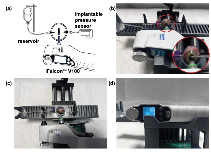
AbstractBackground: Precise intraocular pressure (IOP) quantification is pivotal for investigating glaucoma pathophysiology and establishing reliable animal models. Aim: This study comprehensively evaluated the accuracy, precision, and reproducibility of the novel iFalcon™ V100 rebound tonometer using ex vivo and in vivo feline models, referenced against intracameral manometry. Methods: Four ex vivo feline eyes and one in vivo subject were included. In the ex vivo phase, IOP was hydrostatically modulated between 5 and 75 mmHg via anterior chamber cannulation. Simultaneous readings were recorded from the iFalcon™ V100 and the reference pressure transducer. Agreement was assessed using linear regression, Bland–Altman analysis, and stratified box plots. In the in vivo phase, bilateral measurements were performed by two independent operators using both the iFalcon™ V100 and the iCare tonometer (Tonovet) to evaluate inter-operator reproducibility and inter-device comparability. Results: Linear regression analysis revealed a strong positive correlation between iFalcon™ V100 readings and manometry. Although a slight proportional bias (slope > 1.0) was observed in the high-IOP range—attributed to feline corneal biomechanics—the device maintained excellent linearity. Bland–Altman analysis demonstrated high measurement accuracy with a mean bias of only 0.41 mmHg and 95% limits of agreement (LoA) ranging from −4.17 to 5.00 mmHg. Stratified analysis indicated robust stability within the high-pressure challenge range (30–70 mmHg); however, data acquisition rates decreased during hypotony (< 10 mmHg) due to corneal damping effects. In vivo assessments showed no statistically significant differences between operators (p > 0.05) and demonstrated high consistency with the iCare tonometer. Conclusion: The iFalcon™ V100 rebound tonometer exhibits sub-mmHg mean accuracy and a wide-range linear response in feline models. Despite a minor species-specific proportional bias, its superior repeatability and inter-operator stability validate its utility as a reliable, non-invasive tool for feline ophthalmic research and longitudinal glaucoma monitoring. Keywords: Intraocular pressure, iFalcon™ V100, Rebound tonometer, Feline model. IntroductionIntraocular pressure (IOP) is a fundamental physical parameter governing ocular structural integrity and refractive function, maintained by the dynamic equilibrium of aqueous humor circulation. In veterinary ophthalmology, IOP monitoring serves as a pivotal indicator for evaluating feline ocular health (Ricci et al., 2024). Unlike in humans, feline glaucoma is characterized by a highly insidious onset and is predominantly secondary to underlying pathologies such as chronic uveitis or intraocular neoplasia (Grahn, 2023; Salih et al., 2023). Due to the stoic nature of cats and their evolutionary capacity to mask pain, early signs of visual impairment and ocular discomfort are frequently overlooked clinically. Consequently, most affected felines present with end-stage disease or irreversible blindness at the time of initial diagnosis. Therefore, precise and routine IOP monitoring represents not only the gold standard for diagnosing glaucoma and assessing prognosis but also a critical defense line for preventing irreversible neurodegeneration and preserving the quality of life in feline patients (Yang et al., 2024). Clinically, IOP assessment methodologies predominantly include Goldmann applanation tonometry (GAT)—the reference standard in human ophthalmology—alongside handheld applanation and indentation tonometers (Bader et al., 2023; Michaud et al., 2024; Kiland et al., 2025). However, the application of these conventional modalities in felines presents significant challenges. Applanation techniques typically necessitate topical anesthesia and corneal compression, which often provoke stress responses and blink reflexes in sensitive subjects, leading to artificially elevated artifacts (Kiland et al., 2023). In contrast, rebound tonometry has emerged as a superior alternative due to its unique ballistic mechanism. By employing a lightweight probe that momentarily contacts the cornea to calculate pressure based on deceleration, this technique requires only milliseconds and eliminates the need for topical anesthesia. This feature significantly mitigates fear and stress in feline patients, ensuring data fidelity. Furthermore, the handheld, portable design with disposable probes minimizes the risk of cross-contamination while offering a more comfortable and efficient diagnostic experience for this highly sensitive species (Kovalcuka et al., 2024). In light of these considerations, this study aimed to systematically validate the performance of the novel iFalcon™ V100 rebound tonometer in feline eyes, using high-precision intracameral manometry as the reference standard (Wang et al., 2025; Su et al., 2026a). To enhance tracking accuracy, the iFalcon™ V100 implements a non-linear compensation algorithm based on Hertzian contact theory. By modeling the probe–cornea interaction as a sphere-on-sphere elastic–plastic impact, this algorithm mathematically decouples the true IOP from variations in corneal thickness and stiffness. This study systematically validated whether this mechanical framework could successfully isolate true IOP from species-specific corneal biomechanics. The investigation encompassed dynamic validation in ex vivo models, as well as a comparative evaluation against the clinically established iCare tonometer in anesthetized in vivo feline subjects. Beyond verifying the static accuracy of the new device, a core objective of this study was to assess its capability to track real-time IOP variations under dynamic fluctuation conditions. Through this multidimensional evaluation, we aimed to clarify the reliability of the device in capturing transient pressure characteristics and its concordance with existing technologies, thereby providing a rigorous scientific basis for the refined management of glaucoma and other complex ocular pathologies in veterinary practice. Materials and MethodsAnimal preparationThis study utilized four fresh ex vivo globes and one in vivo feline subject, all obtained from the Laboratory Animal Center. The ex vivo specimens were harvested from animals humanely euthanized for unrelated non-ophthalmic investigations. Before enucleation (for donor eyes) or testing (for the in vivo subject), all eyes underwent slit-lamp biomicroscopy to exclude corneal scarring, ulceration, or other anterior segment pathologies. Following enucleation, the globes were immediately immersed in balanced salt solution (BSS), and all ex vivo experimental procedures were completed within a 2-hour post-mortem window to ensure tissue viability. Experimental setupIn the initial phase of the study, an ex vivo feline ocular manometry model was established. Freshly harvested feline globes underwent micro-dissection to remove residual extraocular muscles and connective tissues while preserving corneal and scleral integrity. To simulate physiological orbital stability and prevent displacement during measurement, the prepared globes were mounted in a custom-designed 3-D-printed holder (Fig. 1). Engineered based on feline ocular anatomical parameters, the holder secured the globe firmly without compressing the posterior pole.
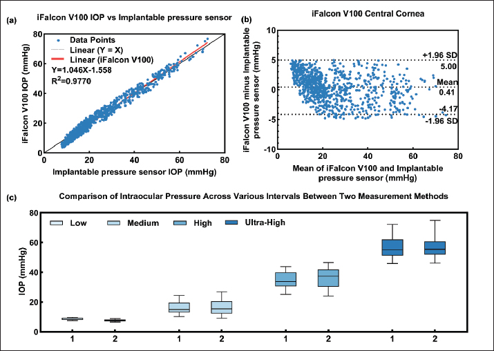
Fig. 1. Experimental setup for ex vivo feline ocular manometry. (a) Schematic diagram of the dual-cannulation system. A reservoir (used for pressure modulation) and a pressure transducer (reference standard) are connected to the globe, with the iFalcon™ V100 rebound tonometer positioned directly anterior to the cornea. (b) Photograph of the actual experimental apparatus showing the mounted feline globe. (c) Superior view (top-down) illustrating the spatial arrangement of the instrumentation. (d) Anterior view (frontal) demonstrating the perpendicular alignment of the tonometer probe with the corneal apex. A dual-channel hydrostatic pressure control system was constructed. On one side, a 23-gauge needle connected to a balanced salt solution reservoir was inserted through the pars plana into the vitreous cavity. The insertion site was sealed with cyanoacrylate adhesive to prevent leakage, and IOP was modulated by adjusting the reservoir height. On the contralateral side, a second needle connected to a high-precision pressure transducer (serving as the reference standard) was inserted through the limbus into the anterior chamber and similarly sealed. The rebound tonometer was horizontally secured on a precision micromanipulator stage and positioned directly anterior to the globe. The probe tip was aligned perpendicularly to the corneal optical center (apex) to ensure positional consistency, with the device set to “feline mode.” Throughout the procedure, the corneal surface was irrigated with balanced salt solution every 1–2 minutes to maintain hydration and prevent desiccation artifacts. The second phase of the study was conducted on the in vivo feline subject under general anesthesia to evaluate the agreement and clinical applicability of the iFalcon rebound tonometer relative to the iCare tonometer. Procedures were performed independently by two experienced operators (Operators 1 and 2). To mitigate the IOP-lowering effect of repeated tonometry (massage effect) and operator bias, a randomized measurement sequence was employed. The order of device usage for each eye was assigned via a random number table. Following the completion of measurements with both devices by the first operator, a 5-minute corneal recovery period was observed before the second operator repeated the protocol. All measurements were obtained with the subject positioned to maintain a neutral head posture, ensuring minimal jugular compression. The mean IOP value for each measurement session was recorded for analysis. Ethical approvalThe experimental protocol was reviewed and approved by the Animal Ethics Committee of Hangzhou Tuoshi Biotechnology Co., Ltd. under the approval number TS2025-11-28-06. ResultsLinear regression analysis (Fig. 2a) demonstrated a strong positive correlation between iFalcon™ V100 readings and reference intracameral manometry across the 5–75 mmHg range in the four ex vivo feline globes. This finding validates the device’s capacity to effectively track true IOP dynamics. However, a regression slope exceeding 1.0 indicated a proportional bias, characterized by a progressive systematic overestimation as IOP elevated. Furthermore, the y-intercept of −1.558 mmHg reflected a minor fixed negative bias, suggesting a slight baseline offset in the measurements.
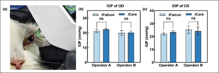
Fig. 2. Quantitative comparison between iFalcon™ V100 and reference intracameral manometry in ex vivo feline eyes. (a) Scatter plot with linear regression. The black solid line indicates the line of equality (Y=X), and the red line represents the linear fit (Y=1.046X − 10558), demonstrating the correlation between the device and the reference standard. (b) Bland–Altman analysis assessing the agreement between methods. The solid line represents the mean bias of 0.41 mmHg (SD=2.34 mmHg), while the dashed lines indicate the 95% limits of agreement, ranging from −4.17 to 5.00 mmHg. (c) Box plots comparing the distribution of IOP. Group 1 represents the reference anterior chamber manometry, and Group 2 represents the iFalcon™ V100 measurements. The boxes indicate the interquartile range (IQR), and the horizontal lines within the boxes denote the medians. IOP levels were stratified as follows: low (5–10 mmHg), medium (> 10–25 mmHg), high (> 25–45 mmHg), and ultra-high (> 45–75 mmHg). Bland–Altman analysis (Fig. 2b) further corroborated the high agreement between the iFalcon™ V100 and reference intracameral manometry. The analysis revealed a mean bias of only 0.41 mmHg (SD=2.34 mmHg), indicating high overall concordance and negligible clinically relevant systematic bias. Notably, the 95% limits of agreement ranged from −4.17 to 5.00 mmHg. These results demonstrate high precision and suggest acceptable interchangeability for individual measurements within the investigated pressure range. To assess performance stability under varying physiological and pathological pressure levels, data were subjected to stratified box plot analysis (Fig. 2c). Measurements were categorized into four intervals: hypotony (5–10 mmHg), medium pressure (> 10–25 mmHg), high pressure (> 25–45 mmHg), and ultra-high pressure (> 45–75 mmHg). The analysis revealed that across all pressure strata, the distribution of iFalcon™ V100 readings demonstrated high concordance with the reference manometry. Notably, even within the high-pressure challenge range (30–70 mmHg), the medians and interquartile ranges (IQR) of both methods remained closely aligned, confirming the tonometer’s excellent reliability across a wide dynamic range. In the in vivo bilateral evaluation of the anesthetized feline subject, measurements obtained by two independent operators using the iFalcon™ V100 showed high agreement with the iCare control group. No statistically significant differences were observed in either inter-operator or inter-device comparisons (p > 0.05) (Fig. 3). This consistency across operators and devices further validates the performance stability and reliability of the iFalcon™ V100 for in vivo applications.
Fig. 3. In vivo IOP assessment in the feline subject. (a) Representative photograph showing the experimental measurement procedure and device positioning . (b) Comparative IOP readings obtained from the left eye (OS) by different operators using iFalcon™ V100 and iCare tonometers. (c) Comparative IOP readings obtained from the right eye (OD) across different operators and devices. DiscussionThrough combined ex vivo and in vivo experiments, this study demonstrates substantial agreement between the iFalcon™ V100 and reference intracameral manometry in feline eyes, validating its efficacy as a non-invasive IOP monitoring tool (Su et al., 2026b). Linear regression analysis revealed a robust positive correlation, confirming the device’s capability to effectively track true IOP dynamics. However, a regression slope slightly exceeding 1.0 indicated a proportional bias, suggesting a minor systematic overestimation in the high-IOP range (Sachdeva et al., 2023; Kuo et al., 2024). This pressure-dependent deviation likely reflects specific biomechanical properties of the feline cornea: as IOP rises, corneal stiffness increases non-linearly, resulting in reduced energy dissipation and a consequent physical enhancement of the probe’s rebound response (Oikawa et al., 2023; Oikawa et al., 2024). This variation underscores the necessity of the device’s embedded Hertzian-theory-based non-linear algorithm. By mathematically accounting for the sphere-on-sphere contact geometry and material depth during the millisecond impact, this framework constrains the mean bias to an extremely low level. These findings substantiate the utility of incorporating fundamental impact mechanics into precision tonometry to eliminate corneal structural confounding. Despite this pressure-related proportional effect, the exceptionally high correlation coefficient indicates that the device maintains superior linearity in feline eyes, rendering the measurement outcomes highly predictable. A pivotal finding of this study is the demonstration of the device’s substantial measurement precision. Bland–Altman analysis revealed 95% limits of agreement ranging from −4.17 to 5.00 mmHg, indicating low random error and high repeatability for individual measurements. Stratified analysis further confirmed that the device maintained excellent stability within the high-pressure challenge range of 30–75 mmHg, with no evidence of sensor saturation. Conversely, the difficulty in obtaining readings observed in the hypotony range (5–10 mmHg) aligns with the physical principles of rebound tonometry: low IOP significantly reduces corneal stiffness, thereby enhancing corneal damping effects and causing excessive dissipation of probe impact energy rather than effective rebound. Furthermore, in vivo results established the clinical applicability of the iFalcon™ V100. Independent operator assessments showed no statistically significant differences (p > 0.05), suggesting that the device design effectively mitigates human error through superior reproducibility. In conclusion, the iFalcon™ V100, characterized by its high precision, wide-range linear response, and robust operational stability, is validated as a reliable tool for feline IOP monitoring and glaucoma research. Notwithstanding the promising findings, several critical limitations of this study warrant consideration. Notably, an elevated measurement failure rate was observed within the 5–10 mmHg interval, suggesting potential constraints in data acquisition capabilities under conditions of hypotony. Consequently, clinicians should exercise caution when evaluating cases of suspected pathological low IOP. Regarding sample size, although the inclusion of four ex vivo globes suffices for preliminary manometric validation, the relatively small cohort inherently restricts a comprehensive analysis of inter-individual biological variability. In addition, the in vivo assessment was limited to a single subject. While this design is adequate for a proof-of-concept comparison, future studies involving larger populations are necessary to generalize these findings (Mills et al., 2023; Lewin et al., 2025). Furthermore, the ex vivo model inherently lacks dynamic in vivo physiological regulatory mechanisms—such as tear film stability and episcleral venous pressure. This discrepancy between experimental and physiological environments implies that measurement biases observed ex vivo could potentially be accentuated or altered in the living eye (Al Sabti et al., 2022; Demirtas et al., 2023). ConclusionThis study comprehensively validates the measurement performance of the iFalcon™ V100 rebound tonometer in feline models. Through comparison with reference intracameral manometry, the device exhibited excellent agreement and sub-mmHg precision (95% LoA < ± 1.1 mmHg), proving its ability to provide highly reliable single measurements. Despite a minor proportional bias in the high-IOP range—due to specific feline corneal biomechanics—and limitations imposed by damping effects at very low IOP (< 10 mmHg), the device maintained superior linearity and stability across a wide dynamic range, particularly in the high-pressure zone critical for glaucoma research. Furthermore, excellent inter-operator reproducibility further confirmed its robustness in actual application. In summary, the iFalcon™ V100 is a precise, reliable, and non-invasive IOP monitoring tool, ideally suited for feline ophthalmic basic research and the long-term monitoring of glaucoma disease models. AcknowledgmentsNot applicable. Conflicts of interestThe authors have no conflicts of interest to declare. FundingThis study received no funding. Authors’ contributionsYC: Conceptualization, methodology, data curation, formal analysis, and original draft preparation. YS: Conceptualization, investigation, methodology, resource provision, and project administration. YL1: Conceptualization, investigation, and methodology. YL2:Review and editing, and resource provision. FS: Review and editing, and resource provision. JF: Conceptualization, investigation, supervision, validation, review and editing, and project administration. All authors have read and approved the final published version of the manuscript. Data availabilityAll data were presented in the study. ReferencesAl Sabti, K. and Raizada, S. 2022. Novel surgical pathway for controlled access to the subretinal space: a case series. Transl. Vis. Sci. Technol. 11(4), 11; doi:10.1167/tvst.11.4.11 Bader, J., Zeppieri, M. and Havens, S. 2023. Tonometry. StatPearls. Demirtas, A., Taylor, E.A., Gludovatz, B., Ritchie, R.O., Donnelly, E. and Ural, A. 2023. An integrated experimental-computational framework to assess the influence of microstructure and material properties on fracture toughness in clinical specimens of human femoral cortical bone. J. Mech. Behav. Biomed. Mater. 145(2023), 106034; doi:10.1016/j.jmbbm.2023.106034 Grahn, B. 2023. Feline glaucoma. Vet. Clin. Small Anim. Pract. 53(2), 367–387; doi:10.1016/j.cvsm.2022.10.003 Kiland, J.A., Terhaar, H.M., Walleck, H.E., Chen, N., Torné, O., Oikawa, K., Wahl, T. and McLellan, G.J. 2025. Comparison of rebound and applanation tonometry in eyes with focal corneal edema. Vet. Ophthalmol. 29(2), e70075; doi:10.1111/vop.70075 Kiland, J.A., Terhaar, H.M., Walleck, H.E., Chen, N., McDaniel, K. and McLellan, G.J. 2023. Comparison of the TONOVET Plus®, TonoVet®, and Tono-Pen Vet™ tonometers in normal cats and cats with glaucoma. Vet. Ophthalmol. 26(5), 414–421; doi:10.1111/vop.13123 Kovalcuka, L., Mālniece, A. and Vanaga, J. 2024. Comparison of Tonovet® and Ricci, C.L., Tonovet plus® tonometers for measuring intraocular pressure in dogs, cats, horses, cattle, and sheep. Vet. World 17(2), 384–392; doi:10.14202/vetworld.2024.384-392 Kuo, D.S. and Rahimy, E. 2024. Agreement of serial iCare HOME2 and Goldmann applanation tonometry. Ophthalmol. Glaucoma 7(5), 440–444; doi:10.1016/j.ogla.2024.04.007 McLellan, G.J. 2025. Comparison of rebound and applanation tonometry in eyes with focal corneal edema. Vet. Ophthalmol. 29(2), e70075; doi:10.1111/vop.70075 Michaud, B. and Lesne, F. 2024. Comparing the intraocular pressure values obtained with a rebound tonometer (TONOVET Plus) and an indentation tonometer (IOPvet) in dogs with and without ocular disease. Vet. Rec. 194(12), e4269; doi:10.1002/vetr.4269 Mills, E.P., Liu, C.C., Emelogu, U., Carter, R.T., Camacho-Luna, P. and Lewin, A.C. 2023. Calibration of a novel rebound tonometer (Tono-Vera® vet) in the porcine eye. Vet. Ophthalmol. 26(3), 268–272; doi:10.1111/vop.13067 Oikawa, K., Torne, O., Sun, D., Moon, A.K.B., Kiland, J.A., Trane, R.M. and McLellan, G.J. 2023. Aqueous humor TGF-β2 and its association with intraocular pressure in a naturally occurring large animal model of glaucoma. Invest. Ophthalmol. Vis. Sci. 64(10), 18; doi:10.1167/iovs.64.10.18 Oikawa, K., Kiland, J.A., Mathu, V., Torné, O., Wickland, C., Neufcourt, S., Mitro, C., Lopez, R. and McLellan, G.J. 2024. Effects of telmisartan on intraocular pressure, blood pressure, and ocular perfusion pressure in normal and glaucomatous cats. Transl. Vis. Sci. Technol. 13(9), 15; doi:10.1167/tvst.13.9.15 Passareli, J.V.G.C., Nascimento, F.F., Kanashiro, G.P., da Costa Zulim, L.F., Giuffrida, R. and Andrade, S.F. 2024. Comparison of three methodologies for measuring intraocular pressure in healthy cats. Vet. World 17(8), 1803–1809; doi:10.14202/vetworld.2024.1803-1809 Sachdeva, R., Iordanous, Y. and Lin, T. 2023. Comparison of intraocular pressure measured by iCare tonometers and Goldmann applanation tonometer. Can. J. Ophthalmol. 58(5), 426–432; doi:10.1016/j.jcjo.2022.06.002 Salih, A., Giannikaki, S., Escanilla, N., Ioannides-Hoey, C.S.F.K. and Best, M. 2023. Etiologies of nontraumatic feline uveitis in the UK: a retrospective observational study of 72 cats. Open Vet. J. 13(9), 1195–1204; doi:10.5455/OVJ.2023.v13.i9.15 Su, Y., Jin, J., Cheng, H. and Fu, J. 2026. Evaluation of the iFalconTMV100 rebound tonometer in ex-vivo rabbit eyes. BMC Vet. Res. 22(1), 101; doi:10.1186/s12917-026-05355-5 Su, Y., Liu, Y., Shi, F. and Fu, J. 2026. Validation of the iFalcon V100 rebound tonometer in canine eyes. Vet. Ophthalmol. 29(2), e70168; doi:10.1111/ vop.70168 Wang, C., Su, Y., Shi, F. and Fu, J. 2025. Comparison of three rebound tonometers in rabbits. Sci. Rep. 15(1), 44840; doi:10.1038/s41598-025-28990-8 Yang, V.Y., Eaton, J.S., Kiland, J.A., Koch, K.E., Oikawa, K., Hetzel, S.J. and McLellan, G.J. 2024. Effects of 0.024% latanoprostene bunod on intraocular pressure and pupil diameter in normal cats and cats with congenital glaucoma. Vet. Ophthalmol. 29(1), e13302; doi:10.1111/vop.13302 | ||
| How to Cite this Article |
| Pubmed Style Chen Y, Su Y, Liu Y, Yang L, Shi F, Fu J. Dynamic accuracy of feline rebound tonometry: Validation via implanted sensors. doi:10.5455/OVJ.2026.v16.i7.61 Web Style Chen Y, Su Y, Liu Y, Yang L, Shi F, Fu J. Dynamic accuracy of feline rebound tonometry: Validation via implanted sensors. https://www.openveterinaryjournal.com/?mno=314201 [Access: July 16, 2026]. doi:10.5455/OVJ.2026.v16.i7.61 AMA (American Medical Association) Style Chen Y, Su Y, Liu Y, Yang L, Shi F, Fu J. Dynamic accuracy of feline rebound tonometry: Validation via implanted sensors. doi:10.5455/OVJ.2026.v16.i7.61 Vancouver/ICMJE Style Chen Y, Su Y, Liu Y, Yang L, Shi F, Fu J. Dynamic accuracy of feline rebound tonometry: Validation via implanted sensors. doi:10.5455/OVJ.2026.v16.i7.61 Harvard Style Chen, Y., Su, . Y., Liu, . Y., Yang, . L., Shi, . F. & Fu, . J. (2026) Dynamic accuracy of feline rebound tonometry: Validation via implanted sensors. doi:10.5455/OVJ.2026.v16.i7.61 Turabian Style Chen, Yulin, Yunhao Su, Yingbo Liu, Lihui Yang, Fushan Shi, and Ji Fu. 2026. Dynamic accuracy of feline rebound tonometry: Validation via implanted sensors. doi:10.5455/OVJ.2026.v16.i7.61 Chicago Style Chen, Yulin, Yunhao Su, Yingbo Liu, Lihui Yang, Fushan Shi, and Ji Fu. "Dynamic accuracy of feline rebound tonometry: Validation via implanted sensors." doi:10.5455/OVJ.2026.v16.i7.61 MLA (The Modern Language Association) Style Chen, Yulin, Yunhao Su, Yingbo Liu, Lihui Yang, Fushan Shi, and Ji Fu. "Dynamic accuracy of feline rebound tonometry: Validation via implanted sensors." doi:10.5455/OVJ.2026.v16.i7.61 APA (American Psychological Association) Style Chen, Y., Su, . Y., Liu, . Y., Yang, . L., Shi, . F. & Fu, . J. (2026) Dynamic accuracy of feline rebound tonometry: Validation via implanted sensors. doi:10.5455/OVJ.2026.v16.i7.61 |


