





| Research Article | ||
Open Vet. J.. 2026; 16(7): 4540-4548
Open Veterinary Journal, (2026), Vol. 16(7): 4540-4548 Research Article Histo-protective efficacy of Withania somnifera (Ashwagandha) root extract against bisphenol A-induced testicular toxicity in ratsFawiziah Khalaf Alharbi*Department of Biology, College of Science, Qassim University, Buraydah, Saudi Arabia *Corresponding Author: Fawiziah Khalaf Alharbi. Department of Biology, College of Science, Qassim University, Buraydah, Saudi Arabia. Email: hrbief [at] qu.edu.sa Submitted: 01/04/2026 Revised: 09/06/2026 Accepted: 19/06/2026 Published: 17/07/2026 © 2025 Open Veterinary Journal
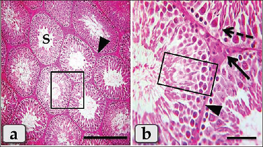
ABSTRACTBackground: Bisphenol A (BPA) is a high-volume synthetic endocrine disruptor widely used in polycarbonate plastics and epoxy resins. Its prevalence in food containers, water bottles, and can linings leads to continuous dietary exposure, raising significant public health concerns. This drives scientific interest in finding effective, natural protective agents. Withania somnifera (Ashwagandha), a prominent adaptogen in Ayurvedic medicine, is renowned for its cytoprotective, antioxidant, anti-inflammatory, and stress-alleviating properties, suggesting potential protective benefits against chemical toxicants. Aim: This study was designed to evaluate the histo-protective efficacy of a standardized W. somnifera (Ashwagandha) root extract (WSRE) against BPA-induced testicular histopathological damage in rats. Methods: Forty adult male Wistar rats were divided into four groups (n=10): Group I (Control) received the vehicle only; Group II (BPA) received 200 mg/kg body weight/day of BPA orally; Group III (Ashwagandha) received 500 mg/kg body weight/day of Ashwagandha orally and Group IV (BPA plus Ashwagandha) received the same doses of both BPA and Ashwagandha at the aforementioned doses. The treatment continued for 60 consecutive days. Following sacrifice, the testes were immediately dissected out, fixed in Bouin’s solution, and processed through dehydration and paraffin embedding. Five-µm-thick sections were obtained and stained with Hematoxylin and Eosin (H&E) to evaluate criteria such as germ cell degeneration, disorganization, vacuolization, and sloughing of spermatogenic cells. Results: Histopathological analysis of the BPA-only group revealed severe testicular injury, including marked distortion of tubule architecture, thinning and disorganization of the germinal epithelium, extensive vacuolization, and sloughing of spermatogenic cells into the lumen. Spermatogenesis was severely disrupted, with a notable absence of mature spermatozoa in many tubules. On the contrary, the BPA plus Ashwagandha co-treatment group showed histoarchitectural preservation. Seminiferous tubules appeared near normal, with well-organized germinal epithelial layers containing all stages of spermatogenic cells. Degeneration, vacuolization, and cellular sloughing were minimal compared to the BPA-only group. Conclusion: Withania somnifera (Ashwagandha) root extract exhibited protective effects against the histopathological damage caused by BPA in rat testes. Maintaining the structural integrity of the germinal epithelium and appearing to preserve the complete spermatogenic cycle, it suggests its potential as a protective phytotherapeutic intervention for male reproductive toxicity. Keywords: Bisphenol A, Phytotherapy, Reproductive toxicity, Testicular histopathology, Withania somnifera. IntroductionBisphenol A (BPA) is a high-production-volume synthetic compound categorized as an endocrine-disrupting chemical (EDC). It is primarily employed in the manufacture of polycarbonate plastics and epoxy resins, making it ubiquitous in consumer goods such as food and beverage containers, water bottles, food storage containers, eyewear lenses, electrical device housings, dental sealants, thermal paper receipts, and metal can linings to prevent contamination and spoiling (Rochester, 2013; Chen et al., 2016; Ullah et al., 2022). While BPA’s functional attributes have led to its global adoption, growing apprehension over its possible role as an endocrine disruptor has prompted heightened regulatory review and a push to find alternatives, especially for applications involving food packaging (Rochester, 2013; Chen et al., 2016). BPA causes toxicity by mimicking endogenous estrogens, attaching to estrogen receptors, and disrupting hormonal signaling pathways. There is substantial evidence that BPA exposure causes testicular dysfunction, which is characterized by oxidative stress, germ cell death, decreased steroidogenesis, and histological changes in the testes. This disruption is especially harmful to the reproductive system (Ma et al., 2021). The production of reactive oxygen species (ROS), which overwhelm cellular antioxidant defenses and cause oxidative stress, is one of the many aspects of BPA’s reproductive toxicity. In the testicular environment, this state of redox imbalance harms cellular macromolecules such as proteins, lipids, and DNA. Simultaneously, BPA disrupts the hypothalamic-pituitary-gonadal (HPG) axis, suppressing the expression of important steroidogenic hormones and enzymes required for spermatogenesis and testosterone synthesis (Kazemi et al., 2016). In contrast to industrial pollutants like BPA, Withania somnifera, also known as Ashwagandha, is a perennial shrub that is valued in Ayurvedic medicine for its adaptogenic and regenerative qualities. This plant’s root extract is a pharmacologically rich matrix with a wide range of bioactive components, including sitoindosides, alkaloids, and withanolides (such as withaferin A and withanolide D), which support its medicinal uses (Singh et al., 2021). These bioactive compounds collectively contribute to the documented therapeutic uses of W. somnifera root extract (WSRE), which include mitigating stress, enhancing energy and endurance, modulating immune function, and exerting broad-spectrum anti-inflammatory and antioxidant effects (Mikulska et al., 2023). Particularly about male reproductive health, WSRE has pharmacological significance. Due to its capacity to revitalize reproductive tissues, preclinical research has demonstrated its function as a spermatogenic agent and testosterone enhancer. Its strong antioxidant ability, which simultaneously scavenges free radicals and upregulates endogenous antioxidant enzymes like glutathione peroxidase (GPx), catalase (CAT), and superoxide dismutase (SOD), is largely responsible for its therapeutic profile (Durg et al., 2023). There is a strong therapeutic connection between the many defensive mechanisms of WSRE and the testicular damage caused by BPA. WSRE’s cytoprotective effects directly target oxidative stress and apoptosis, the main pathophysiology of BPA-induced testicular damage. Theoretically, WSRE can counteract the ROS burst caused by BPA by strengthening the testicular antioxidant defense system, which would stop the subsequent chain reaction of lipid peroxidation and cellular damage (Alahmar and Singh, 2022). There has been a noticeable increase in recent research on the use of natural phytochemicals to reduce EDC toxicity, which is indicative of a move toward preventive and restorative approaches. Research on substances like resveratrol and curcumin has shown encouraging outcomes in protecting testicular architecture from a variety of toxins, setting a solid precedent for the assessment of WSRE against BPA (Jahan et al., 2021). Therefore, the present study was designed to investigate the histo-protective potential of W. somnifera root extract against BPA-induced testicular damage in rats. Based on published literature, we hypothesize that co-administration of WSRE may attenuate BPA-driven damage and preserve the cytoarchitecture of seminiferous tubules. However, as oxidative stress and apoptosis were not directly measured (e.g., via TUNEL assay or biochemical markers), these mechanisms are proposed as potential explanations requiring future verification. The findings aim to provide preliminary evidence for the potential use of this adaptogen in strategies aimed at reducing the reproductive risks associated with environmental endocrine disruptors. Materials and MethodsChemicals and reagentsBPA (purity ≥99%) was purchased from Sigma-Aldrich (St. Louis, MO). A standardized hydroalcoholic extract of W. somnifera root (Ashwagandha), certified to contain a minimum of 5% withanolides, was obtained as a dry powder from Natural Remedies Pvt. Ltd. (Bangalore, India). For oral administration, BPA was first dissolved in absolute ethanol to create a 10% stock solution and subsequently diluted with corn oil to achieve the final experimental concentration, with the ethanol content not exceeding 1% of the final vehicle. This low concentration of ethanol is physiologically negligible and has been demonstrated to cause no adverse effects in rodent studies (Gonzalez-Martin et al., 2005; McKee et al., 2006). The W. somnifera root extract was freshly suspended in a 1% aqueous solution of carboxymethyl cellulose (CMC) before each administration. All other analytical grade chemicals, reagents, and solvents, including ethanol, corn oil, and CMC, were sourced from El-Gomhouria Co. (Cairo, Egypt). Animals and housingForty adult male Wistar albino rats, aged 3 months, with an initial body weight of 200–220 g, were procured from the Laboratory Animal Center at Faculty of Veterinary Medicine, Zagazig University, Zagazig, Egypt. The animals were acclimatized for 1 week before the commencement of the experiment under standard laboratory conditions. They were housed in polypropylene cages with sterilized paddy husk bedding, maintained at a controlled temperature of 22°C ± 2°C and a relative humidity of 55% ± 5%, under a standard 12-hour light/dark cycle. Throughout the study period, all rats were allowed unrestricted access to a standard commercial pelleted rodent diet and filtered water ad libitum. All experimental procedures were conducted in accordance with the guidelines for care and use of laboratory animals and in compliance with the guidelines of the Ethical Committee of the National Research Centre, Egypt. Study design and treatmentsAnimals were divided into four groups (n=10 rats/ group). The treatment protocol lasted for 60 consecutive days and was designed as follows: Group I (Control): Rats received the vehicles only. This consisted of 1 ml/kg/day of corn oil (containing <1% ethanol as a residual solvent) via oral gavage and 1 ml/kg/day of 1% carboxymethyl cellulose (CMC) aqueous solution via oral gavage. Group II (BPA): Rats received a daily oral dose of BPA at 200 mg/kg, dissolved in the corn oil/ethanol vehicle, alongside the 1% CMC vehicle. This dose and duration have been reliably shown to induce significant testicular oxidative stress, hormonal disruption, and histopathological damage in adult rat models (Ullah et al., 2018; Abdel-Wahab, 2021). It is important to note that this BPA dose represents an induced toxicity model to establish reproducible testicular damage and should not be directly compared to real environmental exposure levels in humans. Group III (Ashwagandha): Rats received a daily oral dose of WSRE at 500 mg/kg, suspended in 1% CMC, alongside the corn oil vehicle. This dosage is based on established protocols demonstrating its efficacy in enhancing male reproductive parameters without adverse effects (Gupta et al., 2013; Sengupta et al., 2018). Group IV (BPA + Ashwagandha): Rats received a daily co-administration of BPA (200 mg/kg, orally) followed one hour later by WSRE (500 mg/kg, orally). Sample collectionAt the end of the experimental period, rats were euthanized under deep anesthesia induced by an intraperitoneal injection of ketamine (200 mg/kg body weight). Following this, a midline abdominal incision was made to access the abdominal cavity. Bilateral orchiectomy was then performed to excise the testes. The collected testes were immediately fixed in Bouin’s solution for subsequent histopathological processing and examination. Histological processingSmall pieces from the fixed testes in the Bouin’s solution were transferred and post-fixed in neutral buffered formalin 10% for 48 hours. Then, the specimens were dehydrated in ascending grades of ethanol, cleared in xylene, and embedded in paraffin wax, forming paraffin blocks. Five-µm-thick sections were obtained and stained with Harris’s Hematoxylin & Eosin (H&E) for routine histological studies according to Suvarna et al. (2019). Ethical approvalThe research protocol has been reviewed and approved by the Institutional Animal Care and Use Committee, Zagazig University, Egypt, with an approval number (ZU—IACUC; No. ZU-IACUC/2/F/263/2026). ResultsThe histological examination of the mature male rat’s testes of the control group (G1) clarified normal, intact testicular parenchyma that was mainly consisted of two parts; tubular part (numerous oval or rounded seminiferous tubules) and intertubular part (a considerable amount of highly vascularized interstitial connective tissue) (Fig. 1a). With higher magnification, the seminiferous tubules were lined with intact stratified seminiferous epithelium that was composed of non-divided fewer pyramidal Sertoli cells surrounded with several rows of normal, organized, proliferating highly divided spermatogenic cells rested on a thin basal lamina and represented by spermatogonia, spermatocyte I, spermatocyte II, spermatids and sperms. The highly vascularized intertubular connective tissue appeared to house two types of cells: ovoid or polygonal Leydig cells with spherical nuclei and flat myoid cells with flat nuclei (Fig. 1b).
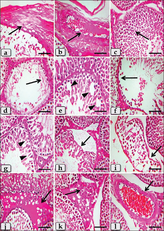
Fig. 1. A photomicrograph of the mature male rat’s testes of the control group (G1), showing normal, intact testicular parenchyma of the tubular part (oval or rounded seminiferous tubules) (S) and intertubular part (a considerable amount of highly vascularized intertubular connective tissue housing leydig and myoid cells) (arrowhead). b) Higher magnification of the inset box of (a) showed normal, intact seminiferous tubules lining stratified seminiferous epithelium; pyramidal Sertoli cells (dashed arrow) surrounded with several rows of normal, organized, proliferated and highly divided spermatogenic cells (inside square) rested on a thin basal lamina, also, showed normal polygonal leydig cells (arrow) and flat myoid cells (arrowhead) in the intertubular part. Stain: All) H&E. Scale bars: a=300 µm, b=50 µm. Meanwhile, the histological examination of the testes of the BPA-treated group (G2) demonstrated severe thickening and fibrosis of the testicular capsule, tunica albuginea (Fig. 2a), with severe subcapsular blood vessels dilatation and congestion (Fig. 2b). At the level of the tubular part, severe degenerative changes of the seminiferous tubules lining epithelium with loss of their normal organization were noticed, in addition, distribution of numerous spermatogenic cells with pyknotic nucleus, also, aggregation of round spermatids filling the lumen of seminiferous tubules with completely loss of any sperms were observed (Fig. 2c). Furthermore, severe coagulative necrosis of spermatogenic cells, especially spermatocytes and spermatids characterized with maintenance of the tubular architecture with loss of cellular details accompanied with completely loss of sperms were clarified (Fig. 2d). Severe hydropic degeneration; vacuolizations; represented in formation of vacuoles with variable shape and size within the cytoplasm of spermatogenic cells, especially spermatogonia and primary spermatocytes were demonstrated (Fig. 2e).
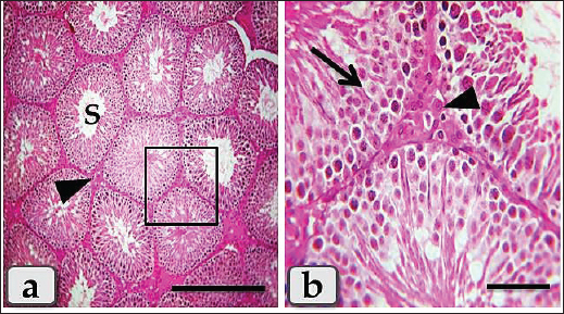
Fig. 2. A photomicrograph of the mature male rat’s testes of the BPA-treated group (G2), showing severe thickening and fibrosis of the testicular capsule (arrow). b) Showed severe subcapsular blood vessel dilatation with congestion (arrow). c) Showed severe degenerative changes of the seminiferous tubules lining epithelium with loss of their normal organization, in addition, distribution of numerous spermatogenic cells with pyknotic nucleus, with severe aggregation of round spermatids at the lumen of seminiferous tubules with complete loss of any sperms (arrow). d) Showed severe coagulative necrosis of spermatogenic cells, especially spermatocytes and spermatids (arrow). e) Showed severe hydropic degeneration; vacuolizations of spermatogenic cells, especially spermatogonia and primary spermatocytes (arrowheads). f) Showed severe germ cell depletions (arrow). g) Showed necrotic Sertoli cells (arrowhead). h) Showed severe damage of the seminiferous tubules’ basal lamina with sloughing of its lining epithelium into the tubular lumen (arrow). i) Showed severe atrophy of the seminiferous tubules (arrow) with increasing of the intertubular spaces. j) Showed severe intertubular edema (arrow). k) Showed severe Leydig cells hyperplasia (arrow). l) Showed severe intertubular blood vessels dilatation with severe congestion; overengorged with blood and severe thickening of the blood vessels’ wall (arrow). Stain: All) H&E. Scale bars: All=50 µm. Some examined sections clarified severe germ cell depletions, and other sections noticed loss of several types of spermatogenic cells, especially secondary spermatocytes, spermatids, and sperms, and only two types of cells were observed; spermatogonia rest on thick basal lamina and primary spermatocytes with a pyknotic nucleus. Also, the lumen of the seminiferous tubules became wider and devoid of any sperm (Fig. 2f). Some examined sections revealed necrotic Sertoli cells (Fig. 2g), severe damage of the seminiferous tubules basal lamina with sloughing of its lining epithelium into the tubular lumen (Fig. 2h). In addition, severe atrophy of the seminiferous tubules was observed with increasing the inter tubular spaces (Fig. 2i). And also, focal coagulative necrosis of round spermatids were noticed (Fig. 2i). At the level of the intertubular part, several pathological changes were assumed resembling severe intertubular edema; increase of the intertubular fluid (Fig. 2j), accompanied with severe leydig cells hyperplasia (Fig. 2k). In addition, severe dilatation of the intertubular blood vessels with severe congestion; engorged with blood were described, accompanied with severe thickening and fibrosis of its wall (Fig. 2l). However, in the BPA group, myoid cells surrounding the seminiferous tubules appeared structurally normal, with no detectable morphological abnormalities compared to the control group. Regarding the histological examination of the testes of the Ashwagandha-treated group (G3), the present investigation revealed intact testicular parenchyma of intact tubular part with normal, organized lining epithelium and normal intertubular part that appeared to look like normal, resembling the testes of the control group without any pathological changes (Fig. 3a,b).
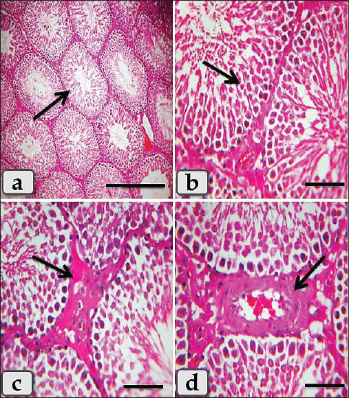
Fig. 3. A photomicrograph of the mature male rat’s testes of the Ashwagandha-treated group (G3), a) showing intact testicular parenchyma of the normal tubular part (S) and intertubular part (arrowhead) that appears normal, resembling the normal control group without any pathological changes. b) Higher magnification of the inset box of Fig. a showed intact seminiferous tubules lining stratified seminiferous epithelium with normal, organized, spermatogenic cells (arrow), with normal intertubular part (arrowhead). Stain: All) H&E. Scale bars: a=300 µm, b=50 µm. Meanwhile, the histological examination of the testes of the BPA plus Ashwagandha-treated group (G4) clarified the preferable and effective therapeutic interference, where most of the examined sections of this group showed normal, intact tubular parts with normal lining epithelium, and intertubular parts that appeared looks like normal (Fig. 4a), but some mild to moderate pathological changes were noticed in very few sections resembling mild degenerative changes of the seminiferous tubules lining epithelium (Fig. 4b), with mild intertubular edema (Fig. 4c), mild leydig cells hyperplasia (Fig. 4c), intertubular blood vessels dilatation and congestion, accompanied with mild thickening and fibrosis of the intertubular blood vessels wall (Fig. 4d).
Fig. 4. A photomicrograph of the mature male rat’s testes of the BPA + Ashwagandha-treated group (G4), a) showing normal, intact testicular parenchyma; tubular part (seminiferous tubules) and intertubular part (arrow) in most of the examined sections. b) Showed mild degenerative changes of the seminiferous tubules lining epithelium (arrow) in some examined sections. c) Showed mild intertubular edema (arrow), with mild Leydig cells hyperplasia. d) Showed mild intertubular blood vessels dilatation with congestion (arrow). Stain: All) H&E. Scale bars: All=50 µm, except a=300 µm. DiscussionThe present study provides compelling histopathological evidence that WSRE offers significant protection against BPA -induced testicular toxicity in rats. The profound architectural preservation observed in the co-treated group (G4), contrasted with the severe degenerative changes in the BPA-only group (G2), provides a robust morphological foundation for proposing the protective effect of this adaptogenic herb. The normal histology observed in the Ashwagandha-only group (G3) indicates good testicular tolerability of the extract under the conditions of this study and suggests that the protective effect seen in the co-treatment group (G4) is likely due to active cytoprotection rather than non-specific stimulation. However, a comprehensive safety assessment would require additional analyses (body/organ weights, hepatic/renal function, and hematological/biochemical parameters) in future studies. The extensive testicular damage in G2 (BPA-treated group), characterized by germinal epithelial disorganization, coagulative necrosis of spermatogenic cells, cytoplasmic vacuolization, Sertoli cell necrosis, and epithelial sloughing, represents a classic histological signature of combined oxidative stress and endocrine disruption. Based on published literature (Rochester, 2013), BPA’s estrogen-mimicking properties enable it to bind estrogen receptors and disrupt the HPT axis, leading to reduced intratesticular testosterone levels. This androgen deprivation directly impairs the progression of meiosis and spermiogenesis, which may explain the “complete loss of any sperms” and selective depletion of advanced germ cells (spermatids, secondary spermatocytes) observed in our study. As suggested by previous reports (Ullah et al., 2019), BPA metabolism generates reactive oxygen species (ROS) that overwhelm endogenous antioxidant defenses, triggering lipid peroxidation of germ cell membranes (manifesting as vacuolization) and activating apoptotic pathways evident through pyknotic nuclei and coagulative necrosis. The severe vascular pathology, including congestion, vessel wall fibrosis, and interstitial edema, may indicate a profound inflammatory response as previously described following BPA exposure, likely mediated by upregulation of pro-inflammatory cytokines and VEGF (Manna et al., 2017). This vascular dysfunction could contribute to a hypoxic microenvironment that exacerbates tubular damage. The observed Leydig cell hyperplasia could represent a compensatory, though ultimately ineffective, attempt to overcome BPA-induced suppression of testosterone synthesis by increasing steroidogenic cell populations. The remarkable histological preservation in the BPA + Ashwagandha group (G4) suggests possible multi-target pharmacological actions of W. somnifera, primarily mediated by its bioactive withanolides. The restoration of organized spermatogenesis and presence of mature spermatozoa raises the hypothesis that Ashwagandha counteracted BPA’s anti-androgenic effects. Based on prior work (Ahmad et al., 2010), Ashwagandha has demonstrated adaptogenic properties that help normalize the HPT axis under stress conditions, potentially supporting Leydig cell function and steroidogenesis to maintain testosterone levels necessary for complete spermatogenesis. However, these hormonal mechanisms were not directly measured in the present study and remain speculative. It is hypothesized that Ashwagandha’s potent antioxidant activity constitutes a primary protective mechanism. Previous studies (Gupta and Kaur, 2016) have shown that the extract and its constituents upregulate endogenous antioxidant enzymes, including SOD, CAT, and GPx, while directly scavenging free radicals. We speculate that by neutralizing the ROS burst induced by BPA, Ashwagandha prevented the lipid peroxidation responsible for the cytoplasmic vacuolization and membrane damage observed in G2. This proposed antioxidant action may be complemented by significant anti-apoptotic properties, as withanolides have been shown in the literature to modulate the Bcl-2/Bax ratio, inhibit cytochrome c release from mitochondria, and suppress caspase-3 activation (Mikulska et al., 2023), thereby potentially preserving germ cell viability. It is important to note that oxidative stress and apoptosis markers were not measured in the current study. The anti-inflammatory properties of Ashwagandha have been reported to contribute to the mitigation of vascular and interstitial pathology (Gupta et al., 2020). By inhibiting nuclear factor-kappa B (NF-κB) translocation and subsequent pro-inflammatory cytokine production, Ashwagandha may theoretically reduce the inflammatory cascade responsible for vascular congestion, edema, and fibrosis. This improved microcirculation and reduced stromal pressure could create a more favorable microenvironment for spermatogenesis. Again, inflammatory markers were not directly assessed here. Furthermore, the preservation of Sertoli cell integrity and blood-testis barrier function, inferred from the absence of epithelial sloughing in G4, is crucial for maintaining the immune-privileged adluminal compartment and is hypothesized to result from the combined antioxidant, anti-apoptotic, and anti-inflammatory effects of Ashwagandha, as described in the literature. In summary, while the histological evidence clearly demonstrates the protective efficacy of Ashwagandha against BPA-induced testicular damage, the proposed mechanisms involving oxidative stress, apoptosis, hormonal regulation, and inflammation remain speculative hypotheses based on published literature. Future studies incorporating direct biochemical and hormonal assays are required to confirm these mechanisms. ConclusionIn this qualitative proof-of-concept study, W. somnifera (Ashwagandha) root extract showed protective effects against BPA-induced testicular toxicity in rats. It preserved the cytoarchitecture of the seminiferous tubules, maintained germinal epithelial integrity, and supported the complete sequence of spermatogenesis. These findings suggest that Ashwagandha may be a promising candidate for further investigation into its potential role in protecting against male reproductive dysfunction caused by environmental endocrine disruptors such as BPA. However, quantitative and mechanistic studies are needed to confirm these observations. Study limitationsThis study was designed as a qualitative histo-protective efficacy investigation to establish proof-of-concept, not as a quantitative or mechanistic study. Consequently, several limitations exist, each of which opens exciting avenues for future research. The histological findings are presented descriptively, as the striking and visually unmistakable differences between treatment groups make statistical analysis informative but not essential for the primary conclusion. The study also lacks quantitative histomorphometric measurements, including tubular diameter, epithelial height, seminiferous tubule count, testicular volume, and Leydig or Sertoli cell numbers. Furthermore, sperm parameters, such as count, motility, and morphology, were not evaluated, meaning functional confirmation of fertility remains an important next step. Hormonal assays, including testosterone, LH, and FSH, were not performed, leaving the role of the HPG axis unexplored and raising the question of whether Ashwagandha acts locally on testicular tissue or systemically. In addition, biochemical markers of oxidative stress (e.g., MDA and GSH), apoptosis (e.g., caspase-3, Bax/Bcl-2 ratio), and inflammation (e.g., IL-1β, IL-6, TNF-α) were not measured to support the proposed antioxidant, anti-apoptotic, and anti-inflammatory mechanisms. Importantly, none of these limitations diminishes the central finding that Ashwagandha root extract dramatically preserves testicular architecture and supports complete spermatogenesis against BPA-induced damage, as clearly demonstrated by the striking histopathological evidence. Rather, they highlight the need for further mechanistic, quantitative, and functional investigations to build upon the solid histological foundation established here. AcknowledgmentThe author would like to thank the Deanship of Graduate Studies and Scientific Research at Qassim University, Saudi Arabia, for financial support (QU-APC-2026). Conflict of interestThe author has no competing interests to declare. Data availabilityAll study data are presented in this manuscript. FundingThe author would like to thank the Deanship of Graduate Studies and Scientific Research at Qassim University, Saudi Arabia, for financial support (QU-APC-2026). Conflict of interestThe author has no competing interests to declare. Authors’ contributionsAll experimental work and manuscript writing were done by the sole author of the manuscript. Data availabilityAll study data are presented in this manuscript. ReferencesAbdel-Wahab, W.M. 2021. Bisphenol A-induced testicular toxicity in rats: the protective role of zinc. Andrologia 53(2), e13934. Ahmad, M.K., Mahdi, A.A., Shukla, K.K., Islam, N., Rajender, S., Madhukar, D., Shankhwar, S.N. and Ahmad, S. 2010. Withania somnifera improves semen quality by regulating reproductive hormone levels and oxidative stress in seminal plasma of infertile males. Fertil. Steril. 94(3), 989–996. Alahmar, A.T. and Singh, R. 2022. Role of antioxidants in ameliorating Bisphenol A-induced oxidative stress in male reproductive functions: a systematic review. Andrologia 54(11), e14597. Chen, D., Kannan, K., Tan, H., Zheng, Z., Feng, Y.L., Wu, Y. and Widelka, M. 2016. Bisphenol analogues other than BPA: environmental occurrence, human exposure, and toxicity—a review. Environ. Sci. Technol. 50(11), 5438–5453. Durg, S., Dhadde, S.B., Vandal, R., Shivakumar, B.S. and Charan, C.S. 2023. Withania somnifera (Ashwagandha) in male reproductive health: a review. J. Ethnopharmacol. 303, 115925. Gonzalez-Martin, M.C., Gonzalez, C., and Fernandez-Briera, A. 2005. Effects of low ethanol ingestion in the rat. The Physiological Society 567P, PC91. Gupta, A. and Kaur, G. 2016. Pharmacological potential of Withania somnifera (L.) Dunal: a review. J. Pharm. Pharmacol. 68(8), 953–969. Gupta, A., Mahdi, A.A., Shukla, K.K., Ahmad, M.K., Bansal, N., Sankhwar, P. and Sharma, V. 2013. Efficacy of Withania somnifera on seminal plasma metabolites of infertile males: a proton NMR study at 800 MHz. J. Ethnopharmacol. 149(1), 208–214. Gupta, A., Singh, S. and Kumar, R. 2020. Withania somnifera (Ashwagandha) and its active constituents: role in inflammation, immunomodulation, and male reproductive health. J. Ethnopharmacol. 257, 112876. Jahan, S., Abid, A., Khalid, F., Ullah, A. and Nawaz, M. 2021. Therapeutic effects of polyphenols in testicular toxicity: a review. Food Chem. Toxicol. 153, 112292. Kazemi, S., Feizi, F., Aghapour, F., Joorsaraee, G.A. and Moghimian, M. 2016. Impact of Bisphenol A on male reproductive toxicity: a review. Int. J. Endocrinol. Metab. 14(3), e38373. Ma, Y., Liu, H., Wu, J., Yuan, L., Wang, Y., Du, X., Wang, R., Marwa, P.W., Petlulu, P., Chen, X. and Zhang, H. 2021. The adverse health effects of bisphenol A and related toxicity mechanisms. Environ. Res. 197, 111087. Manna, P., Jain, S.K. and Saka, S.M. 2017. Bisphenol A induces oxidative stress and inflammation in the testis of adult rats. Toxicol. Ind. Health. 33(6), 499–510. Mckee, R.H., Lammers, J.H., Hoogendijk, E.M., Emmen, H.H., Muijser, H., Barsotti, D.A., Owen, D.E. and Kulig, B.M. 2006. Model studies for evaluating the acute neurobehavioral effects of complex hydrocarbon solvents: i. Validation of methods with ethanol. NeuroToxicology 27(6), 1064–1079. Mikulska, P., Malinowska, M., Ignacyk, M., Szustowski, P., Nowak, J., Pesta, K., Szeląg, M., Szklanny, D., Judasz, E., Kaczmarek, G., Ejiohuo, O.P., Paczkowska-Walendowska, M., Gościniak, A. and Cielecka-Piontek, J. 2023. Ashwagandha (Withania somnifera)—Current research on the health-promoting activities: a narrative review. Pharmaceutics 15(4), 1057. Rochester, J.R. 2013. Bisphenol A and human health: a review of the literature. Reprod. Toxicol. 42, 132–155. Sengupta, P., Agarwal, A., Pogrebetskaya, M., Roychoudhury, S., Durairajanayagam, D. and Henkel, R. 2018. Role of Withania somnifera (Ashwagandha) in the management of male infertility. Reprod. Biomed. Online 36(3), 311–326. Singh, N., Bhalla, M., De Jager, P. and Gilca, M. 2021. An overview of ashwagandha: a Rasayana (rejuvenator) of Ayurveda. Afr. J. Tradit. Complement. Altern. Med. 8(5S), 208–213. Suvarna, S.K., Layton, C. and Bancroft, J.D. 2019. Bancroft’s Theory and Practice of Histological Techniques. 8th ed., London: Elsevier, pp: 83–92. Ullah, A., Pirzada, M., Jahan, S., Ullah, H. and Khan, M.J. 2022. Bisphenol A and its analogues: a comprehensive review on the occurrence and endocrine-disrupting impact on the male reproductive system. Ecotoxicol. Environ. Saf. 248, 114279. Ullah, A., Pirzada, M., Jahan, S., Ullah, H., Shaheen, G., Rehman, H., Siddiqui, M.F. and Butt, M.A. 2018. Bisphenol A and its analogs bisphenol B, bisphenol F, and bisphenol S: comparative in vitro and in vivo studies on the sperms and testicular tissues of rats. Chemosphere 209, 508–516. Ullah, A., Pirzada, M., Jahan, S., Ullah, H. and Khan, M.J. 2019. Bisphenol A analogues bisphenol B, bisphenol F, and bisphenol S induce oxidative stress, disrupt daily sperm production, and damage DNA in rat spermatozoa: a comparative in vitro and in vivo study. Toxicol. Ind. Health. 35(4), 294–303 | ||
| How to Cite this Article |
| Pubmed Style Fawiziah Khalaf Alharbi. Histo-protective efficacy of Withania somnifera (Ashwagandha) root extract against bisphenol A-induced testicular toxicity in rats. Open Vet. J.. 2026; 16(7): 4540-4548. doi:10.5455/OVJ.2026.v16.i7.37 Web Style Fawiziah Khalaf Alharbi. Histo-protective efficacy of Withania somnifera (Ashwagandha) root extract against bisphenol A-induced testicular toxicity in rats. https://www.openveterinaryjournal.com/?mno=316008 [Access: July 15, 2026]. doi:10.5455/OVJ.2026.v16.i7.37 AMA (American Medical Association) Style Fawiziah Khalaf Alharbi. Histo-protective efficacy of Withania somnifera (Ashwagandha) root extract against bisphenol A-induced testicular toxicity in rats. Open Vet. J.. 2026; 16(7): 4540-4548. doi:10.5455/OVJ.2026.v16.i7.37 Vancouver/ICMJE Style Fawiziah Khalaf Alharbi. Histo-protective efficacy of Withania somnifera (Ashwagandha) root extract against bisphenol A-induced testicular toxicity in rats. Open Vet. J.. (2026), [cited July 15, 2026]; 16(7): 4540-4548. doi:10.5455/OVJ.2026.v16.i7.37 Harvard Style Fawiziah Khalaf Alharbi (2026) Histo-protective efficacy of Withania somnifera (Ashwagandha) root extract against bisphenol A-induced testicular toxicity in rats. Open Vet. J., 16 (7), 4540-4548. doi:10.5455/OVJ.2026.v16.i7.37 Turabian Style Fawiziah Khalaf Alharbi. 2026. Histo-protective efficacy of Withania somnifera (Ashwagandha) root extract against bisphenol A-induced testicular toxicity in rats. Open Veterinary Journal, 16 (7), 4540-4548. doi:10.5455/OVJ.2026.v16.i7.37 Chicago Style Fawiziah Khalaf Alharbi. "Histo-protective efficacy of Withania somnifera (Ashwagandha) root extract against bisphenol A-induced testicular toxicity in rats." Open Veterinary Journal 16 (2026), 4540-4548. doi:10.5455/OVJ.2026.v16.i7.37 MLA (The Modern Language Association) Style Fawiziah Khalaf Alharbi. "Histo-protective efficacy of Withania somnifera (Ashwagandha) root extract against bisphenol A-induced testicular toxicity in rats." Open Veterinary Journal 16.7 (2026), 4540-4548. Print. doi:10.5455/OVJ.2026.v16.i7.37 APA (American Psychological Association) Style Fawiziah Khalaf Alharbi (2026) Histo-protective efficacy of Withania somnifera (Ashwagandha) root extract against bisphenol A-induced testicular toxicity in rats. Open Veterinary Journal, 16 (7), 4540-4548. doi:10.5455/OVJ.2026.v16.i7.37 |


