





| Research Article | ||
Open Vet. J.. 2026; 16(7): 4962-4969
Open Veterinary Journal, (2026), Vol. 16(7): 4962-4969 Research Article Hypoxic mesenchymal stem cell exosomes with hyaluronic acid attenuate UVB-induced collagen loss by suppressing TNF-α and p21 in wistar rat skinLis Julianti1,2, Eko Setiawan1,3,4 and Suparmi Suparmi5*1Master’s Study Program of Biomedical Science, Faculty of Medicine, Universitas Islam Sultan Agung, Semarang, Indonesia 2dr.Liez Skin Expert, Sungai Bangkong, Kota Pontianak, Indonesia 3Department of Surgery, Faculty of Medicine, Universitas Islam Sultan Agung, Semarang, Indonesia 4Department of Surgery, Sultan Agung Islamic Hospital, Semarang, Indonesia 5Department of Biology, Faculty of Medicine, Universitas Islam Sultan Agung, Semarang, Indonesia *Corresponding Author: Suparmi Suparmi. Department of Biology, Faculty of Medicine, Universitas Islam Sultan Agung, Semarang, Indonesia. Email: suparmi [at] unissula.ac.id Submitted: 03/04/2026 Revised: 11/06/2026 Accepted: 20/06/2026 Published: 27/07/2026 © 2025 Open Veterinary Journal
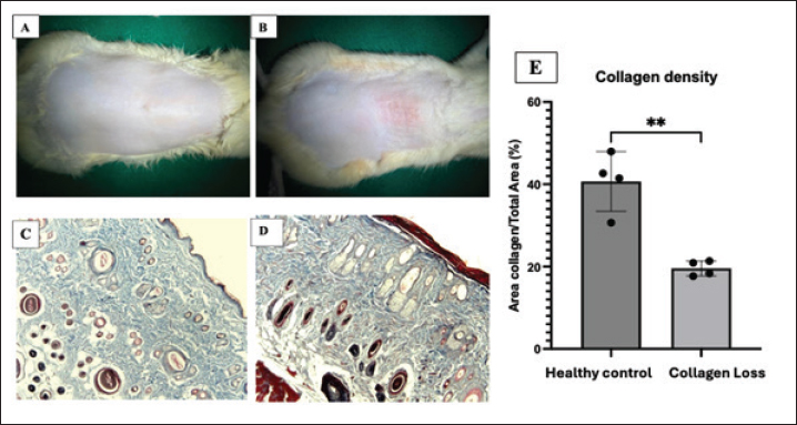
ABSTRACTBackground: Ultraviolet B (UVB) exposure causes collagen loss and photoaging by increasing pro-inflammatory and senescence-associated genes, such as tumour necrosis factor-alpha (TNF-α) and p21, in the skin. Hypoxia-treated mesenchymal stem cell exosomes (hypMSC-Exos) and hyaluronic acid (HA) are proposed as potential therapeutic agents for skin regeneration and repair related to UVB-induced collagen depletion. Aim: This study aimed to assess the impact of hypMSC-Exos and HA on the gene expression of TNF-α and p21 in the skin of Wistar rats exposed to UVB. Methods: Thirty male Wistar rats were randomly assigned to five groups: the healthy control, UVB + NaCl (200 μl of 0.9% NaCl), UVB + HA (200 μl of HA), UVB + hypMSC-Exos (200 μl of an exosome preparation equivalent to 2.5 × 10⁵ cells), and UVB + hypMSC-Exos+HA (200 μl of HA combined with 200 μl of exosomes). UVB irradiation (302 nm, 150 mJ/cm²) was applied to a 2 × 3 cm dorsal area for 8 minutes, five times weekly over 2 weeks to induce collagen depletion. Treatments were administered once after UVB exposure. TNF-α and p21 gene expression in the skin tissue was measured using qRT-PCR. Results: TNF-α gene expression was 0.073 ± 0.003, 0.19 ± 0.018, 0.17 ± 0.02, 0.135 ± 0.022, and 0.067 ± 0.004 for the control, UVB+NaCl, UVB+HA, UVB+ hypMSC-Exos, and UV+ hypMSC-Exos +HA, respectively. The p21 gene expression was 0.085 ± 0.012, 0.38 ± 0.033, 0.32 ± 0.024, 0.30 ± 0.01, and 0.19 ± 0.015 for control, UVB+NaCl, UVB+HA, UVB+ hypMSC-Exos, and UV+ hypMSC-Exos +HA, respectively. UVB exposure increased TNF-α and p21 gene expression compared with healthy skin. Using hypMSC-Exos or HA alone reduced TNF-α and p21 levels compared with the UVB-exposed control group, whereas the combination of both treatments produced the greatest decreases in both genes, with significant differences between groups (p < 0.05). Conclusion: The combination of hypoxic MSC-derived exosomes with hyaluronic acid offers a promising therapeutic strategy for reducing TNF-α and p21 gene expression in UVB-induced collagen loss in Wistar rat skin. This approach utilizes the anti-inflammatory, antioxidant, and regenerative properties of hypMSC-Exos, which are enhanced by the stability and sustained-release features of HA hydrogels. Further research and clinical trials are necessary to fully understand the mechanisms and optimize the therapeutic potential of this treatment. Keywords: Hyaluronic acid, Mesenchymal stem cell, p21, TNF-α, UVB radiation. IntroductionChronic exposure to ultraviolet B (UVB) radiation induces the excessive production of reactive oxygen species (ROS), DNA damage, and low-grade chronic inflammation, which together remodel the dermal extracellular matrix (ECM) and promote cellular senescence (Suman and Suman, 2019; Blackstone et al., 2020; Kaltchenko and Chien, 2025). UVB-driven inflammation is largely followed by the activation of mitogen-activated protein kinase (MAPK) and nuclear factor kappa-light-chain-enhancer of activated B cells (NF-κB) pathways, which upregulate tumour necrosis factor-alpha (TNF-α), interleukin 1 beta (IL-1β), and IL-6, amplify ROS production, and induce matrix metalloproteinases (MMPs) that degrade collagen types I and III. In parallel, DNA damage and ROS promote the expression of cyclin-dependent kinase inhibitors, including p21, thereby enforcing growth arrest and senescence in keratinocytes, fibroblasts, and vascular cells (Meng et al., 2023; Najafabadi et al., 2024). Elevated TNF-α and p21 have been consistently observed in UVB-exposed skin models and are closely associated with increased SA-β-gal activity, collagen loss, and dermal thinning (Wang et al., 2025a,b). Thus, strategies that concurrently suppress TNF-α–driven inflammation and p21-associated senescence are of significant interest in providing superior anti-photoaging benefits. Mesenchymal stem cell-derived exosomes (MSC-Exos) have emerged as promising therapeutic agents for skin regeneration and repair. These nanosized vesicles are rich in bioactive molecules, including microRNAs, proteins, and lipids, which mediate their anti-inflammatory, antioxidant, and regenerative properties (Dong et al., 2023; Gui et al., 2025). The exosomes of hypoxic mesenchymal stem cell-conditioned medium (hypMSC-Exos) exhibit significant antioxidant and anti-inflammatory properties by upregulating key protective factors, such as glutaredoxin 5 (GLRX5) and circular RNAs (circRNAs), which inhibit ferroptosis and inflammation (Zha et al., 2023; Liu et al., 2024), as well as miRNAs and antioxidant proteins, to potentiate anti-senescence and tissue-protective functions (Ha et al., 2020; Zhang et al., 2020). In UVB-irradiated Wistar rats, hypMSC-Exos reduced malondialdehyde (MDA) and MMP-1 levels, indicating reduced oxidative stress and inflammation (Angelina et al., 2025). In UV-induced senescent dermal fibroblasts and photoaged mice, exosomes of hypoxic adipose-derived stem cells significantly decreased SA-β-gal activity and downregulated p16 and p21 while improving collagen density and reducing MMP-1/2/3 expression (Huynh et al., 2025;Wang et al., 2025). However, there remains limited in vivo evidence linking hypMSC-Exos to the coordinated suppression of both TNF-α and p21 in UVB-irradiated skin. Hyaluronic acid (HA) is a major ECM glycosaminoglycan known for its biocompatibility and moisture retention, making it an excellent carrier for therapeutic agents. Experimental models show that restoring HA levels—via oral or topical routes—prevents UV-induced skin dehydration, reduces ECM degradation, and partially normalizes inflammation. Oral HA or collagen-related interventions increase the expression of HA synthases and filaggrin, elevate tissue HA content, and attenuate wrinkle formation and inflammatory responses in UV-irradiated hairless mice. More recently, functionalized HA microneedles have been engineered to enhance dermal penetration; these systems promote collagen regeneration, reduce ROS levels, and improve wrinkle reduction and collagen deposition in UVB-photoaged skin, outperforming conventional HA microneedles (Tang et al., 2024). Combining hypMSC-Exos with bioactive or structural cofactors has proven to be a rational strategy for potentiating anti-aging efficacy. Previous research has demonstrated that combining hypMSC-Exos with alkaline water exerts a synergistic therapeutic effect, reducing oxidative stress markers and NF-κB p65-mediated inflammation more effectively than either treatment alone, highlighting the enhanced anti-inflammatory and antioxidant potential of this strategy (Nasution et al., 2025). Exosomes from human umbilical cord MSCs combined with hydrolyzed collagen oligopeptides synergistically reduced ROS, SA-β-gal, TNF-α, IL-1β, and senescence markers (p16, p21, and p53) while more strongly increasing collagen I/III and decreasing MMP-1/3/9 expression than either agent alone in senescent human skin fibroblasts (Zhu et al., 2024). Similarly, co-delivery of human umbilical cord MSC exosomes with epigallocatechin gallate via microneedles yielded superior protection against UV-induced skin damage, markedly decreasing inflammatory mediators and DNA damage while promoting tissue regeneration (He et al., 2025). These findings support the concept that exosomes can be paired with complementary molecules to achieve synergistic anti-inflammatory and anti-senescence effects. However, research on the use of a combination of hypMSC-Exos and HA in regulating TNF-α and p21 expression during UVB-induced collagen loss remains limited. The present study evaluates whether the subcutaneous injection of hypMSC-Exos, together with HA, can more effectively reduce TNF-α and p21 gene expression in UVB-irradiated Wistar rat skin than either treatment alone, thereby providing mechanistic insight into a potentially synergistic cell-free, HA-supported exosome therapy for photoaging. Materials and MethodsPreparation of hypoxic MSC-derived exosomesHypoxic MSC-derived exosomes (hypMSC-Exos) were obtained from the Stem Cell and Cancer Research (SCCR) in Indonesia, as described by Nasution et al. (2025). Umbilical-cord MSCs were isolated from 19-day-pregnant Wistar rats and characterized by differentiation assays (alizarin red, alcian blue) and flow cytometry for CD45, CD31, CD90, and CD29. For exosome production, confluent MSC cultures were incubated in serum-free medium under hypoxia (1%–5% O2) at 37°C and 5% CO2 for 24–48 hours in a controlled hypoxic chamber; pO2 was monitored (BioSpherix). Exosomes were isolated by tangential flow filtration (Formulatrix). The exosome dose used in this study was chosen based on the dosing range established by Nasution et al. (2025) and from pilot dose-finding experiments performed by the SCCR to identify an efficacious, non-toxic range; full details of the dose selection, including particle concentration (particles/ml), total administered dose, and references to prior dose–response work. HA specification and concentration rationaleStaris Aquarius Hydro (Astella/HAJU Medical)—non-crosslinked HA, 24 mg/ml, supplied as 2 ml syringes (2 ml × 3 per box)—was used for HA treatment. The manufacturer’s product brochure (Astella; see page 8: https://www.tradekorea.com/product/download.do?productfileno=102799) reports the concentration and the mid-dermal “skin booster” indication but does not specify the molecular weight; this information is retained in the product documentation. Rationale for use and dose: a 200-µl subcutaneous dose (24 mg/ml) was selected to provide local HA-mediated enhancement of exosome retention and sustained release without systemic exposure. Non-crosslinked HA was chosen to maximize biocompatibility, hydration, and viscoelastic properties, and to facilitate exosome diffusion within the dermal matrix. Experimental animals and UVB protocolThirty male Wistar rats (38 weeks old, weighing 200–250 g) were sourced from the SCCR. Animals were housed in polypropylene cages (70 × 50 × 30 cm) at 23°C ± 2°C, 60% humidity, and 12-hours light/dark, with ad libitum access to chow and water, and were acclimated for 7 days. Body weights were monitored weekly; no significant weight gain occurred in any group during the study period (data not shown), so the effects of body weight or overweight on TNF-α levels can be considered negligible. Dorsal hair was removed from all rats before UVB exposure to ensure uniform irradiation of the skin. UVB irradiation (302 nm, 150 mJ/cm2) was applied to a 2 × 3 cm dorsal area for 8 minutes, 5 times/week for 2 weeks, to induce collagen loss. The UVB effect was confirmed by gross photos and Masson’s trichrome staining. Treatment groupsOn day 15, rats were randomized into five groups (n=6/group): the healthy control; UVB+NaCl (200 μl of 0.9% NaCl, subcutaneous); the UVB+HA (200 μl of HA, subcutaneous); the UVB+ hypMSC-Exos (200-μl exosome preparation equivalent to 2.5 × 10⁵ cells, subcutaneous); UVB + hypMSC-Exos + HA (200-μl HA, + 200-μl exosomes). Treatments were administered once post-UVB as described. Tissue collection and qRT-PCROn day 20, 2 × 2 cm dorsal skin samples were excised under ketamine/xylazine anaesthesia, preserved in RNAlater, and stored at −80°C. Total RNA was extracted using TRIzol/TRI reagent from ~50 mg tissue. cDNA synthesis and qRT-PCR were performed using iTaq™ Universal SYBR® Green Supermix on a CFX96™ real-time PCR system (Bio-Rad). Cycling conditions were: 95°C for 3 minutes; 40 cycles of 95°C for 10 seconds and 60°C for 30 seconds. Primers were designed using Primer3 to meet the iTaq™ recommendations (primer Tm ≈ 60°C, amplicon size 70–150 bp, primer concentration 300–500 nM) and the company product sheet guidance (iTaq™ Universal SYBR® Green Supermix; Bio-Rad: https://www.bio-rad.com/webroot/web/pdf/lsr/literature/10023652.pdf ), noting that Bio-Rad supplies the supermix and cycling recommendations but does not provide specific primer sequences. Relative expression of TNF-α (and p21/p65 as applicable) was calculated by the 2−ΔΔCt normalized to β-actin. Reactions were run in technical triplicate. Statistical analysisData are presented as mean ± SEM. Normality and homogeneity of variance were assessed before analysis. TNF-α group comparisons used Kruskal–Wallis with Mann–Whitney post hoc; p21 comparisons used one-way ANOVA with LSD post hoc. Significance was set at p < 0.05. Analyses and figures were produced using GraphPad Prism v10.1.0 and IBM SPSS Statistics v31.0.0.0 for Mac. Ethical approvalAll procedures followed the laboratory animal care guidelines, the Helsinki Declaration, and the National Ethical Criteria for Health Research (Indonesian Ministry of Health). Approval was granted by the Ethics Committee, Faculty of Medicine at Universitas Islam Sultan Agung (Document Number: 23/I/2026/Komisi Bioetik). ResultsThe characteristics of rats’ umbilical MSCs exhibited a spindle-shaped, fibroblast-like morphology. The MSCs adhered to the plastic flask surface and reached approximately 80% confluence (Fig. 1A). Osteogenic differentiation was confirmed by the formation of calcium deposits, which stained red with alizarin red (Fig. 1B). Fig. 1C shows the chondrocyte differentiation visualized using alician blue staining. Flow cytometric analysis further validated the mesenchymal nature of the isolated cells, revealing low levels of the hematopoietic markers CD45 and CD31 and high expression of the mesenchymal markers CD90.1 and CD29 (Fig. 1D). The isolation of hypoxic mesenchymal stem cell exosomes (hypMSC-Exos) resulted in 10.00 mg/2 ml, with a concentration of 5 mg/ml. Characteristics of the UVB-induced collagen loss modelMasson’s trichrome staining (100×) revealed intact, smooth, homogeneous skin in the controls, with dense, well-organized, blue-staining collagen and normal adnexal structures (Fig. 2A, C). UVB-exposed skin (Fig. 2B, D) showed decreased homogeneity, epidermal acanthosis, dermal thickening, mild erythema, and reduced blue collagen staining. Collagen fibers appeared thinner, fragmented, disorganized, and separated by widened stromal spaces, disrupting the ECM around adnexa. Image analysis (Fig. 2E) confirmed significant collagen loss: controls had ~40%–45% collagen area, whereas the UVB had ~18%–22%, indicating a substantial reduction after irradiation.
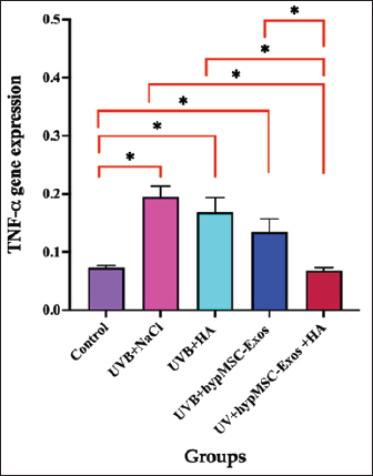
Fig. 2. (A–B) Gross shaved dorsal skin: control (A) and UVB-exposed with mild erythema (B). (C–D) Masson’s trichrome (100×): control shows dense intact blue collagen; UVB shows reduced, fragmented collagen and stromal disorganization. (E) The collagen area fraction significantly decreased in UVB (mean±SD; **p < 0.01). Combination HypMSC-Exos and HA suppress TNF-a mRNA expression in UVB-induced collagen lossUVB exposure induced a robust proinflammatory response in the dorsal skin, as indicated by increased TNF-α mRNA levels measured by qRT-PCR (Fig. 3). Relative TNF-α expression in the UVB+NaCl group was markedly elevated compared with the baseline (non-UVB controls), confirming sustained inflammatory activation 5 days after a single treatment. Therapeutic interventions attenuated TNF-α upregulation to varying degrees. Both the UVB+HA and UVB+ HypMSC-Exos groups demonstrated significant reductions in TNF-α mRNA relative to the UVB+NaCl group, indicating anti-inflammatory effects of HA and HypMSC-Exos when administered individually. The UVB+ HypMSC-Exos group showed a greater decrease than the UVB+HA group, suggesting a stronger modulatory effect of exosome treatment on proinflammatory signalling.
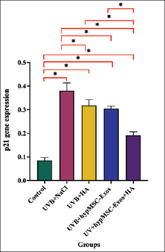
Fig. 3. Relative TNF-α mRNA expression in the dorsal skin (qRT-PCR). Data are mean ± SEM; * p < 0.05 based on the Mann–Whitney test. Combination HypMSC-Exos and HA suppress p21 mRNA expression in UVB-induced collagen lossFigure 4 shows that the treatments modulated UVB-induced inflammation. UVB+NaCl showed the highest p21 gene expression, indicating persistent proinflammatory signalling five days post-injection. All interventions significantly reduced p21 versus UVB+NaCl (p < 0.05). HA alone (UVB+HA) decreased p21, while HypMSC-Exos (UVB+ HypMSC-Exos) showed a greater reduction, indicating stronger immunomodulation. The combined treatment (UVB+HypMSC-Exos+HA) yielded the greatest suppression, which was significantly lower than that of the UVB+NaCl and all other treatments, suggesting additive or synergistic effects.
Fig. 4. Relative p21 expression in the dorsal skin (qRT-PCR). Data are mean ± SEM; * p < 0.05 based on the LSD post hoc test. DiscussionRecent studies have explored the therapeutic efficacy of hypMSC-Exos combined with HA for mitigating inflammation and gene-expression alterations in UVB-induced collagen loss. The macroscopic and histopathological data validate the UVB irradiation protocol as an effective method for inducing dermal collagen loss in the dorsal skin of Wistar rats. The combination of visible erythema and histologic signs—reduced collagen staining intensity, fiber fragmentation, stromal disorganization, and epidermal hyperplasia—supports the model of photoinduced dermal damage suitable for testing interventions aimed at preserving or restoring the dermal collagen structure and function. UV exposure accelerates HA degradation and disrupts the balance between HA synthesis and catabolism, contributing to dryness, barrier impairment, and dermal atrophy (Kang et al., 2018; Tang et al., 2024). HA not only acts as a structural and hydrating component but can also serve as a delivery matrix or co-therapy that enhances the delivery and retention of hypMSC-Exos at the target site, improving their therapeutic efficacy (Khan et al., 2022; Ren et al., 2024). Therapeutic interventions with hypMSC-Exos show a stronger modulatory effect on pro-inflammatory signalling than HA, as evidenced by significant reductions in TNF-α and p21 mRNA levels. Exosomes are small vesicles that carry proteins, lipids, and RNAs, facilitating intercellular communication and modulating various biological processes (Huynh et al., 2025). HypMSC-Exos show enhanced antioxidant and anti-inflammatory effects in UVB-induced skin injury models, more effectively reducing ROS accumulation, TNF-α, IL-1β, and IL-6, and protecting the microvasculature compared with normoxic exosomes (Ha et al., 2020). This result is consistent with previous research, which reported that in Wistar rats exposed to UVB radiation, hypMSC-Exos reduced MDA and MMP-1 levels, indicating reduced oxidative stress and inflammation (Angelina et al., 2025). HypMSC-Exos have been shown to downregulate pro-inflammatory cytokines and fibrosis markers, including TNF-α and p21, via multiple signalling pathways (Zhang et al., 2021; Kim et al., 2025; Ligang et al., 2026). The combined treatment (UVB+ hypMSC-Exos+HA) produced the greatest suppression of TNF-α expression, with levels approaching baseline and significantly lower than those in UVB+NaCl and in each single-treatment group. This additive or synergistic reduction suggests that HA Staris® may enhance the retention or activity of hypMSC-Exos in the dermal microenvironment, thereby improving their anti-inflammatory effects. Combining hypMSC-Exos with HA hydrogels improves their stability and retention at the target site. The use of HA as a carrier for hypMSC-Exos is particularly beneficial for maintaining exosome bioactivity in the harsh environment of UVB-irradiated skin. This sustained-release system ensures continuous exosome delivery, thereby enhancing their uptake by target cells and prolonging their therapeutic effects (Zhao et al., 2025). HypMSC-Exos contain microRNAs that play a pivotal role in cellular communication and tissue repair. For instance, miR-125b in hypoxic exosomes targets and suppresses tumour protein p53 inducible nuclear protein 1 (TP53INP1), alleviating hypoxia-induced apoptosis and promoting cell proliferation (Zhang et al., 2021). In addition, miR-21-5p and miR-214-5p in hypoxic exosomes activate signal transducer and activator of transcription 3 (STAT3) signalling, enhancing cell migration and differentiation (Ha et al., 2020; Gui et al., 2025; Khosravipour et al., 2025; Zhang, 2025). HypMSC-Exos exhibit promising therapeutic potential in mitigating UVB-induced dermal inflammation. Their ability to suppress TNF-α mRNA levels and other proinflammatory cytokines, coupled with their regenerative properties, positions them as effective agents for treating UVB-induced skin damage. The current study is limited by the absence of protein-level validation of the gene expression findings. In addition, therapeutic efficacy was assessed only at the level of gene expression, without an evaluation of long-term functional or clinical outcomes. Further research and clinical trials are necessary to fully elucidate their mechanisms and optimize their therapeutic applications. ConclusionThe combination of hypoxic MSC-derived exosomes with HA presents a promising therapeutic strategy for reducing TNF-α and p21 gene expression in UVB-induced collagen loss of Wistar rat skin. The combination of hypoxic MSC-derived exosomes and HA shows a promising strategy for mitigating UVB-induced skin damage. This approach leverages the anti-inflammatory, antioxidant, and regenerative properties of hypMSC-Exos, which are enhanced by the stability and sustained-release properties of HA hydrogels. Further research and clinical trials are needed to fully understand the mechanisms and optimize the therapeutic potential of this treatment. AcknowledgmentsNone. FundingNone. Authors' contributionsLJ: conceptualisation, methodology, investigation, data curation, software, formal analysis, writing—review and editing; ES: conceptualisation, methodology, supervision, writing—review and editing; SS: conceptualisation, methodology, supervision, software, validation, visualisation, formal analysis, writing—original draft preparation, writing—review and editing. Conflict of interestThe authors declare no conflict of interest. Data availabilityAll data supporting the findings of this study are available within the manuscript. ReferencesAngelina, J., Putra, A., Trisnadi, S., Hermansyah, D., Setiawan, E., Sumarawati, T. and Amalina, N. 2025. Hypoxia-conditioned mesenchymal stem cells (MSC) exosomes attenuate ultraviolet-B (UVB)-mediated malondialdehyde (MDA) and matrix metalloproteinase-1 (MMP)-1 upregulation in collagen loss models. Med. Glas. (Zenica). 22(1), 9–14; doi:10.17392/1923-22-01 Blackstone, B.N., Wilgus, T.A., Roy, S., Wulff, B.C. and Powell, H.M. 2020. Skin Biomechanics and miRNA expression following chronic UVB irradiation. Adv. Wound. Care. 9, 79–89; doi:10.1089/wound.2019.1034 Dong, J., Wu, B. and Tian, W. 2023. Exosomes derived from hypoxia-preconditioned mesenchymal stem cells (hypoMSCs-Exo): advantages in disease treatment. Cell Tissue Res. 392, 621–629; doi:10.1007/s00441-023-03758-6 Gui, Q., Ding, N., Liu, J., Zhao, Y., Du, A., Zhu, J., Wu, H., Wu, M., Wang, Y., Zhu, L., 2025. miR-200a-3p in human umbilical cord mesenchymal stem cell-derived exosomes attenuates UVB-induced skin inflammatory response and oxidative stress via Keap1-Nrf2 Pathway. Stem Cells Int. 2025, 1–21; doi: 10.1155/sci/7831890 Ha, D.H., Kim, H.K., Lee, J., Kwon, H.H., Park, G.H., Yang, S.H., Jung, J.Y., Choi, H., Lee, J.H., Sung, S., Yi, Y.W. and Cho, B.S. 2020. Mesenchymal stem/stromal cell-derived exosomes for immunomodulatory therapeutics and skin regeneration. Cells 9, 1–45; doi:10.3390/cells9051157 He, C., Wang, Z., Jiang, Z., Zhou, Y., Chen, X., Zhang, J., Wang, B., Wang, Q., Wu, T., Li, C. and Jian, Z. 2025. Microneedles combining delivery of hUMSC-derived exosomes and EGCG mitigate UV-induced skin damage. J. Nanobiotechnol. 23 (643), 1–19;, doi: 10.1186/s12951-025-03735-x Huynh, C., Vu, N., Van, T. and Pham, P. 2025. Effects of exosomes from hypoxia-induced adipose-derived stem cells on ameliorating photoaging. Clin. Cosmet. Investig. Dermatol. 18, 1683–1702; doi:10.2147/ccid.s523936 Huynh, T.D., Gargiulo Isacco, C., Ngo, Q.T.M., Nguyen, B.T., Huu Nguyen, T.N., Dang Bui, T.M., Ngo, V.M., Truong, K.Q., Chau, T.V., Truong, H.C., Nguyen, K.D.C., Jirillo, E., Pham, V.H., Santacroce, L. and Tran, T.C. 2025. Exosomes from adipose tissue mesenchymal stem cells, a preliminary study for in vitro and in vivo application. Bioengineering 12, 1–47; doi:10.3390/bioengineering12101129 Kaltchenko, M.V. and Chien, A.L. 2025. Photoaging: current concepts on molecular mechanisms, prevention, and treatment. Am. J. Clin. Dermatol. 26, 321–344; doi:10.1007/s40257-025-00933-z Kang, M.C., Yumnam, S. and Kim, S.Y. 2018. Oral intake of collagen peptide attenuates ultraviolet B irradiation-induced skin dehydration in vivo by regulating hyaluronic acid synthesis. Int. J. Mol. Sci. 19, 1–12; doi:10.3390/ijms19113551 Khan, K., Makhoul, G., Yu, B., Jalani, G., Derish, I., Rutman, A.K., Cerruti, M., Schwertani, A. and Cecere, R. 2022. Amniotic stromal stem cell-loaded hydrogel repairs cardiac tissue in infarcted rat hearts via paracrine mediators. J. Tissue. Eng. Regen. Med. 16, 110–127; doi:10.1002/term.3262 Khosravipour, A., Ebrahimzadeh, M.H., Mousavi Shaegh, S.A., Matin, M. and Ebrahimzadeh-Bideskan, A. 2025. Effects of hypoxic and photobiomodulation preconditioning on mesenchymal stem cell-derived exosomes: an in vitro study with an osteogenic potential approach. Photochem. Photobiol. Sci. 24, 2051–2068; doi:10.1007/s43630-025-00810-y Kim, J., Jo, T., Nam, H., Kim, B.J., Nam, S.M., Kim, J., Choi, J. and Jeong, W. 2025. Adipose-derived exosomes as a preventative strategy against complications in hyaluronic acid filler applications: a comprehensive in vivo analysis. J. Plast. Reconstr. Aesthetic Surg. 102, 498–505; doi:10.1016/j.bjps.2024.07.071 Ligang, Z., Tao, L., Jie, Y., Nini, Z., Li, Y., Guilin, H., Xiaohua, H. and Min, D. 2026. Function of human amniotic mesenchymal stem cell exosomes in repairing submandibular gland epithelial cells after radiation injury in SD rats. Chin. J. Tissue Eng. Res. 30, 3250–3257; doi:10.12307/2026.088 Liu, Y., Wang, Y., Yang, M., Luo, J., Zha, J., Geng, S. and Zeng, W. 2024. Exosomes from hypoxic pretreated ADSCs attenuate ultraviolet light-induced skin injury via GLRX5 delivery and ferroptosis inhibition. Photochem. Photobiol. Sci. 23, 55–63; doi:10.1007/s43630-023-00498-y Meng, Y., Li, C., Liang, Y., Jiang, Y., Zhang, H., Ouyang, J., Zhang, W., Deng, R., Tan, Q., Yu, X. and Luo, Z. 2023. Umbilical cord mesenchymal-stem-cell-derived exosomes exhibit anti-oxidant and antiviral effects as cell-free therapies. Viruses 15, 1–16; doi:10.3390/v15102094 Najafabadi, A.H., Soheilifar, M.H. and Masoudi-Khoram, N. 2024. Exosomes in skin photoaging: biological functions and therapeutic opportunity. Cell. Commun. Signal. 22, 32; doi:10.1186/s12964-023-01451-3 Nasution, I., Putra, A. and Setiawan, E. 2025. Synergistic effects of hypoxia-preconditioned mesenchymal stem cells secretome and alkaline water in alleviating oxidative stress and inflammation in type 2 diabetic rats. Mater. Sociomed. 37, 4–10; doi:10.5455/msm.2024.37.4-10 Ren, Y., Wang, W., Yu, C., Wang, Y., Qiu, Y., Yue, Z., Yu, Q., Lu, J., Che, P., Li, J. and Sun, H. 2024. An injectable exosome-loaded hyaluronic acid-polylysine hydrogel for cardiac repair via modulating oxidative stress and the inflammatory microenvironment. Int. J. Biol. Macromol. 275, 1–13; doi:10.1016/j.ijbiomac.2024.133622 Suman, G. and Suman, S. 2019. Ultraviolet radiation-induced immunomodulation: skin ageing and cancer. In Skin Aging & Cancer. Dwivedi, A., Agarwal, N., Ray, L., Tripathi, A. and Eds. Springer Singapore, pp: 47–58. https://doi.org/10.1007/978-981-13-2541-0_5 Tang, Z., Liu, Z., Zhang, Y., Luo, S., Xu, Y. and Ren, L. 2024. Functional hyaluronic acid microneedles for skin photoaging based on collagen induction and oxidative stress regulation strategies. Int. J. Biol. Macromol. 227, 1–18; doi: 10.1016/j.ijbiomac.2024.134080 Wang, H., Wu, S., Bai, X., Pan, D., Ning, Y., Wang, C., Guo, L., Guo, J. and Gu, Y. 2025. Mesenchymal stem cell-derived exosomes hold promise in the treatment of diabetic foot ulcers. Int. J. Nanomed. 20, 5837–5857; doi:10.2147/ijn.s516533 Wang, Y., Wang, H., Tan, J., Cao, Z., Wang, Q., Wang, H., Yue, S., Li, W. and Wang, D. 2025. Therapeutic effect of mesenchymal stem cells and their derived exosomes in diseases. Mol. Biomed. 6, 34; doi:10.1186/s43556-025-00277-4 Zha, J., Pan, Y., Liu, X., Zhu, H., Liu, Y. and Zeng, W. 2023. Exosomes from hypoxia-pretreated adipose-derived stem cells attenuate ultraviolet light-induced skin injury via delivery of circ-Ash1l. Photodermatol. Photoimmunol. Photomed. 39, 107–115; doi:10.1111/phpp.12857 Zhang, J.W. 2025. Impact of miR-214-5p and miR-21-5p from hypoxic endometrial exosomes on human umbilical cord mesenchymal stem cell function. World. J. Stem. Cells. 17(2), 1–5; doi:10.4252/WJSC.V17.I2.102404 Zhang, P., Li, T., Wu, X., Nice, E., Huang, C. and Zhang, Y. 2020. Oxidative stress and diabetes: antioxidative strategies. Front. Med. 14, 583–600; doi:10.1007/s11684-019-0729-1 Zhang, X.F., Wang, T., Wang, Z.X., Huang, K.P., Zhang, Y.W., Wang, G.L., Zhang, H.J., Chen, Z.H., Wang, C.Y., Zhang, J.X. and Wang, H. 2021. Hypoxic ucMSC-secreted exosomal miR-125b promotes endothelial cell survival and migration during wound healing by targeting TP53INP1. Mol. Ther. Nucleic Acids 26, 347–359; doi:10.1016/j.omtn.2021.07.014 Zhao, X., Shi, Y., Sun, Z., Duan, W., Chang, L., Xu, B., Lai, K., Zhang, J., Tian, B., Tao, W., Mi, Z., Zhang, M., Yang, W., Luo, Z. and Ye, Z. 2025. Injectable hydrogel loaded with exosomes from hypoxic umbilical cord-derived mesenchymal stem cells alleviates intervertebral disc degeneration by reversing nucleus pulposus cell senescence. Regen. Biomater. 1–12; doi:10.1093/rb/rbaf039 Zhu, H., Guo, X., Zhang, Y., Khan, A., Pang, Y., Song, H., Zhao, H., Liu, Z., Qiao, H. and Xie, J. 2024. The combined anti-aging effect of hydrolyzed collagen oligopeptides and exosomes derived from human umbilical cord mesenchymal stem cells on human skin fibroblasts. Molecules 29(7), 1468; doi:10.3390/molecules29071468 | ||
| How to Cite this Article |
| Pubmed Style Julianti L, Setiawan E, Suparmi S. Hypoxic mesenchymal stem cell exosomes with hyaluronic acid attenuate UVB-induced collagen loss by suppressing TNF-α and p21 in wistar rat skin. Open Vet. J.. 2026; 16(7): 4962-4969. doi:10.5455/OVJ.2026.v16.i7.73 Web Style Julianti L, Setiawan E, Suparmi S. Hypoxic mesenchymal stem cell exosomes with hyaluronic acid attenuate UVB-induced collagen loss by suppressing TNF-α and p21 in wistar rat skin. https://www.openveterinaryjournal.com/?mno=316253 [Access: July 27, 2026]. doi:10.5455/OVJ.2026.v16.i7.73 AMA (American Medical Association) Style Julianti L, Setiawan E, Suparmi S. Hypoxic mesenchymal stem cell exosomes with hyaluronic acid attenuate UVB-induced collagen loss by suppressing TNF-α and p21 in wistar rat skin. Open Vet. J.. 2026; 16(7): 4962-4969. doi:10.5455/OVJ.2026.v16.i7.73 Vancouver/ICMJE Style Julianti L, Setiawan E, Suparmi S. Hypoxic mesenchymal stem cell exosomes with hyaluronic acid attenuate UVB-induced collagen loss by suppressing TNF-α and p21 in wistar rat skin. Open Vet. J.. (2026), [cited July 27, 2026]; 16(7): 4962-4969. doi:10.5455/OVJ.2026.v16.i7.73 Harvard Style Julianti, L., Setiawan, . E. & Suparmi, . S. (2026) Hypoxic mesenchymal stem cell exosomes with hyaluronic acid attenuate UVB-induced collagen loss by suppressing TNF-α and p21 in wistar rat skin. Open Vet. J., 16 (7), 4962-4969. doi:10.5455/OVJ.2026.v16.i7.73 Turabian Style Julianti, Lis, Eko Setiawan, and Suparmi Suparmi. 2026. Hypoxic mesenchymal stem cell exosomes with hyaluronic acid attenuate UVB-induced collagen loss by suppressing TNF-α and p21 in wistar rat skin. Open Veterinary Journal, 16 (7), 4962-4969. doi:10.5455/OVJ.2026.v16.i7.73 Chicago Style Julianti, Lis, Eko Setiawan, and Suparmi Suparmi. "Hypoxic mesenchymal stem cell exosomes with hyaluronic acid attenuate UVB-induced collagen loss by suppressing TNF-α and p21 in wistar rat skin." Open Veterinary Journal 16 (2026), 4962-4969. doi:10.5455/OVJ.2026.v16.i7.73 MLA (The Modern Language Association) Style Julianti, Lis, Eko Setiawan, and Suparmi Suparmi. "Hypoxic mesenchymal stem cell exosomes with hyaluronic acid attenuate UVB-induced collagen loss by suppressing TNF-α and p21 in wistar rat skin." Open Veterinary Journal 16.7 (2026), 4962-4969. Print. doi:10.5455/OVJ.2026.v16.i7.73 APA (American Psychological Association) Style Julianti, L., Setiawan, . E. & Suparmi, . S. (2026) Hypoxic mesenchymal stem cell exosomes with hyaluronic acid attenuate UVB-induced collagen loss by suppressing TNF-α and p21 in wistar rat skin. Open Veterinary Journal, 16 (7), 4962-4969. doi:10.5455/OVJ.2026.v16.i7.73 |


