Open Veterinary Journal, (2026), Vol. 16(7): 4340-4350
Research Article
10.5455/OVJ.2026.v16.i7.21
Antihypertensive and cardioprotective effects of rice bran supplementation via suppression of the iNOS and TNF-α axis in DOCA-salt-induced hypertensive rats
Aulanni’am Aulanni’am1*, Muhammad Fikri Nur1, Saidun Fiddaroini1, Akhmad Sabarudin1,
Anna Safitri1, Wibi Riawan2, Dyah Kinasih Wuragil3, Syifa Mustika4and Ahmad Lubab5
1Department of Chemistry, Faculty of Mathematics and Natural Sciences, Brawijaya University, Malang, Indonesia
2Department of Biochemistry, Faculty of Medicine, Brawijaya University, Malang, Indonesia
3Faculty of Veterinary Medicine, Brawijaya University, Malang, Indonesia
4Department of Internal Medicine, Faculty of Medicine, Brawijaya University, Malang, Indonesia
5Department of Mathematics, Faculty Science and Technology, Universitas Islam Negeri Sunan Ampel, Surabaya, Indonesia
*Corresponding Author: Aulanni’am Aulanni’am. Department of Chemistry, Faculty of Mathematics and Natural Sciences, Brawijaya University, Malang, Indonesia Email: aulani [at] ub.ac.id
Submitted: 06/04/2026 Revised: 06/06/2026 Accepted: 15/06/2026 Published: 11/07/2026
© 2026 Open Veterinary Journal
This is an Open Access article distributed under the terms of the Creative Commons Attribution-Non Commercial-No Derivatives License (http://creativecommons.org/licenses/by-nc-nd/4.0/), which permits non-commercial re-use, distribution, and reproduction in any medium, provided the original work is properly cited, and is not altered, transformed, or built upon in any way.
ABSTRACT
Background: Hypertension is a major global health burden and a leading contributor to cardiovascular morbidity, with a prevalence of 34.1% in Indonesia. Although angiotensin-converting enzyme (ACE) inhibitors such as captopril are widely used, their long-term use is limited due to side effects. Rice bran, a nutrient-rich byproduct of rice milling, has been reported to possess antioxidant and antihypertensive potential; however, its cardioprotective effects in hypertensive conditions remain underexplored.
Aim: This study aims to evaluate the antihypertensive and cardioprotective effects of rice bran supplementation in a deoxycorticosterone acetate (DOCA)-salt-induced hypertensive rat model, with a focus on inflammatory and structural changes in the heart.
Methods: Male Wistar rats were divided into five groups: normotensive control (K−), hypertensive control (K+), a group treated with captopril (5 mg/kg BW), and groups supplemented with rice bran at 1% (D1) and 2% (D2) of the daily diet for 4 weeks. Hypertension was induced using DOCA-salt. Serum protein profiles were analyzed by sodium dodecyl sulfate–polyacrylamide gel electrophoresis, cardiac histopathology was assessed using hematoxylin–eosin staining, and tumor necrosis factor-α (TNF-α) and nitric oxide synthase (iNOS) expression were evaluated by immunohistochemistry.
Results: Rice bran supplementation, particularly at 2%, significantly improved cardiac structure, as evidenced by reduced cardiomyocyte hypertrophy, decreased necrosis, and restoration of myocardial organization. SDS–PAGE analysis revealed the reappearance of the ~40 kDa protein band and a reduction in the ~150 kDa band in the treated group, indicating the restoration of cardiac protein expression patterns. Inflammatory markers were significantly suppressed, with TNF-α reduced by 74.9% (D1) and 76.8% (D2), exceeding the reduction observed with captopril (54.1%). Similarly, iNOS expression decreased most effectively in the D2 group (29.52% compared with the hypertensive control).
Conclusion: Rice bran supplementation provides significant antihypertensive and cardioprotective effects in DOCA-salt-induced hypertensive rats, primarily through reduced inflammatory responses and improved cardiac structural integrity. These findings support the potential of rice bran as a functional dietary intervention for managing hypertension-induced cardiac injury.
Keywords: ACE inhibitor, Histopathology, Hypertension, Immunohistochemistry, Rice bran.
Introduction
Hypertension represents a major global health burden and remains a leading contributor to cardiovascular morbidity and mortality across both developed and developing countries. Clinically defined by a sustained elevation in blood pressure (≥140/90 mmHg), this condition markedly increases the risk of myocardial infarction, stroke, heart failure, and chronic kidney disease. Despite the availability of established pharmacological interventions, hypertension continues to be underdiagnosed and inadequately controlled worldwide, underscoring a critical need for more effective and sustainable management strategies (Nurmahdi et al., 2017). In Indonesia, the prevalence of hypertension has reached 34.1%, highlighting its significance as a national public health concern with profound implications for long-term organ damage, particularly affecting the heart, brain, and kidneys (Wahidin et al., 2025).
At the mechanistic level, hypertension is a multifactorial disorder driven by the interplay of oxidative stress, sodium imbalance, and chronic low-grade inflammation. Central to its regulation is the renin–angiotensin–aldosterone system (RAAS), in which angiotensin-converting enzyme (ACE) catalyzes the conversion of angiotensin I to angiotensin II, a potent vasoconstrictor that elevates vascular resistance and promotes aldosterone-mediated sodium and water retention. This cascade ultimately increases blood volume and blood pressure, positioning the RAAS axis as a primary therapeutic target in antihypertensive treatment (Li et al., 2017; Vn Dr and Vn Dr, 2021). Current standard therapies, including ACE inhibitors such as captopril, effectively attenuate RAAS activity; however, their long-term use is frequently limited by adverse effects, including persistent cough, hypotension, renal dysfunction, and electrolyte imbalance (Gan et al., 2018). These limitations highlight the necessity for alternative strategies that are both effective and exhibit improved safety profiles.
In this context, functional food-based interventions have emerged as a promising approach for the management of hypertension. Rice bran, a nutrient-dense byproduct of rice milling, remains underutilized despite its rich composition of proteins, lipids, dietary fiber, and phytochemicals. Previous studies have suggested that rice bran possesses antioxidant and ACE-inhibitory activities, attributed to its content of compounds such as γ-oryzanol, tocotrienols, and ferulic acid (Dang and Vasanthan, 2019). These bioactive constituents are proposed to modulate oxidative stress and inflammatory signaling pathways, which are critically involved in hypertension-associated organ damage. However, existing studies remain largely limited to in vitro or biochemical evaluations, and comprehensive in vivo evidence elucidating the cardioprotective mechanisms of rice bran in hypertensive conditions is still lacking.
Therefore, this study aims to evaluate the antihypertensive and cardioprotective effects of rice bran supplementation in a DOCA-salt-induced hypertensive rat model. Specifically, this work integrates serum protein profiling (SDS–PAGE), histopathological assessment, and immunohistochemical analysis of key inflammatory markers (TNF-α and iNOS) to provide a multilevel understanding of the therapeutic potential of rice bran in modulating hypertension-induced cardiac injury.
Materials and Methods
Experimental animals and study design
Male Wistar rats (Rattus norvegicus) were used and acclimatized for 1 week under standard laboratory conditions with free access to food and water. Animals were fed standard AD II chow containing 12% water, 15% crude protein, 3%–7% crude fat, 6% fiber, a maximum of 7% ash, 0.9%–1.1% calcium, and 0.6%–0.9% phosphorus. The number of experimental animals was determined using the Federer equation (t−1)(n−1) ≥ 15, where t represents the number of treatment groups, and n represents the number of animals per group. The rats were randomly divided into five groups: (1) normotensive control (K−), (2) hypertensive control (K+), (3) captopril-treated group (5 mg/kg body weight, BW), (4) rice bran-treated group at 1% of total daily feed (D1), and (5) rice bran-treated group at 2% of total daily feed (D2).
Induction of hypertension (DOCA salt model)
Hypertension was induced using the deoxycorticosterone acetate (DOCA)-salt (Tokyo Chemical Industries, Lot: OZLZC-HG) model over a 5-week period. Rats received subcutaneous injections of DOCA dissolved in corn oil in the cervical region twice weekly (10 injections total). The first five injections were administered at 20 mg/kg BW, followed by five injections at 10 mg/kg BW (Riyadi et al., 2020). During the induction period, animals were provided with 2% (w/v) a NaCl solution as drinking water. Systolic blood pressure was measured weekly using a CODA™ Tail-Cuff Blood Pressure System (Kent Scientific) until the end of the experiment at necropsy.
Therapeutic interventions
Rice bran administration
Rice bran was administered at doses of 1% (D1) and 2% (D2) of the total daily feed (25 g/day). The required amount of rice bran was suspended in 1.5 ml of distilled water and delivered once daily via oral gavage for 4 weeks (Nur et al., 2026).
Captopril administration
Captopril was administered orally at a dose of 5 mg/kg BW once daily for 4 weeks. The solution was freshly prepared in distilled water before administration.
Serum protein analysis by SDS–PAGE
Serum protein profiles were analyzed using SDS–PAGE. Samples were mixed with reducing sample buffer (1:1, v/v) and heated in boiling water for 10 minutes to ensure protein denaturation. A volume of 15 µl of each sample was loaded onto a gel consisting of a 12% separating gel and 5% stacking gel.
Electrophoresis was performed at a constant voltage of 150 V for 60 minutes. Following separation, gels were stained with Coomassie brilliant blue for 30 minutes and subsequently destained for approximately 17 hours to enhance band visualization. Protein bands were documented using a Bio-Rad gel documentation system, and molecular weight estimation was conducted using Image Lab software (Ramadani et al., 2024).
Histopathological analysis (Hematoxylin–Eosin staining)
Cardiac tissues were fixed in 10% paraformaldehyde, processed, and embedded in paraffin. Tissue sections were mounted on glass slides and subjected to deparaffinization followed by rehydration through a graded ethanol series (100%, 95%, 90%, 80%, and 70%) for 5 minutes each, and rinsed with distilled water.
Sections were stained with hematoxylin for 10 minutes, followed by washing under running tap water for 30 minutes. Eosin staining was then performed for 5 minutes. After staining, tissues were dehydrated through ascending ethanol concentrations, cleared in xylene for 5 minutes, and mounted with Entellan (Merck, Lot: HX43836061) and a coverslip. Histological observations were conducted under a light microscope (Tuska et al., 2025).
Immunohistochemical analysis of TNF-α and iNOS
Cardiac tissue sections were prepared similarly to histopathological analysis and subjected to immunohistochemical staining. After deparaffinization and rehydration, slides were washed with phosphate-buffered saline (PBS) and incubated with hydrogen peroxide (H2O2) (Scytek Laboratories, Lot: 82573) for 20 minutes to block endogenous peroxidase activity.
Blocking was performed using 5% (w/v) bovine serum albumin (BSA) in PBS for 30 minutes at room temperature. Sections were then incubated with primary antibodies against TNF-α (Santa Cruz Biotechnology Cat#:sc-52746) and iNOS. (Santa Cruz Biotechnology Cat#:sc-7271). After washing, slides were incubated with a biotin-labeled secondary antibody (Scytek Laboratories, Lot: 81223), followed by streptavidin–horseradish peroxidase (SA–HRP) (Scytek Laboratories, Lot: 82493). Signal visualization was achieved using 3,3′-diaminobenzidine (DAB) (Scytek Laboratories, Lot: 82932), producing a brown precipitate at antigen sites. Counterstaining was performed using Mayer’s hematoxylin. Slides were examined under a microscope, and quantitative analysis was conducted using ImageJ software 1.54 g (Panjaitan et al., 2025).
Ethical approval
All experimental procedures were approved by the Research Ethics Committee of Brawijaya University (KEP-216-UB/2024).
Results
Body weight analysis
Body weight was monitored at four time points throughout the experimental period: at baseline (before induction), following DOCA-salt induction, and after 2 and 4 weeks of treatment (Table 1). At baseline, mean body weights ranged from 140.0 g to 172.0 g across all groups. All groups demonstrated an overall increase in body weight from baseline to study end, with final body weights ranging from 177.5 g to 255.0 g.
Table 1. Average body weight (g) of experimental animals.
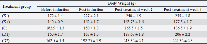
The normotensive control group achieved the highest final body weight (255.0 g), consistent with unimpeded growth under normal physiological conditions. In contrast, the hypertensive control group exhibited the lowest final body weight (177.5 g) and the smallest overall weight gain (37.5 g), which is in accordance with previous reports describing attenuated growth in DOCA-salt hypertensive rat models, likely attributable to hemodynamic stress and sodium–water imbalance. The captopril-treated group showed a transient reduction in body weight following DOCA-salt induction (162.5 to 150.0 g), before recovering progressively to 186.5 g by the end of the study. Groups receiving rice bran supplementation at 1% and 2% of total daily feed reached final body weights of 206.0 g and 224.3 g, respectively, both of which exceeded that of the hypertensive control group. No sustained or abnormal body weight loss was observed in any experimental group, indicating that neither DOCA-salt induction nor rice bran supplementation produced overt adverse effects on the overall health of the animals.
SDS–PAGE analysis of serum protein profile
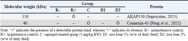
Serum protein profiling by SDS–PAGE revealed distinct alterations in band patterns across experimental groups, reflecting the molecular impact of DOCA-salt-induced hypertension and subsequent therapeutic interventions (Fig. 1, Table 2). Notably, a protein band at approximately 40 kDa was detected exclusively in the captopril-treated (C) and 2% rice bran-treated (D2) groups, while it was absent in the normotensive control (K−), hypertensive control (K+), and 1% rice bran-treated (D1) groups. In contrast, a prominent band at approximately 150 kDa was observed in the hypertensive control and, to a lesser extent, in the D1 group but was markedly reduced or absent in the captopril and D2 groups.
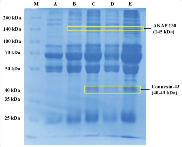
Fig. 1. SDS–PAGE gel of rat serum: (M) protein marker, (A) normotensive group rat serum, (B) hypertensive group rat serum, (C) captopril group rat serum, (D) 1% rice bran therapy group rat serum, (E) 2% rice bran therapy group rat serum.
Table 2. Serum protein band distribution identified by SDS–PAGE across experimental groups.
Histopathological analysis of cardiac tissue
Histopathological evaluation was conducted to elucidate the structural impact of DOCA-salt-induced hypertension and to assess the extent of myocardial recovery following therapeutic intervention. Cardiac tissue sections were stained using hematoxylin–eosin (H&E) and examined at 100× and 400× magnification (Fig. 2). The analysis focused on key morphological parameters, including cardiomyocyte hypertrophy, nuclear integrity, myofibrillar organization, and vascular alterations.
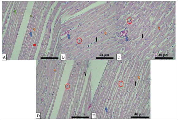
Fig. 2. Histopathological analysis of rat cardiac tissue stained with hematoxylin–eosin (H&E) at 400× magnification. (A) Normotensive control (K−); (B) hypertensive control (K+); (C) captopril-treated group (5 mg/kg BW); (D) rice bran 1% (D1); (E) rice bran 2% (D2). Arrows indicate representative histological features: green=cardiomyocytes; yellow=muscle fibers; blue=capillaries; red=striations; orange=endothelial cells; black=myocardial hypertrophy. Red circles denote areas of necrosis.
The normotensive control group (K−) exhibited preserved myocardial architecture, characterized by well-organized cardiomyocytes arranged in branching fascicles with centrally located nuclei and clearly defined transverse striations. Intercalated discs were uniformly distributed, ensuring effective mechanical and electrical coupling between adjacent cells (Lowrie, 2020). Capillary networks were intact, with no evidence of vascular leakage or interstitial disruption, confirming physiological cardiac homeostasis.
In contrast, the hypertensive group (K+) displayed extensive structural remodeling consistent with chronic pressure overload. Cardiomyocytes exhibited pronounced hypertrophy, as indicated by increased fiber diameter, nuclear enlargement, and hyperchromatic nuclei, reflecting enhanced protein synthesis and sustained mechanical stress. These alterations are hallmarks of compensatory hypertrophic signaling driven by angiotensin II and oxidative stress. At the tissue level, interstitial hemorrhage and vascular congestion were evident, suggesting endothelial dysfunction and increased vascular permeability.
TNF-α expression in cardiac tissue
Tumor necrosis factor-α (TNF-α) expression in cardiac tissue was evaluated using immunohistochemistry (IHC), in which antigen–antibody binding was visualized as a brown precipitate following 3,3′-diaminobenzidine (DAB) development. The intensity and distribution of staining served as a semiquantitative indicator of myocardial inflammatory status (Fig. 3, Table 3).
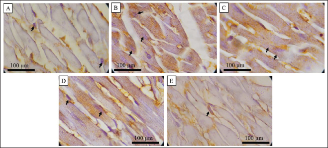
Fig. 3. Immunohistochemical detection of TNF-α expression in rat cardiac tissue at 1000× magnification. (A) Normotensive control (K−); (B) hypertensive control (K+); (C) captopril-treated group (5 mg/kg BW); (D) rice bran 1% (D1); (E) rice bran 2% (D2). Arrows indicate TNF-α–positive cardiomyocytes, visualized as brown DAB staining.
Table 3. TNF-α expression in the hypertensive rat heart.
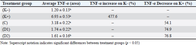
Statistical analysis confirmed significant differences in TNF-α expression among groups (one-way ANOVA, p < 0.0001). Although data were normally distributed (Shapiro–Wilk, p > 0.05), variance heterogeneity necessitated further analysis using the Kruskal–Wallis test, which validated the observed intergroup differences. Pairwise comparisons demonstrated significant reductions in TNF-α expression in both rice bran-treated groups compared to the hypertensive control, with the greatest effect observed in the D2 group (p=0.0001).
iNOS expression in cardiac tissue
Inducible nitric oxide synthase (iNOS) expression in cardiac tissue was evaluated by immunohistochemistry (IHC), where antigen–antibody interactions were visualized as brown deposits following DAB chromogen development. The extent of staining was quantified to reflect the level of iNOS expression across experimental groups (Fig. 4, Table 4).
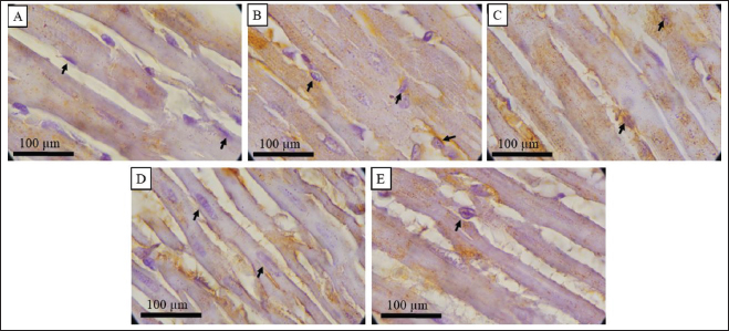
Fig. 4. Immunohistochemical analysis of iNOS expression in rat cardiac tissue at 1000× magnification. (A) Normotensive control (K−); (B) hypertensive control (K+); (C) captopril-treated group (5 mg/kg BW); (D) rice bran 1% (D1); (E) rice bran 2% (D2). Arrows indicate iNOS-positive cardiomyocytes, visualized as brown DAB staining.
Table 4. iNOS expression in the hypertensive rat heart.
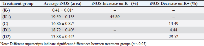
Rice bran supplementation exhibited a dose-dependent effect. The 1% rice bran group (D1) showed a reduction in iNOS expression; however, this decrease was not statistically significant compared with the hypertensive control (p > 0.05), suggesting that the lower dose was inadequate to effectively disrupt the ROS–NF-κB–iNOS axis. In contrast, the 2% rice bran group (D2) demonstrated a significant reduction in iNOS expression (p < 0.05), with the greatest decrease of 29.52% relative to the hypertensive group (Kang et al., 2025). This group also exhibited the lowest iNOS levels among all hypertensive treatment groups, indicating superior efficacy.
Discussion
The selective reappearance of the unidentified ~40 kDa band in the C and D2 groups suggests a therapy-dependent restoration of specific cardiac-associated proteins that are otherwise suppressed under hypertensive conditions. Based on its molecular weight, this band is possibly connexin-43 (Cx43), a critical gap junction protein (40–43 kDa) responsible for maintaining electrical coupling and synchronized contraction in cardiomyocytes (Peng et al., 2022). However, given the absence of confirmatory techniques such as immunoblotting or proteomic identification, this assignment should be interpreted as putative. Nevertheless, the observed pattern aligns with established pathophysiological mechanisms, in which hypertension-induced oxidative stress and inflammatory signaling—particularly elevated TNF-α—lead to downregulation and disorganization of Cx43, thereby impairing intercellular communication and contributing to cardiac dysfunction (Mentari and Machrina, 2023).
In the hypertensive control group, the absence of the ~40 kDa band likely reflects suppression or degradation of such protective proteins under conditions of sustained oxidative stress. DOCA-salt induction is known to enhance mineralocorticoid receptor activation, promoting NADPH oxidase-driven reactive oxygen species (ROS) generation. This oxidative milieu activates the NF-κB signaling pathway, resulting in increased production of proinflammatory cytokines, including TNF-α (Suprayitno, 2025). Elevated TNF-α has been shown to directly disrupt gap junction integrity by inhibiting Cx43 expression and promoting its internalization or degradation, thereby exacerbating electrical and structural remodeling in the myocardium.
The lack of reappearance of this band in the D1 group indicates that the lower dose of rice bran was insufficient to counteract the oxidative–inflammatory cascade to a level required for restoration of protein expression. In contrast, the re-emergence of the ~40 kDa band in the D2 group suggests a threshold-dependent therapeutic effect, in which higher rice bran supplementation more effectively attenuates ROS accumulation and inflammatory signaling, thereby permitting recovery of proteins involved in cardiac structural and functional integrity.
Conversely, the presence of an unidentified ~150 kDa band predominantly in the hypertensive control group, with reduced intensity in D1 and near-complete disappearance in C and D2 groups, is indicative of proteins associated with stress-responsive signaling. This band is possibly A-kinase anchoring protein 150 (AKAP150), a scaffolding protein implicated in the regulation of PKA/PKC signaling and β-adrenergic pathways (Suprayitno, 2025). Similar to the 40 kDa band, this assignment remains putative based on molecular weight correspondence. Under hypertensive conditions, elevated TNF-α and ROS levels are known to dysregulate intracellular signaling networks, leading to increased expression or activation of AKAP150, which in turn contributes to maladaptive cardiac remodeling and hyperactivation of adrenergic signaling pathways.
The pronounced intensity of the ~150 kDa band in the hypertensive group reflects a state of heightened inflammatory and oxidative stress, whereas its attenuation in the captopril and D2 groups suggests effective suppression of these pathological processes. Captopril, through inhibition of ACE, reduces angiotensin II levels, thereby limiting downstream ROS production and inflammatory activation. Similarly, rice bran at higher doses appears to exert a comparable effect, likely through combined antioxidant and anti-inflammatory mechanisms, ultimately leading to downregulation of stress-associated signaling proteins.
Taken together, the reciprocal pattern observed between the ~40 kDa and ~150 kDa bands highlights a coordinated molecular shift from a pathological to a protective state following effective intervention. While the hypertensive condition is characterized by suppression of structural proteins and upregulation of stress-associated signaling components, therapeutic administration—particularly at higher rice bran doses—reverses this trend, restoring protein expression patterns associated with cardiac homeostasis. These findings provide molecular-level support for the cardioprotective effects observed in subsequent histopathological and immunohistochemical analyses, reinforcing the role of rice bran supplementation in modulating key pathways involved in hypertension-induced cardiac injury.
Notably, nuclear damage was prominent, with widespread karyopyknosis and karyolysis, as shown by the red circle in Figure 2B–E, indicating irreversible cellular injury and necrosis. These degenerative changes are closely associated with excessive ROS generation and inflammatory activation. In particular, increased inducible iNOS expression leads to overproduction of nitric oxide (NO), which reacts with superoxide to form peroxynitrite. This highly reactive species induces lipid peroxidation, protein nitration, and cytoskeletal disruption, ultimately compromising cardiomyocyte integrity (Guo et al., 2023). The combined effect of oxidative and nitrosative stress therefore underlies the severe structural deterioration observed in the hypertensive myocardium.
Administration of captopril (C) resulted in partial attenuation of these pathological features. While cardiomyocyte hypertrophy, indicated by the black arrow in Figure 2B, and nuclear abnormalities remained detectable, their severity was reduced compared with the untreated hypertensive group. This partial recovery reflects the pharmacological inhibition of the RAAS, leading to decreased angiotensin II levels and subsequent reduction in oxidative stress and inflammatory signaling. However, the persistence of structural abnormalities suggests that RAAS inhibition alone may be insufficient to fully reverse established myocardial damage, particularly under conditions of sustained oxidative burden.
Rice bran supplementation demonstrated a dose-dependent improvement in cardiac morphology. In the 1% rice bran group (D1) (Fig. 2D), cardiomyocyte diameter was moderately reduced, and myofibrillar organization appeared more coherent compared to the hypertensive control. Nuclear morphology shifted toward a less hyperchromatic and more regular profile, indicating attenuation of hypertrophic signaling and reduced cellular stress. The decreased incidence of karyopyknosis and karyolysis, indicated by the red circle in Figure 2D, further suggests partial protection against necrotic progression. However, residual structural abnormalities indicate that this dose provides only limited suppression of the underlying oxidative–inflammatory cascade.
The most pronounced histological recovery was observed in the 2% rice bran group (D2) (Fig. 2E). Cardiomyocyte size was markedly reduced, approaching that of the normotensive control, and myofibrillar arrangement was restored with improved alignment and intercellular organization. Nuclear morphology was largely normalized, with reduced hyperchromatism and restoration of a typical ovoid shape, indicating regression of hypertrophic remodeling. In addition, interstitial hemorrhage and vascular congestion were substantially diminished, suggesting improved endothelial stability and reduced inflammatory damage. The marked reduction in karyopyknosis and karyolysis, indicated by the red circle in Figure 2B and E, further supports the protective effect of high-dose rice bran against cardiomyocyte necrosis.
Mechanistically, these structural improvements can be attributed to the combined attenuation of haemodynamic stress, oxidative damage, and inflammatory signaling. Rice bran supplementation likely modulates RAAS activity, thereby reducing angiotensin II-mediated vasoconstriction and pressure overload, while simultaneously exerting antioxidant effects that limit ROS accumulation. This dual action suppresses NF-κB activation and downstream proinflammatory mediators, including TNF-α and iNOS, ultimately preserving cellular integrity and preventing structural degeneration.
Overall, the modulation of iNOS expression across treatment groups reinforces the central role of the ROS–NF-κB signaling axis in hypertension-induced cardiac injury. The superior performance of 2% rice bran compared with both the hypertensive control and lower dose intervention underscores its potential as an effective multi-target therapeutic strategy. By simultaneously attenuating oxidative stress and inflammatory signaling, rice bran supplementation provides a more comprehensive protective effect against myocardial damage than single-pathway pharmacological approaches.
The normotensive control group (K−) (Fig. 3A) exhibited minimal TNF-α expression (1.20% ± 0.13%), reflecting basal inflammatory homeostasis. In contrast, DOCA-salt-induced hypertensive rats (K+) (Fig. 3B) demonstrated a marked elevation in TNF-α expression (6.93% ± 0.53%), corresponding to a 477.6% increase relative to the control group. This substantial upregulation confirms the establishment of a robust inflammatory phenotype. Mechanistically, DOCA-salt induction enhances mineralocorticoid receptor activation, which stimulates NADPH oxidase-mediated ROS production. The resulting oxidative stress activates the NF-κB signaling pathway, a central regulator of proinflammatory gene expression, thereby promoting TNF-α synthesis (Sarbini et al., 2012; Rasyid, 2019). Elevated TNF-α further amplifies inflammatory signaling, contributing to endothelial dysfunction, mitochondrial damage, and disruption of cardiomyocyte contractile integrity (Setiawan and Ernawati, 2024).
Therapeutic intervention significantly attenuated TNF-α expression, although with distinct efficacy profiles. Captopril treatment reduced TNF-α levels to 3.18% ± 0.22% (−54.1% vs. hypertensive group), indicating effective suppression of RAAS-mediated inflammatory signaling. Histologically, this was reflected by reduced staining intensity and a more localized distribution pattern. However, residual TNF-α expression suggests that RAAS inhibition alone does not fully abrogate inflammation, likely due to persistent oxidative stress independent of angiotensin II signaling.
In contrast, rice bran supplementation demonstrated superior anti-inflammatory efficacy. The 1% rice bran group (D1) (Fig. 3D) reduced TNF-α expression to 1.74% ± 0.22% (−74.9%), while the 2% group (D2) (Fig. 3E) further decreased it to 1.61% ± 0.16% (−76.8%). Notably, TNF-α levels in the D2 group were statistically indistinguishable from the normotensive control (p ≥ 0.05), indicating near-complete restoration of inflammatory homeostasis (Munawaroh et al., 2024). Although the difference between D1 and D2 was not statistically significant, the consistent trend toward lower expression in D2 suggests a dose-dependent biological effect approaching a saturation threshold.
The enhanced efficacy of rice bran relative to captopril can be attributed to its multi-target mechanism. Beyond potential modulation of the RAAS axis, rice bran exerts antioxidant effects that directly reduce ROS accumulation, thereby limiting NF-κB activation and downstream TNF-α production. This dual modulation—simultaneous attenuation of oxidative stress and inflammatory signaling—provides a broader therapeutic impact compared with single-target pharmacological intervention. Consequently, suppression of TNF-α is more pronounced, leading to improved preservation of cardiac cellular integrity.
From a mechanistic perspective, the observed reduction in TNF-α aligns with the suppression of the ROS–NF-κB axis. Excessive ROS generated under hypertensive conditions activates NF-κB, which in turn upregulates TNF-α, establishing a positive feedback loop that perpetuates inflammation. Rice bran supplementation interrupts this cycle by reducing oxidative stress, thereby preventing NF-κB activation and limiting cytokine production. Similar anti-inflammatory effects have been reported in other plant-derived systems rich in phenolic and flavonoid compounds, which act as ROS scavengers and NF-κB inhibitors (Herbani and Tilaqza, 2025).
Collectively, these findings demonstrate that rice bran supplementation effectively mitigates hypertension-induced cardiac inflammation, with efficacy exceeding that of captopril. The ability to restore TNF-α expression to near-baseline levels highlights its potential as a multi-functional therapeutic strategy targeting both oxidative and inflammatory pathways in hypertensive cardiomyopathy.
The normotensive control group (K−) (Fig. 4A) exhibited minimal iNOS expression (0.41% ± 0.01%), consistent with basal physiological conditions in which iNOS is tightly regulated and expressed at negligible levels. Under normal homeostasis, the transcription factor NF-κB remains inactive, thereby limiting transcription of proinflammatory genes, including iNOS (Mestecky et al., 2015). This low expression profile reflects the absence of oxidative and inflammatory stimuli in healthy myocardial tissue.
In contrast, the hypertensive group (K+) (Fig. 4B) demonstrated a marked elevation in iNOS expression (19.59% ± 0.13%), representing a significant increase compared with the normotensive control (p < 0.05). This upregulation confirms the induction of a pronounced inflammatory and oxidative stress response following DOCA-salt administration. Mechanistically, mineralocorticoid-driven activation of NADPH oxidase enhances ROS generation, which subsequently activates NF-κB signaling. As a downstream effect, NF-κB induces transcription of the iNOS gene, resulting in excessive nitric oxide (NO) production (Shobako and Ohinata, 2020).
Although NO plays a physiological role in vascular regulation, its overproduction under pathological conditions leads to the formation of peroxynitrite through reaction with superoxide radicals. This potent oxidant promotes lipid peroxidation, protein nitration, and mitochondrial dysfunction, thereby exacerbating cardiomyocyte injury and contributing to structural and functional deterioration observed in hypertensive myocardium.
Therapeutic interventions modulated iNOS expression with varying efficacy. Captopril administration significantly reduced iNOS levels compared with the hypertensive group (p < 0.05), reflecting suppression of RAAS-mediated oxidative stress and partial inhibition of NF-κB activation. However, residual iNOS expression indicates that inhibition of angiotensin II alone is insufficient to fully suppress downstream inflammatory signaling.
The pronounced suppression of iNOS in the D2 group highlights the importance of sufficient therapeutic dosing to achieve effective modulation of oxidative and inflammatory pathways. Mechanistically, rice bran likely reduces ROS accumulation, thereby preventing NF-κB activation and subsequent transcription of the iNOS gene. This upstream inhibition results in decreased pathological NO production and limits the formation of reactive nitrogen species such as peroxynitrite. Consequently, cellular damage associated with nitrosative stress is attenuated, contributing to improved myocardial integrity.
Overall, the modulation of iNOS expression across treatment groups reinforces the central role of the ROS–NF-κB signaling axis in hypertension-induced cardiac injury. The superior performance of 2% rice bran compared with both the hypertensive control and lower-dose intervention underscores its potential as an effective multi-target therapeutic strategy. By simultaneously attenuating oxidative stress and inflammatory signalling, rice bran supplementation provides a more comprehensive protective effect against myocardial damage than single-pathway pharmacological approaches.
Conclusion
This study demonstrates that DOCA-salt-induced hypertension leads to pronounced cardiac injury characterized by myocardial hypertrophy, structural disorganization, altered serum protein profiles, and significant upregulation of inflammatory mediators, particularly TNF-α and iNOS, driven by activation of the ROS–NF-κB signaling axis. Therapeutic intervention with rice bran supplementation effectively attenuated these pathological changes in a dose-dependent manner, with the 2% dose showing the most prominent effects, including restoration of myocardial architecture, normalization of protein band patterns, and substantial suppression of inflammatory markers.
Acknowledgment
The authors would like to thank the Directorate of Research and Community Engagement at the University of Brawijaya, Indonesia, for funding this research and publications through the Penguatan Ekosistem Riset Guru Besar (No: 01047.2/UN10.A0501/B/KS/2025) grant from Universitas Brawijaya.
Funding
Penguatan Ekosistem Riset Guru Besar No: 01047.2/UN10.A0501/B/KS/2025, grant from Universitas Brawijaya.
Authors’ contributions
Aulanniam Aulanniam: Conceived and designed the experiment, resources, data analyzed and interpretation, funding acquisition, and writing. Muhammad Fikri Nur: investigation, data interpretation, visualization, formal analysis, methodology, software, writing and editing. Saidun Fiddarioni: Formal analysis, validation, interpretation of the data, writing, formal analysis and editing. Akhmad Sabarudin: Conceived and designed the experiment, methodology, and supervision. Anna Safitri: Conceived and designed the experiment, methodology, and supervision. Wibi Riawan: Methodology, software, validation software, and supervision. Dyah Kinasih Wuragil: Conceived and designed the experiment, methodology, and supervision. Syifa Mustika: Methodology and validation software. Ahmad Lubab: Statistic analysis, data validation, and Supervision.
Conflict of interest
The authors declare that there is no conflict of interest.
Data availability
All data supporting the findings of this study are available within the manuscript.
References
Dang, T.T. and Vasanthan, T. 2019. Modification of rice bran dietary fiber concentrates using enzyme and extrusion cooking. Food Hydrocolloids 89, 773–782; doi: 10.1016/j.foodhyd.2018.11.024
Gan, Z., Huang, D., Jiang, J., Li, Y., Li, H. and Ke, Y. 2018. Captopril alleviates hypertension-induced renal damage, inflammation, and NF-κB activation. Braz. J. Med. Biol. Res. 51(11), 11; doi:10.1590/1414-431x20187338
Guo, Y., You, Y., Shang, F.F., Wang, X., Huang, B., Zhao, B., Lv, D., Yang, S., Xie, M., Kong, L., Du, D., Luo, S., Tian, X. and Xia, Y. 2023. INOS aggravates pressure overload-induced cardiac dysfunction via activation of the cytosolic-mtDNA-mediated cGAS-STING pathway. Theranostics 13(12), 4229–4246; doi:10.7150/thno.84049
Herbani, M. and Tilaqza, A. 2025. Studi in vitro Potensi Anti Inflamasi Ekstrak Etanol, Etil Asetat, Dekokta Dan Infusa Rambut Jagung (Zea Mays). Jurnal. Ilmiah. Biosaintropis. (Bioscience-Tropic). 10(2), 37–45; doi:10.33474/ejbst.v10i2.613
Kang, J.H., Kim, J.H., Gim, J.A. and Lee, M.Y. 2025. INOS in macrophage polarization: pharmacological and regulatory insights. Int. J. Mol. Sci. 26(24), 12056; doi:10.3390/ijms262412056
Li, X.C., Zhang, J. and Zhuo, J.L. 2017. The vasoprotective axes of the renin-angiotensin system: physiological relevance and therapeutic implications in cardiovascular, hypertensive and kidney diseases. Pharmacol. Res. 125, 21–38; doi:10.1016/j.phrs.2017.06.005
Lowrie. 2020. Histology—an essential textbook , 1st ed. Stuttgart, Germany: Thieme Medical Publishers.
Mentari, C. and Machrina, Y. 2023. Efek Hepatoprotektor Ekstrak Etanol Kulit Melinjo terhadap Ekspresi Gen Alanine Aminotransferase 1 Hepar pada Kondisi Hiperurisemia. AVERROUS Jurnal. Kedokteran. Dan. Kesehatan. Malikussaleh 9(1), 11–23; doi:10.29103/averrous.v9i1.10936
Mestecky, J., Strober, W., Russell, M.W., Cheroutre, H., Lambrecht, B.N. and Kelsall, B.L., Eds. 2015. Mucosal immunology, 4th ed. Amsterdam, Netherlands: Elsevier; doi:10.1016/C2010-1-65194-2
Munawaroh, F., Hanif, A.A., Ragil Kemuning, A., Mutmainah, I., Rachmawati, Y. and Muharam Nurdin, N. 2024. Efek asam klorogenat pada ekspresi mRNA IL-6 dan CD86 lobus frontal tikus model diabetes mellitus. Plexus Med. J. 3(4), 154–162; doi:10.20961/plexus.v3i4.1815
Nur, M.F., Ramadani, D., Baharuddin, M.F., Khairana, A.D., Fiddaroini, S., Sabarudin, A., Safitri, A., Riawan, W., Wuragil, D.K., Nurmahdi, H. and Aulanni’am, A. 2026. Antihypertensive effect of rice bran derived amino acids through modulation of iNOS expression and renal histopathology in DOCA salt induced hypertensive rats. Jurnal. Penelitian. Pendidikan. IPA. 12(1), 62–71; doi: 10.29303/jppipa.v12i1.13491
Nurmahdi, H., Prasetyawan, S., Wenno, M.R. and Aulann’Am, A. 2017. Antihypertension effect from Bakasang’s peptide extract based on MDA levels in sera and iNOS expression in cardiac tissue of rats hypertensive model. Int. J. Pharm. Clin. Res. 9(2), 2; doi:10.25258/ijpcr.v9i2.8295
Panjaitan, T.F.C., Fiddaroini, S., Suprayitno, E., Aulanni’Am, A. and Hardoko, H. 2025. Potential analysis of snakehead fish albumin extract (Channa striata) as immunostimulant and anti-inflammatory in indomethacin-induced inflammatory bowel disease (IBD) Rats. Sci. Technol. Indonesia 10(3), 725–740; doi:10.26554/sti.2025.10.3.725-740
Peng, B., Xu, C., Wang, S., Zhang, Y. and Li, W. 2022. the role of connexin hemichannels in inflammatory diseases. Biology 11(2), 237; doi:10.3390/biology11020237
Ramadani, D., Wijaya, A.B., Srihardyastutie, A., Wuragil, D.K., Rosandi, R., Rudijanto, A. and Aulanni’Am, A. 2024. Characterization of TPO enzyme and its immunogenecity in new zealand white rabbits (Oryctolagus cuniculus). Jurnal. Penelitian. Pendidikan. IPA. 10(12), 10645–10652; doi:10.29303/jppipa.v10i12.9539
Rasyid, A. 2019. Mekanisme imunodepresi pascastroke. Majalah Kedokteran Neurosains Perhimpunan Dokter Spesialis Saraf Indonesia 36(3), 217–225; doi:10.52386/neurona.v36i3.78
Riyadi, P.H., Tanod, W.A., Sulistiyati, D.T., Aulanni’am, A. and Suprayitno, E. 2020. Effects of Nile tilapia (Oreochromis niloticus) viscera hydrolyzate on blood pressure, TNF-α and IL-6 expression in rats (Rattus norvegicus) induced by DOCA-salt. Indian. J. Anim. Res. 55(1), 19–24; doi: 10.18805/ijar.b-1195
Sarbini, D., Sargowo, D. and Rohman, M.S. 2012. Hibiscus sabdariffa Linn terhadap NF-ĸβ, TNF-α dan ICAM-1 pada human umbilical vein endothelial cells (HUVECs) Cultured yang dipapar low density lipoprotein (LDL) Teroksidasi. J. Exp. Life. Sci. 1(2), 102–110; doi:10.21776/ub.jels.2011.001.02.07
Setiawan, B. and Ernawati. 2024. Peran Nutrasetikal dalam Mitigasi Inflamasi Kronis: tinjauan Pustaka. CALVARIA Med. J. 2(1), 29–35; doi:10.30742/cmj.v2i1.44
Shobako, N. and Ohinata, K. 2020. Anti-hypertensive effects of peptides derived from rice bran protein. Nutrients 12(10), 3060; doi:10.3390/nu12103060
Suprayitno, E. 2025. Profil Kadar Nitric Oxide (NO) dan Patologis Jantung Tikus Wistar Jantan (Rattus novergicus) Hipertensi dengan Pemberian Hidrolisat Kolagen Kulit Ikan Gabus (Channa Striata). JFMR-J. Fisheries Mar. Res. 9(1), 89–100; doi:10.21776/ub.jfmr.2025.009.01.9
Tuska, H.S.A., Hariati, A.M., Sanusi, A., Ciptadi, G., Rumhayati, B., Susanto, H., Residiwati, G. and Aulanni’Am, A. 2025. Reproductive toxicity and histopathological effects of heavy metal exposure in male Nile tilapia from the Brantas River, Indonesia. Biodiversitas J. Biol. Diversity 26(10), 26; doi:10.13057/biodiv/d261031
Vn Dr, V. and Vn Dr, K. 2021. A study on correlates of chronic kidney disease. Int. J. Adv. Res. Med. 3(1), 226–228; doi:10.22271/27069567.2021.v3.i1d.144
Wahidin, M., Mustikawati, I.S., Handayani, R. and Letelay, A.M. 2025. Overview of hypertension prevalence and its main risk factors in Indonesia – a district-level data analysis. Amerta Nutr. 9(3), 438–442; doi:10.20473/amnt.v9i3.2025.438-442








