





| Research Article | ||
Open Vet. J.. 2026; 16(7): 4510-4525
Open Veterinary Journal, (2026), Vol. 16(7): 4510-4525 Research Article Prevalence and multivariable risk factor analysis of equine piroplasmosis in Punjab, PakistanShoaib Ahmad1, Aneela Z. Durrani1*, Jawaria A. Khan1, Furqan Awan¹ and Muhammad U. Z. Khan21Department of Veterinary Medicine, University of Veterinary and Animal Sciences, Lahore, Pakistan 2Institute of Microbiology, University of Agriculture, Faisalabad, Pakistan *Corresponding Author: Aneela Z. Durrani. Department of Veterinary Medicine, University of Veterinary and Animal Sciences, Lahore, Pakistan. Email: aneela [at] uvas.edu.pk Submitted: 08/04/2026 Revised: 11/06/2026 Accepted: 23/06/2026 Published: 17/07/2026 © 2025 Open Veterinary Journal
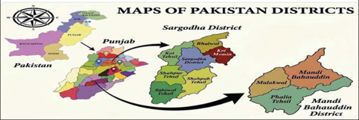
AbstractBackground: Equine piroplasmosis (EP), caused by intraerythrocytic parasites (Babesia caballi and Theileria equi), is an emerging equine tick-borne disease of great veterinary significance to equine health globally. Aim: The present study aimed to investigate the molecular prevalence and associated risk factors of EP in Punjab, Pakistan. Methods: A total of 600 equines (n=600), consisting of horses (n1=200), mules (n2=200), and donkeys (n3=200), were examined year-round in the districts of Sargodha and Mandi Bahauddin, Punjab, Pakistan, using microscopy, enzyme-linked immunosorbent assay (ELISA), and polymerase chain reaction (PCR). Results: Overall PCR-confirmed positivity % was 45.0% (270/600), while ELISA and microscopy detected 56% (336/600) and 33.0% (198/600) positivity, respectively. There was no noticeable difference in positivity % of PCR between equine species (p > 0.05). Analysis of district-level data showed significantly higher PCR positivity in Sargodha than in Mandi Bahauddin (p=0.049). Seasonal variation was significant (p < 0.001), with PCR positivity highest in spring (62.8%) and lowest in winter (20.0%), whereas ELISA seropositivity peaked during summer (65.8%) and was lowest during winter (17.3%). Univariate analysis revealed significant differences in PCR positivity across gender, age group, gelding status, and stress (p < 0.05). Multivariable logistic regression identified stress as a consistent independent predictor of PCR-confirmed infection across all species, while seasonal effects remained significant in several species-specific models (p < 0.05). Conclusion: The current study demonstrated that stress and seasonal factors play an important role in the epidemiology of EP. PCR, ELISA, and microscopy were useful diagnostic tools; however, PCR remained the most definitive method for EP detection. Keywords: Equine piroplasmosis, PCR, Prevalence, Risk factors. IntroductionEquines play an important role in equestrian sports and agricultural activities in Pakistan (Mahmood et al., 2024a; Rehan et al., 2025). Equine piroplasmosis (EP) is a tick-borne hemoprotozoan infection that affects representatives of the genus Equus, which includes horses, donkeys, and mules (Tirosh-Levy et al., 2020). Theileria equi and Babesia caballi are intraerythrocytic apicomplexan parasites that cause this disease and are transmitted mainly by ixodid ticks of the genera Dermacentor, Hyalomma, and Rhipicephalus (Mandal, 2025; İnci et al., 2026). Theileria equi is believed to be more pathogenic and widespread in geographical location than B. caballi, especially in endemic areas (Dorrego et al., 2023; Fernandes et al., 2025). Infection is characterized by a wide clinical spectrum, ranging from acute febrile illnesses to chronic or subclinical infection. The clinical features of EP include fever, anemia, icterus, anorexia, lethargy, and poor performance (Mendoza et al., 2024; Mohd Rajdi et al., 2025). Cases that are severe can lead to hemolysis and systemic complications (Obeagu, 2024). Recovered animals are often persistent reservoirs of infection, and this is a source of tick-transmission in endemic environments (Rendle et al., 2024). The EP is common in Asia, Africa, and South and Central America (Mlangeni, 2016; Zhao et al., 2020; Raza et al., 2024). Conversely, some nations have eliminated the disease due to the management of its entry into the country (Gupta et al., 2026). In regions with high endemicity, the nature of climatic conditions, the ecology of the vectors, and the practice of managing the tick population will contribute to the transmission dynamics (Zubair et al., 2024). EP is not well known in South Asia, especially in Pakistan, although ecological factors are favorable to the tick survival and to large-scale equine rearing. Pakistan has a prolonged subtropical summer season with favorable warm and humid conditions (Mahmood et al., 2024b) that support tick survival and proliferation, potentially increasing the transmission risk of EP in equine populations. It is reported to be endemic to a significant degree, though existing data are geographically limited and often based on traditional diagnostic methods (Bakht and Sabir, 2025). Several methods are used to diagnose EP; however, it is difficult to make an accurate diagnosis because parasitemia fluctuates and clinical presentation varies. The microscopic detection of protozoa in Giemsa-stained blood smears serves as a confirmatory test only during the acute phase of disease but is limited in chronic infections (Couturier and Theel, 2024; Leow et al., 2025) and is impractical for large-scale screening. Likewise, enzyme-linked immunosorbent assay (ELISA) is recommended as the preferred diagnostic method during the latent stages of disease due to its high sensitivity, but it fails to distinguish between past and active infection in endemic areas (Chidzwondo and Mutapi, 2024; Freimane et al., 2025). Thus, polymerase chain reaction (PCR) has greater sensitivity and specificity, can detect low parasitemia, and can differentiate between species (Facile et al., 2025; Wang et al., 2025). Equine traits and stable management affect the virulence of infection (Jafari et al., 2026). This study was designed to establish EP molecular prevalence in Punjab, Pakistan, and to compare diagnostic modalities and identify factors that are associated with PCR-confirmed infection. Materials and MethodsStudy designStudy area and durationThis study was conducted between April 2024 and April 2025 in the two districts, Sargodha (32.08 °N, 72.67 °E) and Mandi Bahauddin (32.58 °N, 73.49 °E), located in the subtropical semi-arid climatic zone of Punjab, Pakistan (Fig. 1).
Fig. 1. Graphical representation of the study districts (Sargodha and Mandi Bahauddin). Study animals and sample sizeA total sample of 600 equines (n=600) was randomly sampled, including horses (n1=200), mules (n2=200), and donkeys (n3=200), from government and private studs (breeding/non-breeding), veterinary hospitals, and field areas of Sargodha and Mandi Bahauddin districts of Punjab. An equal number of samples were collected from each district (n=300). Demographic characteristicsThe study included equal representation of horses, donkeys, and mules, with balanced sex distribution (50% males; 50% females). Age categories were uniformly represented. Body condition scores ranged from poor to good. Questionnaire and epidemiological dataA structured and pre-tested questionnaire was used to collect data on host characteristics, breed, sex, age, management practices, veterinary care, health status, and tick exposure. Geographic coordinates of sampling sites were recorded and mapped using Geographic Information Systemtools. Inclusion and exclusion criteriaEquines from different management systems and clinical backgrounds were included in the study. Animals with a history of tick infestation, fever (>104°F), anemia, lymphadenitis, excessive weight loss, spontaneous bleeding disorders, and without anti-piroplasm treatment within the previous month were preferentially sampled. Equines affected by other major infectious or non-infectious diseases were excluded. Sampling for prevalence estimation was performed irrespective of clinical status. Stress assessment criteriaStress status was assessed through owner interviews, management evaluation, and clinical examination. Animals showing poor body condition, excessive workload, transportation stress, overcrowding, heat exposure, nutritional deficiencies, physical exhaustion, or weakness were categorized as stressed. Animals not exhibiting these conditions were categorized as non-stressed (Hockenhull and Whay, 2014; Baxley et al., 2024). Sample collectionApproximately 5 ml of jugular blood was aseptically collected into EDTA vacutainer tubes and transported to the laboratory using ice boxes under proper cold conditions. Thin blood smears were prepared immediately, while the remaining samples were stored at −20°C until DNA extraction. Verbal informed consent was obtained from animal owners prior to sample collection. Diagnostic methodsMicroscopyThin blood smears were air-dried, methanol-fixed, and stained with 10% Giemsa for 30 minutes. Slides were examined under 100× oil immersion microscopy. Identification of B. caballi was based on paired pyriform merozoites, whereas tetrad (Maltese cross) formations were considered characteristic of T. equi (Wise et al., 2013). Serological testing (competitive ELISA)Commercial competitive ELISA kits for the detection of antibodies against T. equi and B. caballi (Biostone Animal Health LLC, USA) were used according to the manufacturer’s instructions and previously described methodology (Raza et al., 2024). Optical density (OD) values were measured at 450 nm using a microplate ELISA reader, and percent positivity (PP) was calculated as follows:
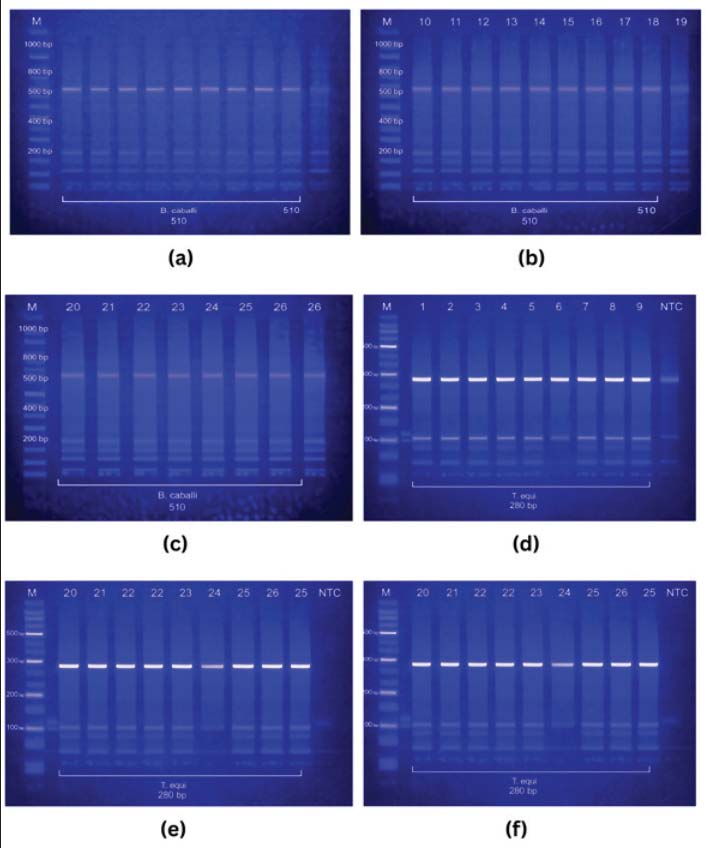
Samples with PP values ≥60% were considered positive, whereas samples with PP values <60% were considered negative according to the kit guidelines. DNA extractionGenomic DNA was extracted from EDTA blood using commercial extraction kits (GeneAll® Exgene™ and Zokeyo Genomic DNA Kit) according to manufacturer protocols. DNA quantification and purity analysisDNA concentration and purity were assessed using a NanoDrop spectrophotometer, and the samples were stored at −20°C until analysis (de Souza Ferreira et al.). On average, each sample produced about 40 ng/μl of DNA, with purity ratios ranging from 1.80 to 1.90, indicating that the samples contain sufficient DNA for PCR amplification. Polymerase chain reactionA multiplex PCR assay targeting the conserved 18S rRNA gene was used to detect T. equi and B. caballi simultaneously (Bhoora et al., 2009). BeqF1/BeqR1 primers amplified a ~280 bp fragment of T. equi, whereas B. caballi-specific primers amplified a ~510 bp fragment (Sumbria et al., 2015). The sequences of the primers were as described under: Theileria equiForward: 5′-TTCGTTGACTGCGCTTGGCG-3′ Reverse: 5′-CTAAGAAGCGGAAATGAAA-3′ (~280 bp) Babesia caballi Forward: 5′-GAGGGACTTTGGGTCATAA-3′ Reverse: 5′-GGTACTCGATCGGTAGGA-3′ (~510 bp) Working primers were maintained at 4°C, while stock solutions were kept at −20°C. PCR was performed in a 25 μl reaction containing 2× Multiplex PCR Master Mix (Zokeyo Biotech), 0.25 μM of each primer, and 30–100 ng genomic DNA, with positive, negative, and internal amplification controls included in each run (Kanavedee and Mahamooth, 2023). Thermocycling conditions consisted of 95°C for 3 minutes; 35 cycles of 95°C for 15 seconds, 59°C for 30 seconds, and 72°C for 30 seconds; followed by 72°C for 5 minutes (Nasoohian et al., 2025). Amplicons were resolved on 2% agarose gel and visualized under UV illumination. Samples yielding ~280 and ~510 bp products were considered positive for T. equi and B. caballi, respectively, while simultaneous amplification indicated mixed infection (Mekata et al., 2025). Statistical analysisData were analyzed using IBM SPSS Statistics Version 26.0. Associations between diagnostic positivity and explanatory variables were evaluated using chi-square tests. Variables with p < 0.05 were included in multivariable binary logistic regression models to estimate adjusted odds ratios and 95% confidence intervals (CIs). PCR-confirmed positivity was used as the primary outcome variable for risk-factor analysis. Agreement among PCR, ELISA, and microscopy was assessed using Cohen's kappa statistic (Landis and Koch 1977). Statistical significance was set at p < 0.05. Ethical approvalThe study was conducted in accordance with ARRIVE guidelines (Percie du Sert et al., 2020) and approved by the Institutional Animal Ethics Review Committee, University of Veterinary and Animal Sciences, Lahore (Approval No. 0104; April 2024). ResultsMolecular setection of EPRepresentative PCR amplification products are presented in Figure 2. Positive amplification of the 18S rRNA gene produced fragments of approximately 280 bp for T. equi and 510 bp for B. caballi. Samples showing both bands were classified as mixed infections. The amplification profiles confirmed the successful detection of both piroplasm species in equine blood samples.
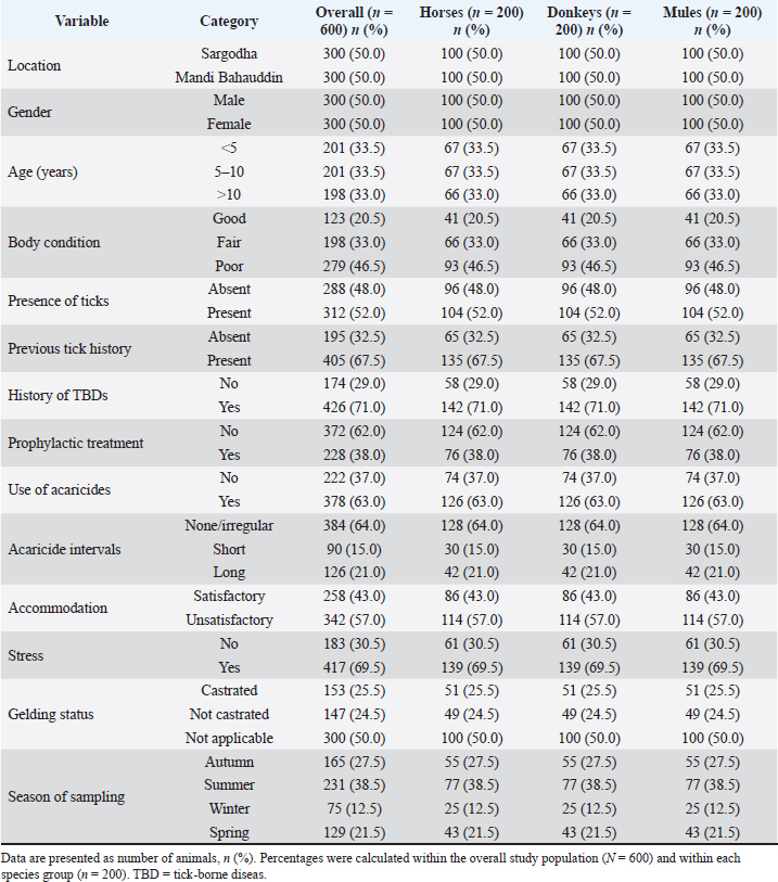
Fig. 2. Multiplex PCR amplification of Babesia caballi (510 bp) and Theileria equi (280 bp) 18S rRNA genes fragments in equine blood samples. M=DNA ladder; NTC=negative control. Demographic characteristicsThe baseline demographic and management characteristics of the sampled equines are presented in Table 1, both overall and stratified by species. Table 1. Baseline demographic and management characteristics of sampled equines (overall and stratified by species).
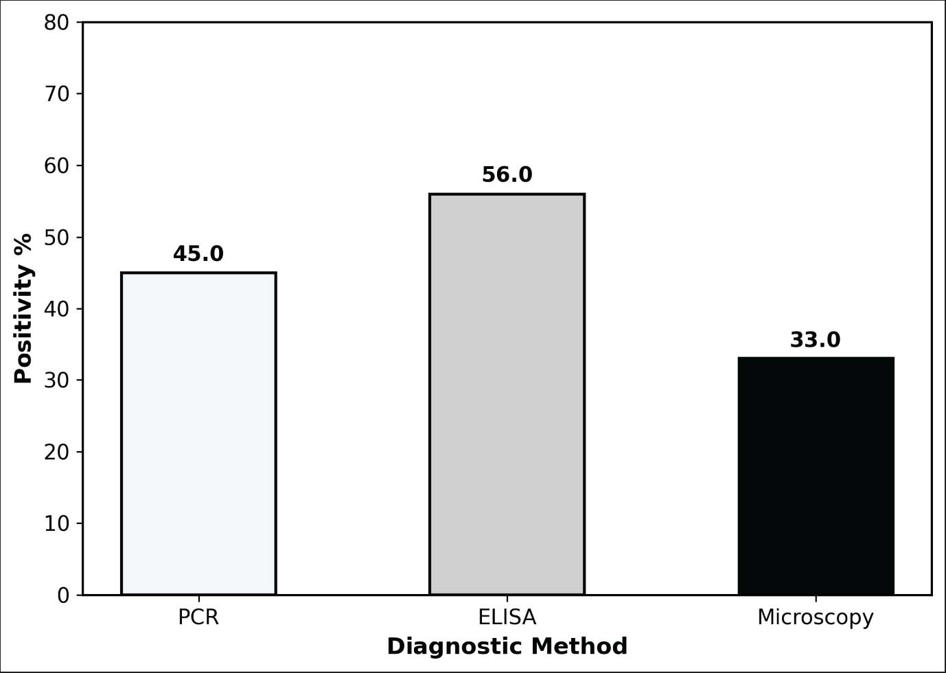
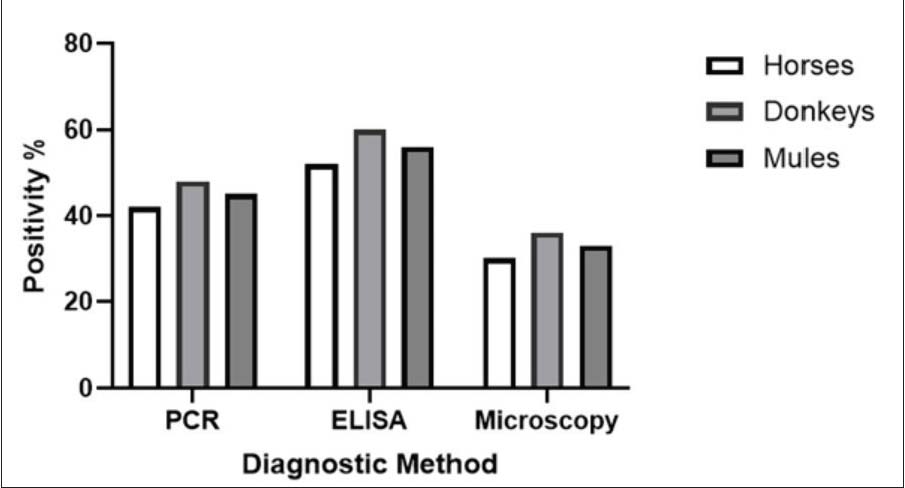
Overall and species-wise positivity of EP by siagnostic methodsOverall positivity for EP varied by diagnostic method, with ELISA demonstrating the highest detection rate (56.0%; 336/600), followed by PCR (45.0%; 270/600) and microscopy (33.0%; 198/600). Species-wise PCR positivity was 42.0% (84/200) in horses, 48.0% (96/200) in donkeys, and 45.0% (90/200) in mules, with no statistically significant difference among species (p=0.38). Numerically higher positivity was consistently observed in donkeys across all diagnostic methods. Detailed diagnostic findings are presented in Table 2. Table 2. Detection of equine piroplasmosis by diagnostic methods overall and stratified by species.
Overall detection rates of EP by PCR, ELISA, and microscopy in sampled equines are shown in Figure 3.
Fig. 3. Overall detection rates of equine piroplasmosis by PCR, ELISA, and microscopy. Species-wise detection rates of EP by PCR, ELISA, and microscopy in horses, donkeys, and mules are shown in Figure 4.
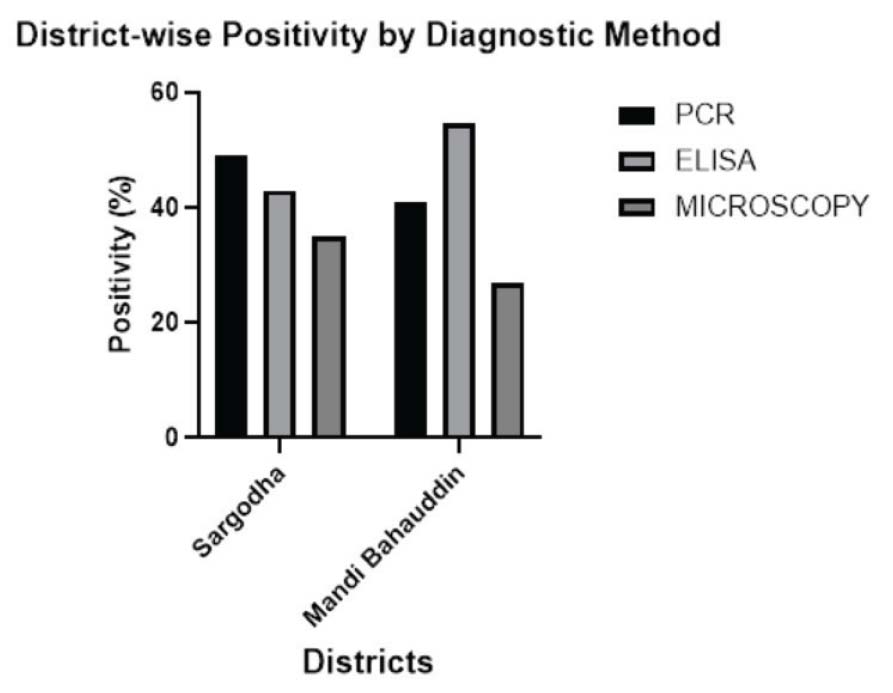
Fig. 4. Species-wise detection rates of equine piroplasmosis by PCR, ELISA, and microscopy in horses, donkeys, and mules. Spatial sistribution of EP by diagnostic methodsSpatial analysis revealed significant variation in diagnostic positivity between Sargodha and Mandi Bahauddin districts. PCR-confirmed positivity was significantly higher in District Sargodha (49.0%; 147/300) compared with Mandi Bahauddin (41.0%; 123/300) (p=0.049). Similarly, microscopy positivity was significantly higher in District Sargodha (35.0%; 105/300) than in Mandi Bahauddin (27.0%; 81/300) (p=0.034). In contrast, ELISA seropositivity was significantly higher in Mandi Bahauddin (54.7%; 164/300) compared with Sargodha (43.0%; 129/300) (p=0.004). Species-wise spatial analysis demonstrated similar trends across horses, donkeys, and mules; however, most species-specific differences were not statistically significant (p > 0.05). A significant location-specific difference in ELISA positivity was observed only in horses (p=0.016). District-wise positivity of EP detected by PCR, ELISA, and microscopy in Sargodha and Mandi Bahauddin districts is shown in Figure 5.
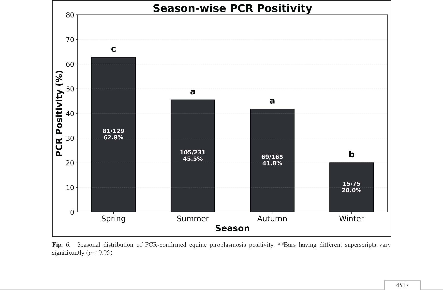
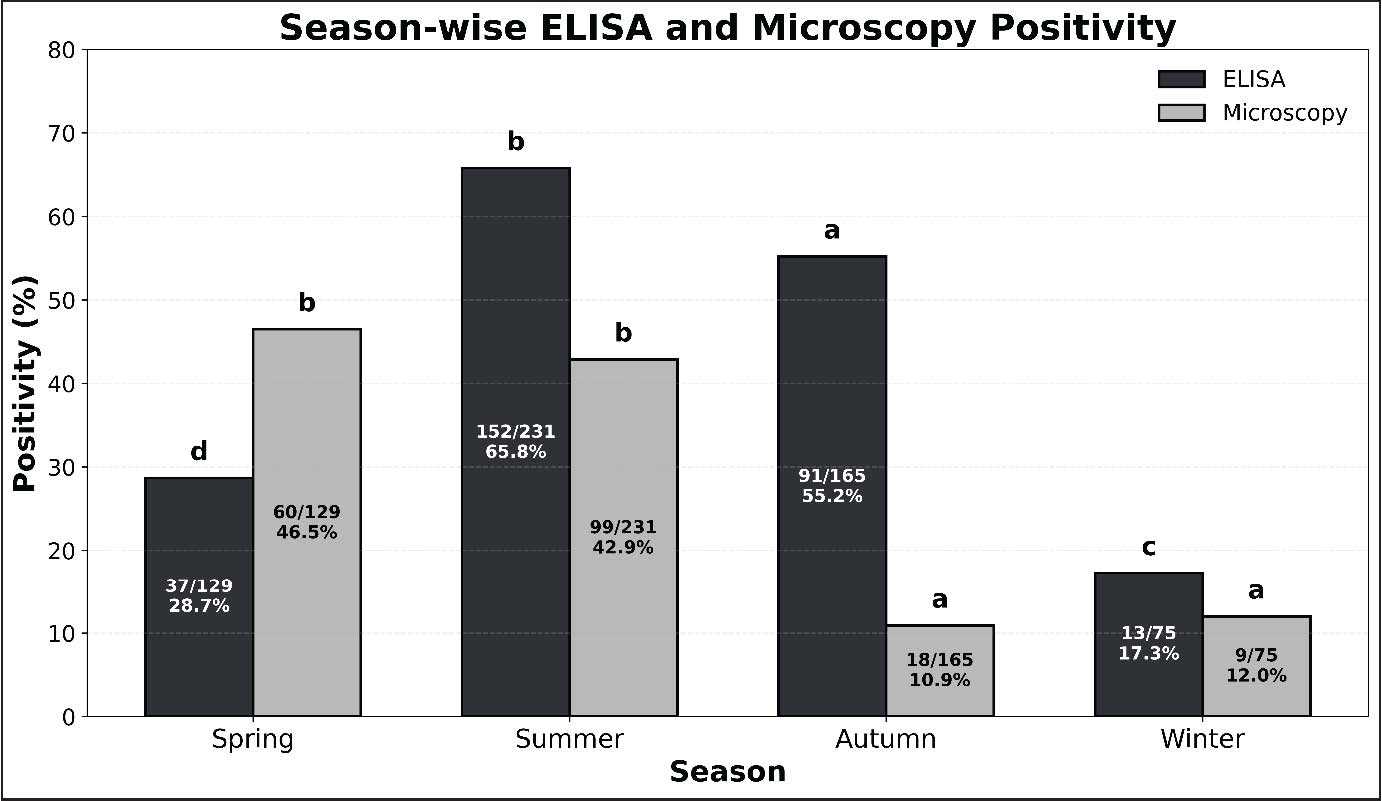
Fig. 5. District-wise positivity of equine piroplasmosis detected by PCR, ELISA, and microscopy in Sargodha and Mandi Bahauddin districts. Seasonal distribution of infectionAll diagnostic methods demonstrated significant seasonal variation in EP positivity (p < 0.001). PCR-confirmed positivity was highest during spring (62.8%; 81/129) and lowest during winter (20.0%; 15/75), while summer (45.5%; 105/231) and autumn (41.8%; 69/165) exhibited intermediate positivity rates. Similarly, ELISA seropositivity peaked during summer (65.8%; 152/231), followed by autumn (55.2%; 91/165), and was lowest during winter (17.3%; 13/75). Microscopy positivity was highest during spring (46.5%; 60/129) and summer (42.9%; 99/231), whereas substantially lower positivity was recorded during autumn (10.9%; 18/165) and winter (12.0%; 9/75). The association between season and positivity was highly significant for all diagnostic methods (p < 0.001) (Figs. 6 and 7).
Fig. 6. Seasonal distribution of PCR-confirmed equine piroplasmosis positivity. a−dBars having different superscripts vary significantly (p < 0.05).
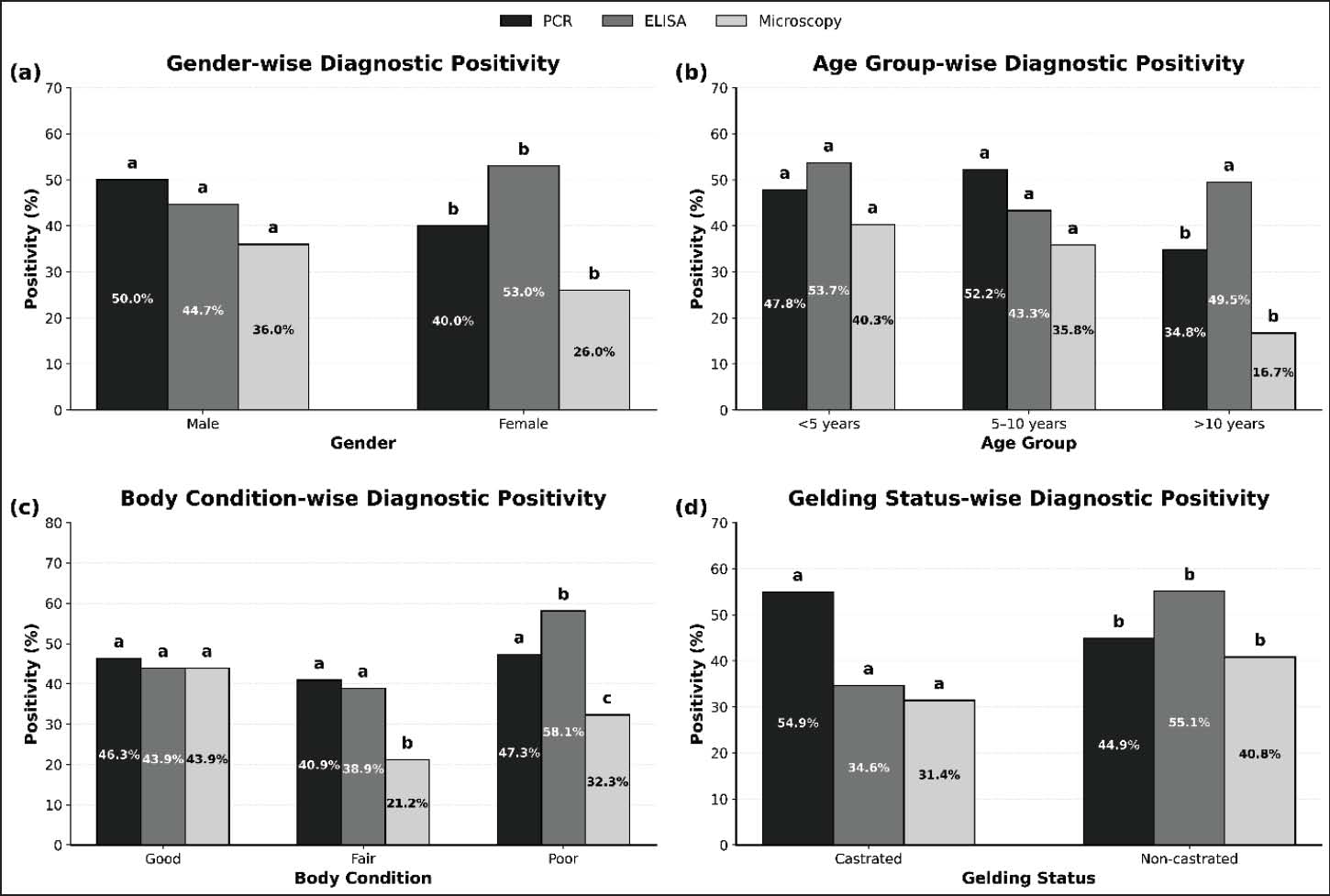
Fig. 7. Seasonal variation in ELISA and microscopy positivity of equine piroplasmosis. a−dBars having different superscripts vary significantly (p < 0.05). Univariable association of host factors with diagnostic positivityGender, age group, body condition, and gelding status were significantly associated with diagnostic positivity. PCR and microscopy positivity were significantly higher in males than females (p=0.014 and p= 0.008, respectively), whereas ELISA seropositivity was significantly higher in females (p=0.041). Age group was significantly associated with PCR and microscopy positivity (p < 0.001), whereas no significant association was observed between age and ELISA positivity (p > 0.05). Body condition was significantly associated with microscopy and ELISA positivity (p < 0.001), whereas PCR positivity did not differ significantly among body condition categories. Gelding status showed a significant association across all diagnostic methods. Non-castrated equines demonstrated significantly higher positivity compared with castrated animals in PCR, ELISA, and microscopy (p ≤ 0.011). Diagnostic positivity of EP according to gender, age group, body condition, and gelding status is shown in Figure 8.
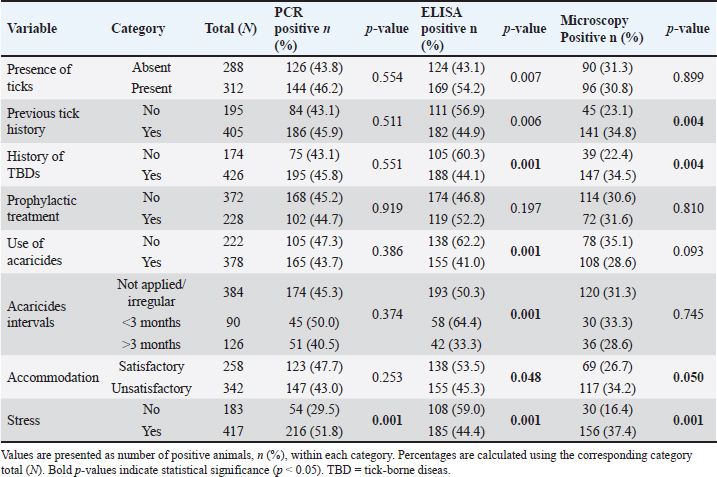
Fig. 8. Diagnostic positivity of equine piroplasmosis according to (a) gender, (b) age group, (c) body condition, and (d) gelding status. a−dBars having different superscripts vary significantly (p < 0.05). Univariable association of tick exposure and management factors with diagnostic outcomesTick exposure and management factors showed variable associations with diagnostic positivity (Table 3). Presence of ticks at sampling was significantly associated with ELISA positivity (169/312, 54.2%) compared with tick-free animals (124/288, 43.1%; p=0.007), whereas no significant association was observed with PCR or microscopy (p > 0.05). Previous tick history and history of tick-borne diseases were significantly associated with ELISA and microscopy positivity (p ≤ 0.006), but not with PCR positivity. Use of acaricides and the acaricide application interval were significantly associated with ELISA positivity (p=0.001), while prophylactic treatment showed no significant association with any diagnostic method (p > 0.05). Accommodation status was significantly associated with ELISA positivity (p=0.048) and showed a borderline association with microscopy positivity (p=0.050) (Table 3). Table 3. Association of tick exposure, management factors, and stress exposure with diagnostic positivity of equine piroplasmosis (N=600)
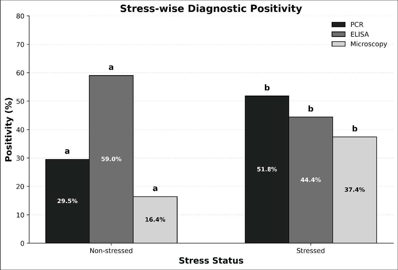
Stress demonstrated a strong association across all diagnostic methods (p=0.001) (Table 3; Fig. 9). PCR positivity was significantly higher in stressed equines (216/417, 51.8%) than in non-stressed equines (54/183, 29.5%). Similarly, microscopy positivity was higher among stressed animals (156/417, 37.4%) than among non-stressed animals (30/183, 16.4%). In contrast, ELISA seropositivity was higher in non-stressed equines (108/183, 59.0%) than in stressed equines (185/417, 44.4%). Similar trends were observed in species-stratified analyses.
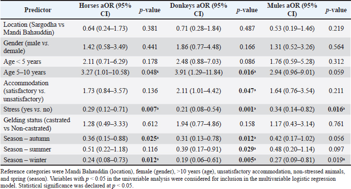
Fig. 9. Effect of stress on diagnostic positivity of equine piroplasmosis. a−dBars having different superscripts vary significantly (p < 0.05). Multivariable logistic regression for predictors of PCR positivity (species-wise models)Multivariable binary logistic regression models were constructed separately for horses, donkeys, and mules to identify independent predictors of PCR-confirmed EP. The dependent variable was PCR positivity, while location, gender, age group, accommodation status, stress, gelding status, and season of sampling were included as explanatory variables. Across the species-specific multivariable logistic regression models, stress remained a significant independent predictor of PCR-confirmed EP in horses, donkeys, and mules (p ≤ 0.016). Seasonal effects were also observed, particularly in relation to autumn and winter, which were associated with significantly lower odds of PCR positivity compared with spring in several species-specific models. Age group (5–10 years) was significantly associated with PCR positivity in horses and donkeys, while satisfactory accommodation was associated with increased odds of PCR positivity in donkeys. No significant associations were observed for location, gender, or gelding status after adjustment (Table 4). Table 4. Species-wise multivariable logistic regression analysis for predictors of PCR positivity
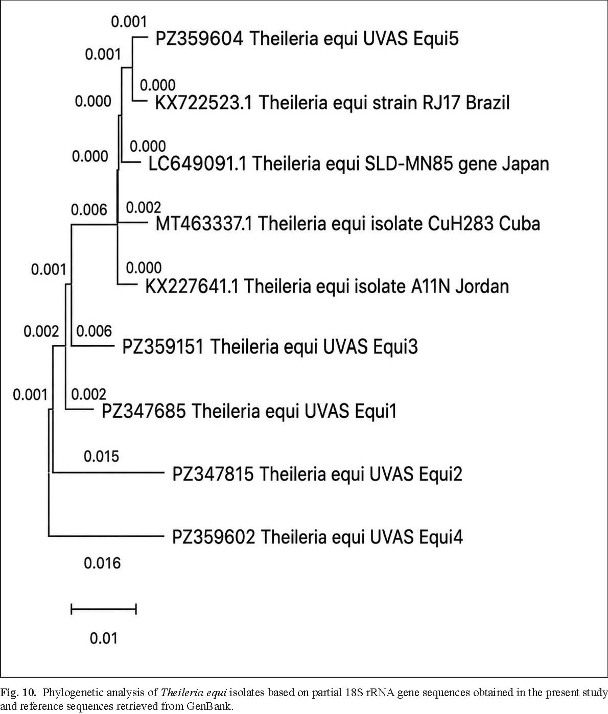
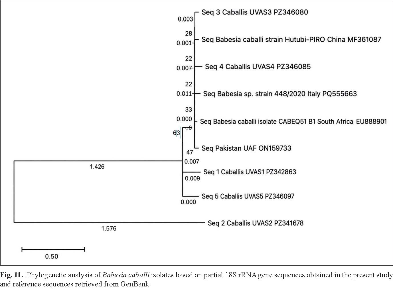
Agreement Between Diagnostic MethodsAgreement between PCR and ELISA was low and statistically non-significant in horses, donkeys, and mules (p > 0.05). In contrast, PCR and microscopy demonstrated significant agreement across all species (p < 0.001). Detailed species-wise agreement statistics are presented in Table 3. Molecular characterization and phylogenetic analysisRepresentative PCR-positive amplicons were subjected to sequencing and molecular characterization. Phylogenetic analysis demonstrated close genetic similarity between local isolates and previously reported global strains of T. equi and B. caballi, indicating relative genetic conservation among circulating isolates. Phylogenetic analysis of T. equi isolates based on partial 18S rRNA gene sequences obtained in the present study and reference sequences retrieved from GenBank is shown in Fig. 10.
Fig. 10. Phylogenetic analysis of Theileria equi isolates based on partial 18S rRNA gene sequences obtained in the present study and reference sequences retrieved from GenBank. Phylogenetic analysis of B. caballi isolates based on partial 18S rRNA gene sequences obtained in the present study and reference sequences retrieved from GenBank is shown in Fig. 11.
Fig. 11. Phylogenetic analysis of Babesia caballi isolates based on partial 18S rRNA gene sequences obtained in the present study and reference sequences retrieved from GenBank. DiscussionThe present study demonstrated high PCR-confirmed positivity (45%) and ELISA seropositivity (56%) for EP in equines from Punjab, Pakistan, indicating the endemic circulation of T. equi and B. caballi in the region. Similar prevalence patterns have been reported from other tropical and subtropical countries where climatic conditions favor tick survival and transmission (Axt et al., 2024; Fernandes et al., 2025; Gupta et al., 2026). These findings support the recognition of EP as an important tick-borne disease of equines in warm climatic regions Higher positivity detected by PCR and ELISA compared with microscopy in the present study reflects the improved ability of molecular and serological methods to detect low parasitemia and subclinical infections. Similar observations have been reported previously, particularly in carrier equines where parasitemia fluctuates below microscopic detection limits (Mendoza et al., 2024; Wang et al., 2025). Seropositive but PCR-negative equines may represent chronically exposed carrier animals with intermittent parasitemia, which can contribute to silent transmission within equine populations. Therefore, combined molecular and serological approaches remain important for accurate diagnosis and surveillance of EP (Tirosh-Levy et al., 2020; Giubega et al., 2022). The World Organization for Animal Health also recommends molecular and serological assays for EP surveillance because they can detect carrier animals (Pitt and Gunn, 2024). No significant species-specific differences in PCR-confirmed positivity were observed among horses, donkeys, and mules, suggesting comparable exposure risk under similar environmental and management conditions. Previous studies have similarly reported comparable EP prevalence among equids maintained under shared ecological settings (Gupta et al., 2026). The slightly higher positivity observed in donkeys and mules may be associated with greater outdoor exposure, increased tick contact, and management-related factors, particularly in working equids. Similar trends have also been reported in Pakistan (Raza et al., 2024). Spatial variation observed between Sargodha and Mandi Bahauddin may reflect regional differences in environmental conditions, tick abundance, vegetation, humidity, and animal management practices. Previous studies have demonstrated that climatic and ecological variations significantly influence tick distribution and pathogen transmission dynamics (Basit et al., 2020; İnci et al., 2026). Differences between PCR and ELISA positivity across districts may further indicate variation between active infection and cumulative exposure, as antibodies may persist even after clearance of active parasitemia (Mahdy et al., 2023). Marked seasonal variation observed in the present study highlights the important role of climatic conditions in EP transmission. PCR-confirmed positivity was highest during spring (62.8%), whereas ELISA seropositivity peaked during summer (65.8%). The increased positivity during spring and summer may be attributed to favorable environmental conditions that support tick survival, reproduction, and pathogen transmission (Makwarela et al., 2025; Mollong et al., 2025). Similar seasonal peaks of EP positivity have also been reported from other endemic regions (Wise et al., 2013). Several host and management-related factors were associated with EP positivity. Multivariable logistic regression identified stress as the most consistent independent predictor of PCR-confirmed infection across horses, donkeys, and mules. In addition, seasonal effects, age group, and accommodation status influenced infection risk in specific species. Stress may reflect compromised welfare, excessive workload, transportation stress, nutritional deficiencies, and poor management conditions that predispose equines to infection. Similar observations have been reported in previous studies evaluating stress-related susceptibility to hemoprotozoan diseases (Mendoza et al., 2024). Likewise, previous tick exposure and poor acaricide practices were associated with increased seropositivity, emphasizing the importance of effective tick control programs. Integrated tick management strategies are considered essential for reducing infection pressure and improving equine health in endemic regions (Makwarela et al., 2025). Overall, the findings indicate that EP remains endemic in Punjab, Pakistan, and is strongly influenced by seasonal, environmental, and management-related factors. Improved tick control practices, enhanced animal welfare, and regular molecular surveillance may help reduce disease burden and minimize economic losses in working equids. LimitationsAlthough EP is a tick-borne disease, tick species identification and characterization were not included in the present study design. However, several previous studies have reported the distribution and ecology of tick species in Pakistan (Iqbal et al., 2013; Ghafar et al., 2020; Rahman et al., 2022). Additionally, only a limited number of representative PCR-positive samples were sequenced and analyzed phylogenetically. Further investigation of seropositive but PCR-negative equines may provide a better understanding of carrier status and infection dynamics. RecommendationsRegular tick control, proper use of acaricides, improved housing, reduced stress, and better management practices should be encouraged to minimize EP in endemic areas. Routine screening and increased awareness among equine owners and caretakers may further support early detection and effective disease control. ConclusionThe present study demonstrated substantial molecular and serological positivity of EP among horses, donkeys, and mules in Punjab, Pakistan. PCR-confirmed infection was significantly associated with season and stress, indicating their important role in disease occurrence. Differences among PCR, ELISA, and microscopy further highlighted the value of molecular and serological tools for accurate detection and surveillance of EP under field conditions. AcknowledgmentsThe authors express their sincere gratitude to the Department of Veterinary Medicine, University of Veterinary and Animal Sciences, Lahore, Pakistan, and Dr. Khalid Mahmood Rana (RV&FC) for their valuable support during this study. Conflict of InterestThe authors disclose that there is no conflict of interest in this research study. FundingNo funds were received during the course of this research study Authors' contributionsShoaib Ahmad and Aneela Z. Durrani conceptualized and designed the study. Shoaib Ahmad performed field sampling, laboratory investigations, and data analysis. Aneela Z. Durrani and Jawaria A. Khan supervised the study and contributed to manuscript preparation and revision. Furqan Awan and Muhammad U. Z. Khan contributed to data collection, statistical analysis, and interpretation of results. All authors reviewed and approved the final manuscript. Data availabilityThe datasets generated and analyzed during the current study are available from the corresponding author on reasonable request. ReferencesAxt, C.W., Springer, A., Strube, C., Jung, C., Naucke, T.J., Müller, E. and Schäfer, I. 2024. Molecular and serological detection of vector-borne pathogens responsible for equine piroplasmosis in Europe between 2008 and 2021. Microorganisms 12(4), 816. Bakht, P. and Sabir, M.J. 2025. Pakistan Journal of Veterinary and Animal Research. Pak. J. Vet. Anim. Res. 1(1), 6–12. Basit, A., Ali, M., Hussain, G., Irfan, S., Saqib, M., Iftikhar, A., Mustafa, I., Mukhtar, I. and Anwar, H. 2020. Effect of equine piroplasmosis on hematological and oxidative stress biomarkers in relation to different seasons in District Sargodha, Pakistan. Pak. Vet. J. 40(1), 43–48. Baxley, B.H., Rodriguez-Mori, H. and Anderson, N.C. 2024. Welfare and stress of horses used for Equine-assisted services: a systematic review. Appl. Anim. Behaviour. Sci. 280, 106424. Bhoora, R., Franssen, L., Oosthuizen, M.C., Guthrie, A.J., Zweygarth, E., Penzhorn, B.L., Jongejan, F. and Collins, N.E. 2009. Sequence heterogeneity in the 18S rRNA gene within Theileria equi and Babesia caballi from horses in South Africa. Vet. Parasitology. 159(2), 112–120. Chidzwondo, F. and Mutapi, F. 2024. Challenge of diagnosing acute infections in poor resource settings in Africa. AAS. Open. Res. 4, 28. Couturier, M.R. and Theel, E.S. 2024. Immunological and molecular approaches for the diagnosis of parasitic infections. Manual Mol. Clin. Lab. Immunol. 1, 470–488; doi: 10.1002/9781683674023.ch47 de Souza Ferreira, M.R., Galvão, A.P.O., Valença, M.M. and Martins, D.B.G. 2022. Storage and purification adaptations for the isolation of total RNA from the dura mater. Arquivos de Neuro-psiquiatria, 80(12), 1213–1219; doi: 10.1055/s-0042-1758865 Dorrego, A., Camino, E., Gago, P., Buendia-Andres, A., Acurio, K., Gonzalez, S., De Juan, L. and Cruz-Lopez, F. 2023. Haemato-biochemical characterization of equine piroplasmosis asymptomatic carriers and seropositive, real-time PCR negative horses. Vet. Parasitology 323, 110046. Facile, V., Magliocca, M., Dini, F.M., Imposimato, I., Mariella, J., Freccero, F., Urbani, L., Rinnovati, R., Sel, E. and Gallina, L. 2025. Molecular diagnosis and identification of equine piroplasms: challenges and insights from a study in Northern Italy. Animals 15(3), 437. Fernandes, T.A., Paulino, P.G., Dos Santos Juliano, D., Rabello, C.A., De Oliveira, N.V.B., De Souza Santana, M., Peckle, M., Massard, C.L., Da Costa Angelo, I., Jacob, J.C.F. and Santos, H.A. 2025. Epidemiology and genetic diversity of Theileria equi and Babesia caballi in draft horses in the Distrito Federal, Brazil. Trop. Anim. Health Prod. 57(2), 72. Freimane, Z., Dobler, G., Karelis, G., Chitimia-Dobler, L., Savicka, O. and Zavadska, D. 2025. Seroprevalence of Tick-Borne Encephalitis Virus in Latvia Using Standard and Subtype-Specific NS1-Based ELISA Assays. Pathogens 14(11), 1115. Ghafar, A., Abbas, T., Rehman, A., Sandhu, Z.U.D., Cabezas-Cruz, A. and Jabbar, A. 2020. Systematic review of ticks and tick-borne pathogens of small ruminants in Pakistan. Pathogens 9(11), 937. Giubega, S., Dreghiciu, C., Ilie, M.S. and Darabus, G. 2022. Diagnostic methods used for the detection of Theileria equi: review of the last decade. Cluj. Vet. J. 27(2), 16–24. Gupta, K.K., Gupta, N., Kumar, S., Srivastava, M. and Kumar, P. 2026. Equine piroplasmosis: an emerging tick-borne threat to equine health. Trop. Anim. Health Prod. 58(1), 29. Hockenhull, J. and Whay, H.R. 2014. A review of approaches to assessing equine welfare. Equine Vet. Educ. 26(3), 159–166. İnci, A., Özdarendeli, A., Yıldırım, A., Sohel, M.H., Özübek, S., Orkun, O., Yukarı, B.A., Kılıç, A.U., Vatansever, Z. and Düzlü, O. 2026. A Compendium Review of the Global Epidemiology of Ticks and Tick-borne Diseases: regional Insights from Türkiye. Turkiye. Parazitolojii. Dergisi. 49(Suppl 1), 1–66. Iqbal, A., Sajid, M.S., Khan, M.N. and Khan, M.K. 2013. Frequency distribution of hard ticks (Acari: ixodidae) infesting bubaline population of district Toba Tek Singh, Punjab, Pakistan. Parasitology. Res. 112(2), 535–541. Jafari, H., Abebe, B.K., Cong, L., Ahmed, Z., Zhaofei, W., Sun, M., Muhatai, G., Chuzhao, L. and Dang, R. 2026. Genomic insights into the adaptive traits and stress resistance in modern horses. Stress. Biol. 6(1), 5. Kanavedee, R. and Mahamooth, T.N. 2023. Automated high-throughput extraction system of sRNA and high-resolution melting quantitative PCR (HRM-qPCR) analysis for viroid detection. Heliyon 9(12); doi: 10.1016/j.heliyon.2023.e22613 Landis, J.R. and Koch, G.G. 1977. An application of hierarchical kappa-type statistics in the assessment of majority agreement among multiple observers. Biometrics 363–374; doi: 10.2307/2529786 Leow, E.H., Ganesan, I., Chong, S.L., Yap, C.J.Y., Chao, S.M., Wang, F. and Ng, Y.H. 2025. Adenine phosphoribosyltransferase (APRT) deficiency: an increasingly recognized disease. Int. Urology Nephrology 57(8), 2519–2529. Mahdy, O.A., Nassar, A.M., Elsawy, B.S.M., Alzan, H.F., Kandil, O.M., Mahmoud, M.S. and Suarez, C.E. 2023. Cross-sectional analysis of Piroplasma species-infecting camel (Camelus dromedaries) in Egypt using a multipronged molecular diagnostic approach. Front. Vet. Sci. 10, 1178511. Mahmood, K., Ali Channa, A., Ghafoor, A. and Riaz, A. 2024a. Factors affecting the efficiency of equine embryo transfer (EET) in polo mares under subtropical conditions of Pakistan. PLos One 19(2), e0298066. Mahmood, K., Hassan, M., Channa, A.A., Ghafoor, A. and Riaz, A. 2024b. Comparative analysis of breeding patterns and reproductive efficiency of mares in subtropical conditions of Pakistan. Vet. Med. Sci. 10(5), e1582. Makwarela, T.G., Seoraj-Pillai, N. and Nangammbi, T.C. 2025. Tick control strategies: critical insights into chemical, biological, physical, and integrated approaches for effective hard tick management. Vet. Sci. 12(2), 114. Mandal, S.C. 2025. Veterinary protozoology. In Textbook of veterinary parasitology. Singapore: Springer Nature Singapore, pp: 473–571; doi: 10.1007/978-981-99-2924-5_8 Mekata, H., Yamamoto, M., Matsuu, A., Maeda, K., Isawa, H., Yoshii, K., Umeki, K. and Okabayashi, T. 2025. Development of a Double-Antigen Sandwich ELISA for Oz Virus and a Seroepidemiological Survey in Wild Boars in Miyazaki, Japan. Pathogens 14(12), 1288. Mendoza, F.J., Pérez-Écija, A., Kappmeyer, L.S., Suarez, C.E. and Bastos, R.G. 2024. New insights in the diagnosis and treatment of equine piroplasmosis: pitfalls, idiosyncrasies, and myths. Front. Vet. Sci. 11, 1459989. Mohd Rajdi, N.Z.I., Shaari, R., Mohamad, M.A., Tan, L.P., Reduan, M.F.H., Rahman, M.S.A. and Goni, M.D. 2025. A case report of equine piroplasmosis in a postpartum indigenous pony mare in Malaysia. Adv. Anim. Vet. Sci. 13(8), 1834–1843; 10.17582/journal.aavs/2025/13.8.1834.1843 Mlangeni, M.A. 2016. Molecular epidemiology of dourine, equine piroplasmosis and ehrlichiosis from donkeys and horses in South Africa (Doctoral dissertation, North-West University, South Africa. Mollong, E., Lébri, M., Marie-Magdeleine, C., Lagou, S.M., Naves, M. and Bambou, J.C. 2025. Sustainable management of tick infestations in cattle: a tropical perspective. Parasites. &. Vectors. 18(1), 62. Nasoohian, N., Fallahzadeh, E., Sabour, S., Soleiman-Meigooni, S. and Azimi, T. 2025. Carbapenem resistance in enterotoxigenic Bacteroides fragilis isolated from colonoscopy candidates. AMB Expr. 15, 186; doi: 10.1186/s13568-025-01980-9 Obeagu, E.I. 2024. Oxidative Stress and Hemolysis: implications for Sickle Cell Anemia. Int. J. Curr. Res. Chem. Pharm. Sci. 11(8), 29–37. Percie du Sert N., Hurst, V., Ahluwalia, A., Alam, S., Avey, M.T., Baker, M., Browne, W.J., Clark, A., Cuthill, I.C. and Dirnagl, U. 2020. The ARRIVE guidelines 2.0: updated guidelines for reporting animal research. J. Cerebral. Blood. Flow. &. Metab. 40(9), 1769–1777. Pitt, S.J. and Gunn, A. 2024. The one health concept. Br. J. Biomed. Sci. 81, 12366. Rahman, A., Kashif, M., Nasir, A., Idrees, A., Jamil, M., Ehsan, M. and Sana, M. 2022. A review of tick and tick control strategies in Pakistan. Pak. J. Med. Health. Sci. 16, 652–655. Rajdi, N.Z.I.M., Shaari, R., Mohamad, M.A., Peng, T.L., Reduan, M.F.H., Rahman, M.S.A. and Goni, M.D. 2025. A case report of equine piroplasmosis in a postpartum indigenous pony mare in Malaysia. Adv. Anim. Vet. Sci. 13(8), 1834–1843. Raza, A., Ijaz, M., Mehmood, K., Ahmed, A., Javed, M.U., Anwaar, F., Rasheed, H. and Ghumman, N.Z. 2024. Theileria equi infection in working horses of Pakistan: epidemiology, molecular characterization, and hematobiochemical analysis. J. Parasitology. 110(1), 79–89. Rehan, A., Tunio, M.T., Badar, S.T. and Mahmood, K., 2026. Factors affecting the reproductive efficiency of American Mammoth Jackstock Donkey Mares (Equus asinus) under Subtropical Conditions of Pakistan. Pakistan J. Zool. 58(3), 1221; doi: 10.17582/journal.pjz/20250420180209 Rendle, D., Hughes, K., Bowen, M., Bull, K., Cameron, I., Furtado, T., Peachey, L., Sharpe, L. and Hodgkinson, J. 2024. BEVA primary care clinical guidelines: equine parasite control. Equine. Vet. J. 56(3), 392–423. Sumbria, D., Singla, L.D., Sharma, A., Bal, M.S. and Kumar, S. 2015. Multiplex PCR for detection of Trypanosoma evansi and Theileria equi in equids of Punjab, India. Vet. Parasitol. 211(3-4), 293–299; doi: 10.1016/j.vetpar.2015.05.018 Tirosh-Levy, S., Gottlieb, Y., Fry, L.M., Knowles, D.P. and Steinman, A. 2020. Twenty years of equine piroplasmosis research: global distribution, molecular diagnosis, and phylogeny. Pathogens 9(11), 926. Wang, T., Chen, X., Yan, X., Su, Y., Gao, W., Liu, C. and Wang, W. 2025. Progress in serology and molecular biology of equine parasite diagnosis: sustainable control strategies. Front. Vet. Sci. 12, 1663577. Wise, L.N., Kappmeyer, L.S., Mealey, R.H. and Knowles, D.P. 2013. Review of equine piroplasmosis. J. Vet. Internal Med. 27(6), 1334–1346. Zhao, S., Wang, H., Zhang, S., Xie, S., Li, H., Zhang, X. and Jia, L. 2020. First report of genetic diversity and risk factor analysis of equine piroplasm infection in equids in Jilin, China. Parasites. &. Vectors. 13(1), 459. Zubair, A.S., Mcalpine, L.S. and Gobeske, K.T. 2024. Virology, ecology, epidemiology, pathology, and treatment of eastern equine encephalitis. J. Neurological. Sci. 457, 122886. | ||


| How to Cite this Article |
| Pubmed Style Ahmad S, Durrani AZ, Khan JA, Awan F, Khan MUZ. Prevalence and multivariable risk factor analysis of equine piroplasmosis in Punjab, Pakistan. Open Vet. J.. 2026; 16(7): 4510-4525. doi:10.5455/OVJ.2026.v16.i7.35 Web Style Ahmad S, Durrani AZ, Khan JA, Awan F, Khan MUZ. Prevalence and multivariable risk factor analysis of equine piroplasmosis in Punjab, Pakistan. https://www.openveterinaryjournal.com/?mno=316749 [Access: July 15, 2026]. doi:10.5455/OVJ.2026.v16.i7.35 AMA (American Medical Association) Style Ahmad S, Durrani AZ, Khan JA, Awan F, Khan MUZ. Prevalence and multivariable risk factor analysis of equine piroplasmosis in Punjab, Pakistan. Open Vet. J.. 2026; 16(7): 4510-4525. doi:10.5455/OVJ.2026.v16.i7.35 Vancouver/ICMJE Style Ahmad S, Durrani AZ, Khan JA, Awan F, Khan MUZ. Prevalence and multivariable risk factor analysis of equine piroplasmosis in Punjab, Pakistan. Open Vet. J.. (2026), [cited July 15, 2026]; 16(7): 4510-4525. doi:10.5455/OVJ.2026.v16.i7.35 Harvard Style Ahmad, S., Durrani, . A. Z., Khan, . J. A., Awan, . F. & Khan, . M. U. Z. (2026) Prevalence and multivariable risk factor analysis of equine piroplasmosis in Punjab, Pakistan. Open Vet. J., 16 (7), 4510-4525. doi:10.5455/OVJ.2026.v16.i7.35 Turabian Style Ahmad, Shoaib, Aneela Z. Durrani, Jawaria A. Khan, Furqan Awan, and Muhammad U. Z. Khan. 2026. Prevalence and multivariable risk factor analysis of equine piroplasmosis in Punjab, Pakistan. Open Veterinary Journal, 16 (7), 4510-4525. doi:10.5455/OVJ.2026.v16.i7.35 Chicago Style Ahmad, Shoaib, Aneela Z. Durrani, Jawaria A. Khan, Furqan Awan, and Muhammad U. Z. Khan. "Prevalence and multivariable risk factor analysis of equine piroplasmosis in Punjab, Pakistan." Open Veterinary Journal 16 (2026), 4510-4525. doi:10.5455/OVJ.2026.v16.i7.35 MLA (The Modern Language Association) Style Ahmad, Shoaib, Aneela Z. Durrani, Jawaria A. Khan, Furqan Awan, and Muhammad U. Z. Khan. "Prevalence and multivariable risk factor analysis of equine piroplasmosis in Punjab, Pakistan." Open Veterinary Journal 16.7 (2026), 4510-4525. Print. doi:10.5455/OVJ.2026.v16.i7.35 APA (American Psychological Association) Style Ahmad, S., Durrani, . A. Z., Khan, . J. A., Awan, . F. & Khan, . M. U. Z. (2026) Prevalence and multivariable risk factor analysis of equine piroplasmosis in Punjab, Pakistan. Open Veterinary Journal, 16 (7), 4510-4525. doi:10.5455/OVJ.2026.v16.i7.35 |


