





| Research Article | ||
Open Vet. J.. 2026; 16(7): 4526-4539
Open Veterinary Journal, (2026), Vol. 16(7): 4526-4539 Research Article Protective effects of Rosmarinus officinalis against methotrexate-induced hepatorenal toxicity in Wistar rats: Biochemical, histopathological, and immunohistochemical evaluationLana J. Muhammed* and Nadia A. SalihDepartment of Basic Sciences, College of Veterinary Medicine, University of Sulaimani, Sulaymaniyah, Iraq *Corresponding Author: Lana Jamal. Department of Basic Sciences, College of Veterinary Medicine, University of Sulaimani, Sulaymaniyah, Iraq.Email: Lanaajamall96 [at] gmail.com Submitted: 08/04/2026 Revised: 10/06/2026 Accepted: 20/06/2026 Published: 17/07/2026 © 2025 Open Veterinary Journal
ABSTRACTBackground: Methotrexate (MTX) is one of the most frequently used chemotherapeutic and immunosuppressive agents; however, its clinical use is limited to hepatotoxicity and other organ toxicities associated with oxidative stress and inflammatory responses. Aim: The current study was conducted to evaluate the potential protective effect of the ethanolic extract of Rosmarinus officinalis against MTX-induced hepatorenal injury in Wistar rats. Methods: Hepatorenal toxicity was induced in Wistar rats via a single intraperitoneal administration of MTX (20 mg/kg). Rosemary extract was orally administered at doses of 100 and 200 mg/kg body weight. Liver function biomarkers (alanine aminotransferase, aspartate aminotransferase, and alkaline phosphatase), renal function parameters (urea and creatinine), and hematological indices were determined alongside oxidative stress markers (superoxide dismutase, catalase, glutathione peroxidase, glutathione, and Malondialdehyde) and levels of tumor necrosis factor-alpha (TNF-α). Hematoxylin and eosin staining and immunohistochemical profiling were used for histopathological evaluation. Results: MTX caused a significant increase in liver enzymes, oxidative markers, and TNF-α while lowering antioxidant defenses. Histopathological examination revealed hepatocyte degeneration, vascular congestion, and inflammatory infiltration. Treatment with R. officinalis extract alleviated these changes in a dose-dependent manner, resulting in improved biochemical parameters and reduced tissue injury. Conclusion: Rosmarinus officinalis extract may exert protective effects against MTX-induced hepatorenal injury through antioxidant and anti-inflammatory mechanisms. However, further studies are required to confirm these effects and validate their therapeutic potential. Keywords: Antioxidant, Hepatotoxicity, Methotrexate, Rosmarinus officinalis, TNF-α. IntroductionMethotrexate (MTX) is a common antifolate drug used to treat malignancies and autoimmune diseases. Most of its therapeutic activity is attributed to the inhibition of dihydrofolate reductase, leading to the disruption of folate metabolism and repression of DNA production in rapidly dividing cells (Bedoui et al., 2019; Hamed et al., 2022). However, MTX accumulates in metabolically active tissue and is associated with a number of serious side effects, most notably hepatotoxicity (Roghani et al., 2020; Ali et al., 2024). MTX-induced hepatotoxicity is mainly mediated through oxidative stress and inflammatory mechanisms. It increases the generation of reactive oxygen species (ROS), which in turn causes lipid peroxidation, depletion of antioxidant defenses, and hepatocellular damage, reflected by elevated alanine aminotransferase (ALT), aspartate aminotransferase (AST), and alkaline phosphatase (ALP) levels (Almalki et al., 2021; Ali et al., 2024). Moreover, the release of pro-inflammatory cytokines [e.g., tumor necrosis factor-alpha (TNF-α)] contributes to hepatocyte apoptosis and inflammatory cell infiltration through activation of the Nuclear Factor kappa B (NF-κB) pathway (Al-Khawalde et al., 2022; El-Dessouki et al., 2025). Natural compounds with antioxidant and anti-inflammatory properties have been investigated as protective agents against drug-induced toxicity. Rosemary (Rosmarinus officinalis L.), which belongs to the Lamiaceae family, is rich in bioactive components such as rosmarinic acid and carnosic acid, which possess excellent ability to counteract both oxidative stress and inflammation (Lesnik et al., 2021; Li Pomi et al., 2023). Although previous studies have demonstrated its hepatoprotective activity, limited information is available regarding its action against MTX-induced hepatorenal toxicity in Wistar rats (Jafaripour et al., 2021; Dogra et al., 2025). Thus, the current work sought to investigate the hepatoprotective and nephroprotective properties of R. officinalis extract against MTX-induced hepatorenal injury in Wistar rats through biochemical, oxidative stress, histopathological, and immunohistochemical approaches. Materials and MethodsPlant material and preparation of the extractAerial parts of R. officinalis were collected in August 2025 from a clean, uncontaminated area of Sitak, Sulaimani Governorate, Iraq. A taxonomist at the Department of Plant Sciences, University of Sulaimani, authenticated and deposited plant materials for future reference. The plant material was cleaned and washed with distilled water, followed by shade drying at room temperature (25°C ± 2°C) for 10–14 days. Dried plant parts were then powdered. Approximately 50 g of powdered material was extracted with 70% ethanol (600 ml) by maceration for 72 hours with occasional agitation. The extract was filtered and concentrated under vacuum at 40°C using a rotary evaporator, then lyophilized to obtain the dried extract. The final extract was stored at −20°C until use. The obtained extract was used as a crude ethanolic extract without further fractionation or purification. The extract was freshly prepared in normal saline and orally administered at doses of 100 and 200 mg/kg body weight. No phytochemical characterization or quantification of individual bioactive constituents was performed; therefore, the observed biological activity reflects the overall effects. Experimental animalsThirty-six adult male Wistar rats (age, 8–12 weeks; weight, 180–220 g) were obtained from the Animal House, College of Veterinary Medicine, University of Sulaimani. All animals were housed under standard laboratory conditions (22°C–24°C, 50%–60% humidity, 12-hour light/dark cycle) and maintained with ad libitum access to food and water. Animals were acclimatized for 1 week before the experiment. All experimental procedures were conducted under the approval of the Institutional Animal Ethics Committee at the University of Sulaimani, in accordance with the National Institutes of Health guidelines for the care and use of laboratory animals. Data were anonymized before analysis. To minimize the potential influence of hormonal fluctuations associated with the estrous cycle on oxidative stress and inflammatory parameters, male rats were selected. Experimental designAnimals were randomly divided into six groups (n=6 per group): randomization was performed using a simple random allocation method.
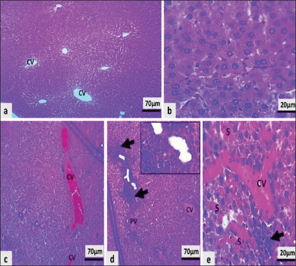
The experiment lasted for 28 days. Rosmarinus officinalis extract was administered orally once daily for 28 consecutive days. MTX was administered intraperitoneally on day 21 to induce toxicity. Thus, rosemary treatment was continued as a protective regimen before and after MTX administration. MTX was administered intraperitoneally according to established experimental models of MTX-induced toxicity, whereas rosemary extract was administered orally to mimic its intended therapeutic use and common route of exposure. The sample size (n=6 per group) was selected based on previous comparable studies investigating MTX-induced toxicity in rats and was considered sufficient to detect biologically relevant differences among groups. Although different administration routes were used, this approach is consistent with previously published MTX-toxicity models and was selected to better reflect the intended therapeutic application of R. officinalis extract (Hamed et al., 2022; Kutbi, 2024). Biochemical analysisBlood samples were collected, and serum was separated for analysis of liver function biomarkers, including ALT, AST, and ALP, as well as renal function parameters (urea and creatinine), using commercial diagnostic kits according to the manufacturers’ instructions. Assessment of oxidative stress markers (OS markers)Oxidative stress was assessed by measuring antioxidant enzymes, including superoxide dismutase (SOD), catalase (CAT), and glutathione peroxidase (GPx), as well as the non-enzymatic antioxidant reduced glutathione (GSH), using standard spectrophotometric methods. Lipid peroxidation was evaluated by measuring Malondialdehyde (MDA) level according to the method of Ohkawa et al. (1979) . The SOD, CAT, and GPx activities were determined according to established spectrophotometric methods, as described by Hamam et al. (2022) and Irato and Santovito (2021). Determination of inflammatory marker (TNF-α)Serum levels of TNF-α were measured using a specific rat TNF-α Enzyme-Linked Immunosorbent Assay (ELISA) kit according to the manufacturer’s instructions. ELISA analysis was performed according to the manufacturer’s instructions and protocols for cytokine quantification. Histopathological examinationLiver and kidney tissues were collected and preserved in 10% neutral buffered formalin after euthanasia. Tissues were processed, embedded in paraffin blocks, and sectioned at 4 to 5 µm thickness. Sections were stained with hematoxylin and eosin (H&E) according to standard histological procedures (Bancroft and Gamble, 2008). Histopathological changes were evaluated using a light microscope. Immunohistochemical analysisTNF-α expression in liver and kidney tissues was evaluated by immunohistochemical staining. The staining intensity and distribution were assessed semi-quantitatively using the H-score method (Fedchenko and Reifenrath, 2014). Statistical analysisData are presented as mean ± SD. A one-way analysis of variance was performed, followed by appropriate post hoc tests. Statistical significance was set at p < 0.05. The sample size was determined on the basis of comparable published animal studies. Ethical approvalAll experimental procedures were approved by the Institutional Animal Ethics Committee at the University of Sulaimani in accordance with the National Institutes of Health guidelines for the care and use of laboratory animals. ResultsHistopathological findingsLiver sections from the control group showed a normal hepatic architecture with intact hepatocytes and a well-defined central vein (Fig. 1A–C). The MTX-treated group showed prominent hepatic lesions with vascular congestion, hepatocellular degeneration, narrowed sinusoids, and inflammatory cell infiltration (Fig. 1D–F).
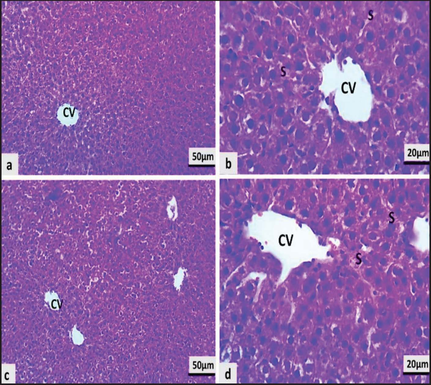
Fig. 1. Representative photomicrographs of H&E-stained liver tissue section (400) (A–C) Control group showing normal hepatic architecture with intact hepatocytes and central veins (CV). (D–F) The MTX-treated group showed marked vascular congestion, hepatocellular degeneration, sinusoidal narrowing, and inflammatory cell infiltration (arrows). Treatment with R. officinalis extract alone preserved hepatic architecture. Low-dose group: mild hepatocyte swelling (Fig. 2A–B). The high-dose group: near-normal hepatic structure with only minimal sinusoidal congestion (Fig. 2C–D).
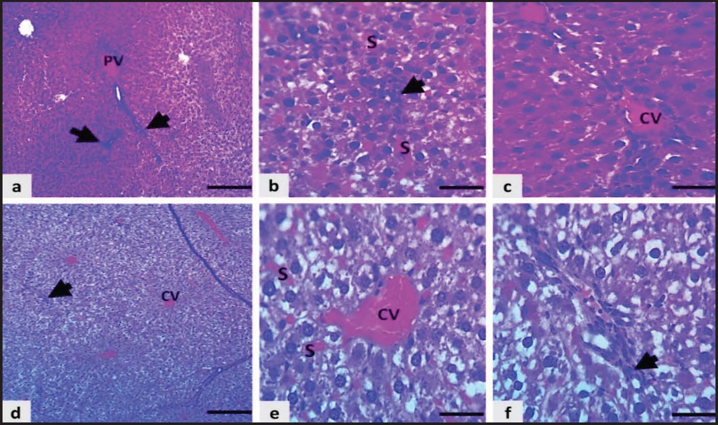
Fig. 2. Photomicrographs of liver sections of H&E (400) from the rosemary-treated groups. (A andB) Low-dose group showing preserved hepatic architecture with mild hepatocyte swelling. (C and D) The high-dose group showed near-normal hepatic structure with minimal sinusoidal congestion. Histopathological damage was dose-dependently alleviated in the MTX plus rosemary cotreated groups. The MTX + low-dose group showed moderate vascular congestion and inflammatory infiltration (Fig. 3A–C), whereas the MTX + high-dose group demonstrated reduced congestion and improved hepatic architecture, approaching normal morphology (Fig. 3D–F).
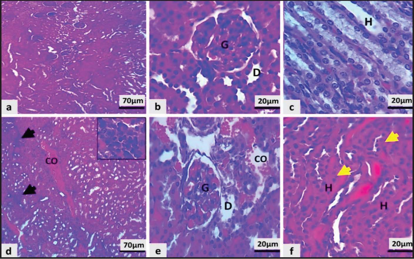
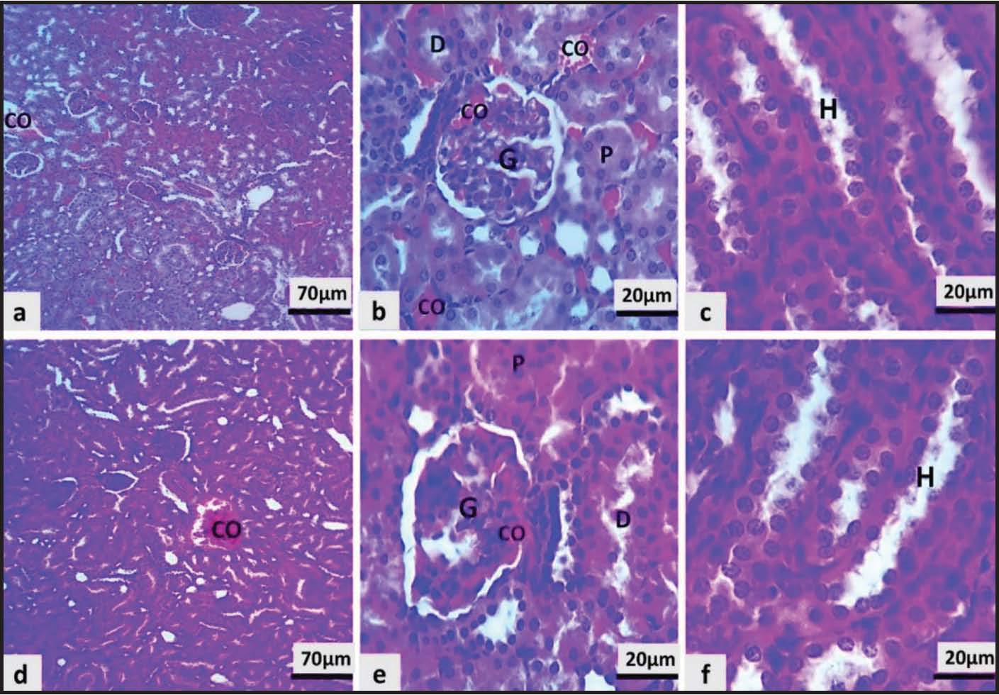
Fig. 3. Liver sections (H&E, 400) of the MTX and rosemary cotreated groups. (A–C) Moderate vascular congestion and inflammatory infiltration with MTX+ low-dose rosemary (D–F) MTX + high-dose rosemary reduced congestion and improved hepatic architecture. Kidney sections from the control group exhibited normal renal architecture (Fig. 4A–C). On the contrary, extensive renal injury with vascular congestion, tubular degeneration, and infiltration of inflammatory cells was observed in MTX-treated rats (Fig. 4D–F).
Fig. 4. Photomicrographs of kidney tissue sections (H&E 400). (A–C) Control group with normal renal architecture. (D–F) MTX-treated group showing vascular congestion, tubular degeneration, and inflammatory infiltration (arrows). Rats treated with rosemary extract showed preserved renal histology. The low-dose group demonstrated mild tubular swelling (Fig. 5A–C), whereas the high-dose group showed near-normal renal structure (Fig. 5D–F).
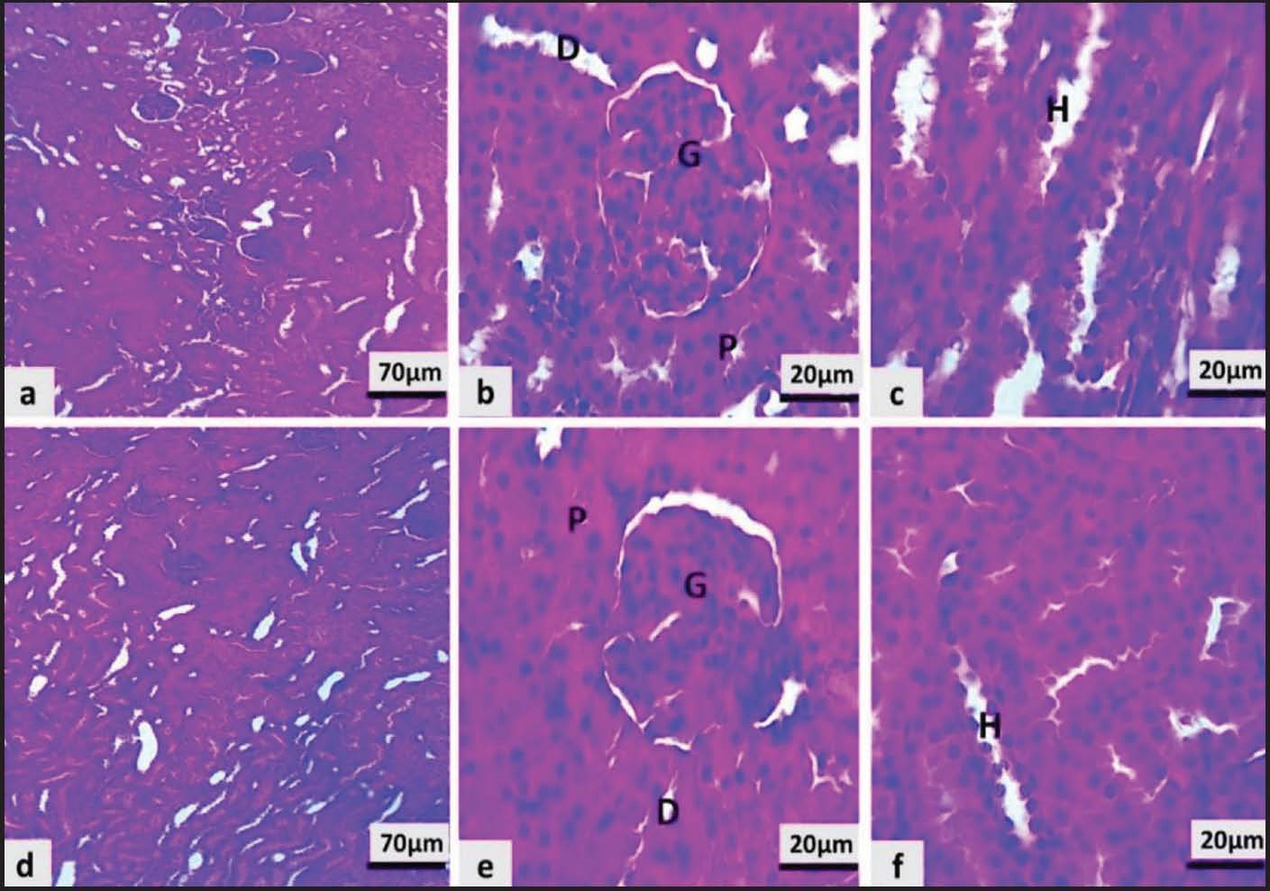
Fig. 5. Kidney sections (H&E 400) from the rosemary-treated groups. (A–C) The low-dose group showed preserved renal architecture with mild tubular swelling. (D–F) The high-dose group showed near-normal histological features. The combined treatment of MTX and RO markedly improved renal impairment. The MTX + low-dose group exhibited moderate tubular degeneration and vascular congestion (Fig. 6A–C), whereas the MTX + high-dose group exhibited significant improvement, as evidenced by decreased pathological changes and restoration of renal architecture (Fig. 6D–F).
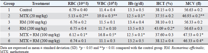
Fig. 6. Kidney sections (H&E 400) of MTX and rosemary cotreated groups. (A–C) Low-dose group with moderate tubular degeneration and vascular congestion. (D–F) The high-dose group showed reduced pathological changes and improved renal structure. Hematological parametersAs shown in Table 1, there were significant changes in hematological parameters in the MTX group compared with the control group. Significant reductions in the Red Blood Cells (RBCs) count, hemoglobin (Hb), and mean corpuscular volume (MCV) (p < 0.05 to 0.01), consistent with impaired erythropoiesis. On the other hand, white blood cell (WBC) counts were markedly elevated. Table 1. Hematological parameters of rats treated with MTX and extract of R. officinalis.
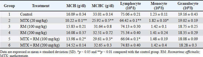
Erythrocytic indices and leukocyte differentials showed that MTX treatment significantly reduced Mean Corpuscular Haemoglobin (MCH), Mean Corpuscular Haemoglobin Concentration (MCHC), and lymphocyte count while increasing monocytes compared with the control group (Table 2). Table 2. Differential and erythrocytic hematological parameters of rats treated with MTX and extract of R. officinalis.
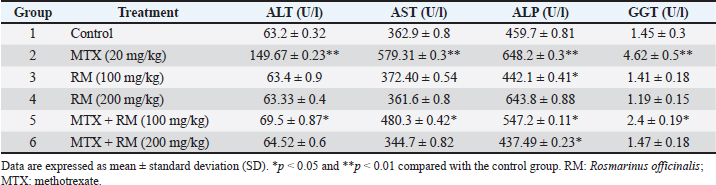
Rosmarinus officinalis extract treatment improved these parameters in a dose-dependent manner. Most hematological values returned to near-normal levels in the high-dose co-treatment group without a significant difference compared with the control group (Tables 1 and 2). Hepatic enzyme activityMTX administration significantly increased ALT, AST, ALP, and GGT levels compared with those in the control group (p < 0.01), indicating hepatocellular injury (Table 3). Treatment with rosemary extract significantly reduced these enzyme levels in a dose-dependent manner, with the high-dose co-treatment group showing values comparable to the control. Table 3. Biochemical assessment of hepatic enzyme activities in MTX-intoxicated rats treated with extract of R. officinalis.
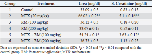
Parameters of renal functionMTX significantly increased urea and serum creatinine levels compared with the control group (Table 4), indicating renal impairment. Treatment with R. officinalis extract dose-dependently improved renal function, with the high-dose co-treatment group showing near-normal values. Table 4. Evaluation of renal function biomarkers in rats exposed to MTX and treated with extract of R. officinalis.
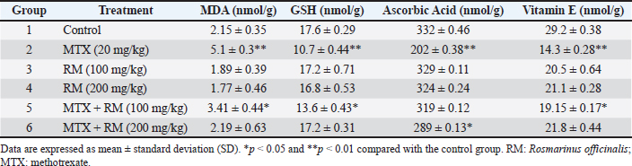
Nonenzymatic antioxidant parametersCompared with the control group, MTX administration significantly increased MDA levels and decreased GSH, ascorbic acid, and vitamin E levels (Table 5), indicating oxidative stress. Treatment with R. officinalis extract significantly restored these parameters, particularly at the higher dose. Table 5. Changes in non-enzymatic antioxidant profiles in MTX-induced hepatotoxic rats treated with extract of R. officinalis.
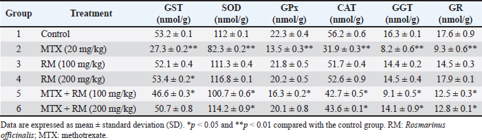
Enzymatic antioxidant parametersMTX significantly reduced antioxidant enzyme activities, including GST, SOD, GPx, CAT, GGT, and GR (Table 6). Treatment with R. officinalis extract restored these enzymatic activities in a dose-dependent manner, with near-normal values in the high-dose co-treatment group. Table 6. Enzymatic antioxidant parameters in rats treated with MTX and Extract of R. officinalis.
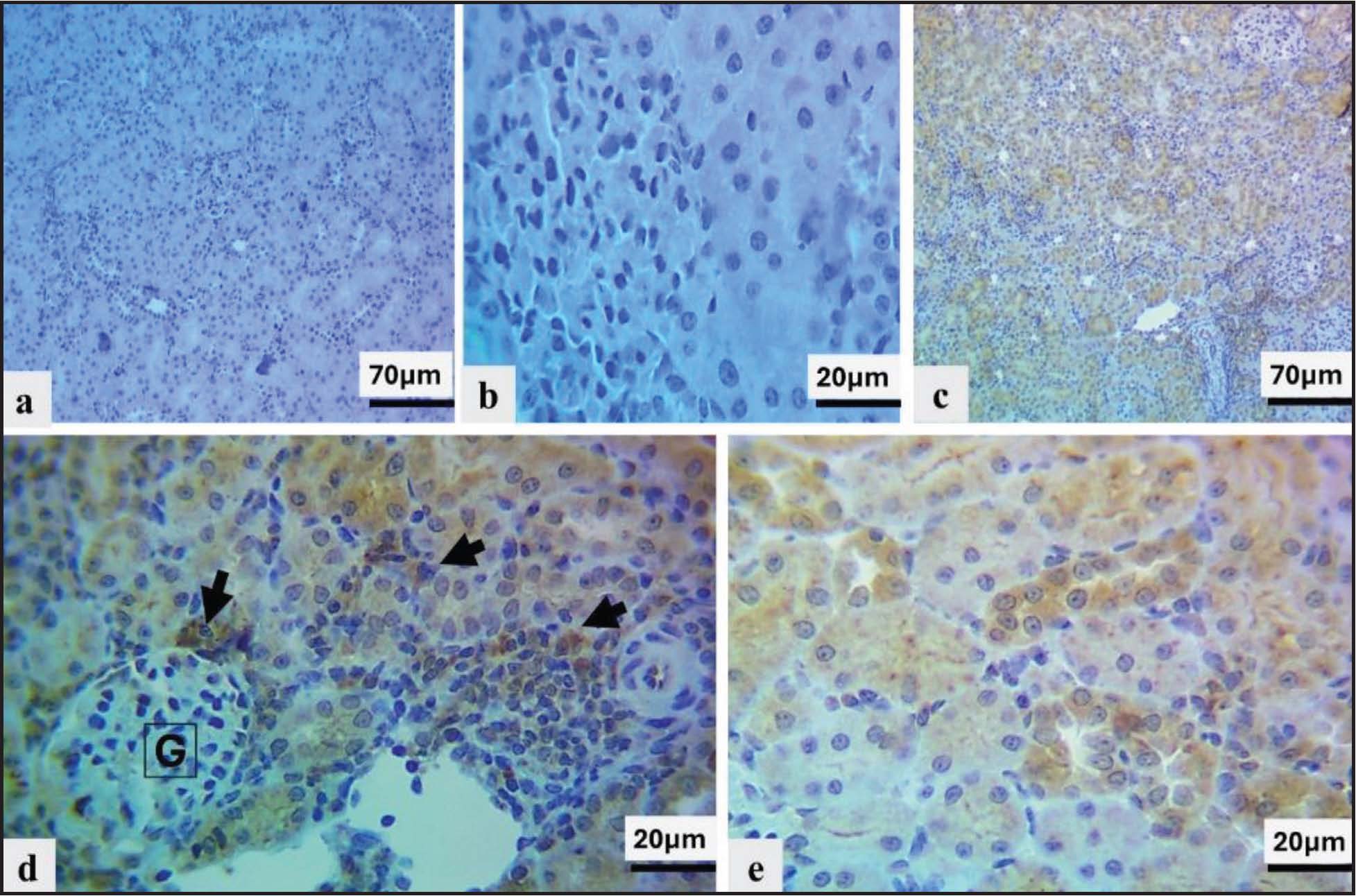
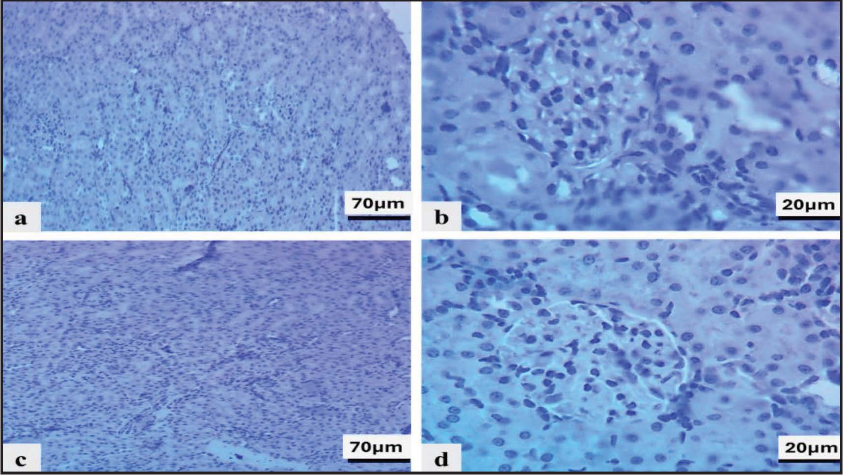
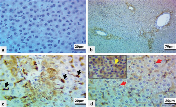
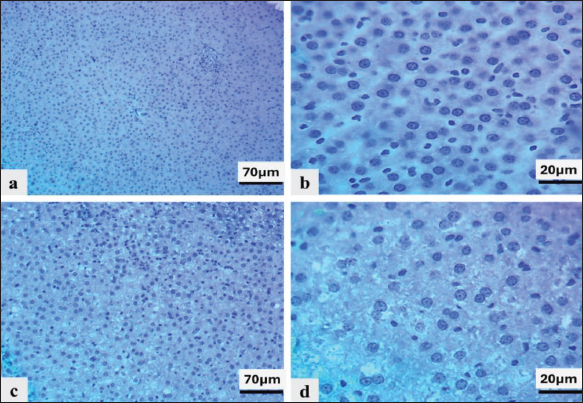
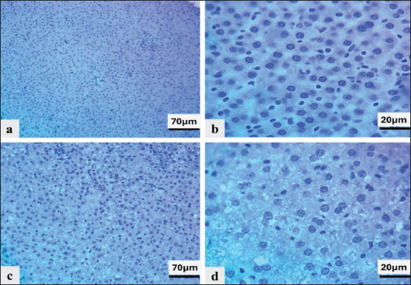
Immunohistochemical findingsImmunohistochemical evaluation revealed no expression of TNF-α in the control and rosemary-treated groups (H-score=0), as shown in kidney sections (Figs. 8a–b, 9a–d) and liver sections (Figs. 11a–b, 12a–d). On the contrary, more robust and diffuse TNF-α immunoreactivity was detected in the hepatic and renal tissues of MTX-treated animals by significantly higher H-scores (kidney: Fig. 8c–d; liver: Fig. 11c–d).
Fig. 8. Immunohistochemical staining of TNF-α in kidney tissue. (a and b) Control group showing no immunoreactivity (H-score=0). (c and d) MTX-treated group showing diffuse moderate to strong immunostaining in glomeruli (G), renal tubules, and inflammatory cells (arrows) (H-score=16, 80%).
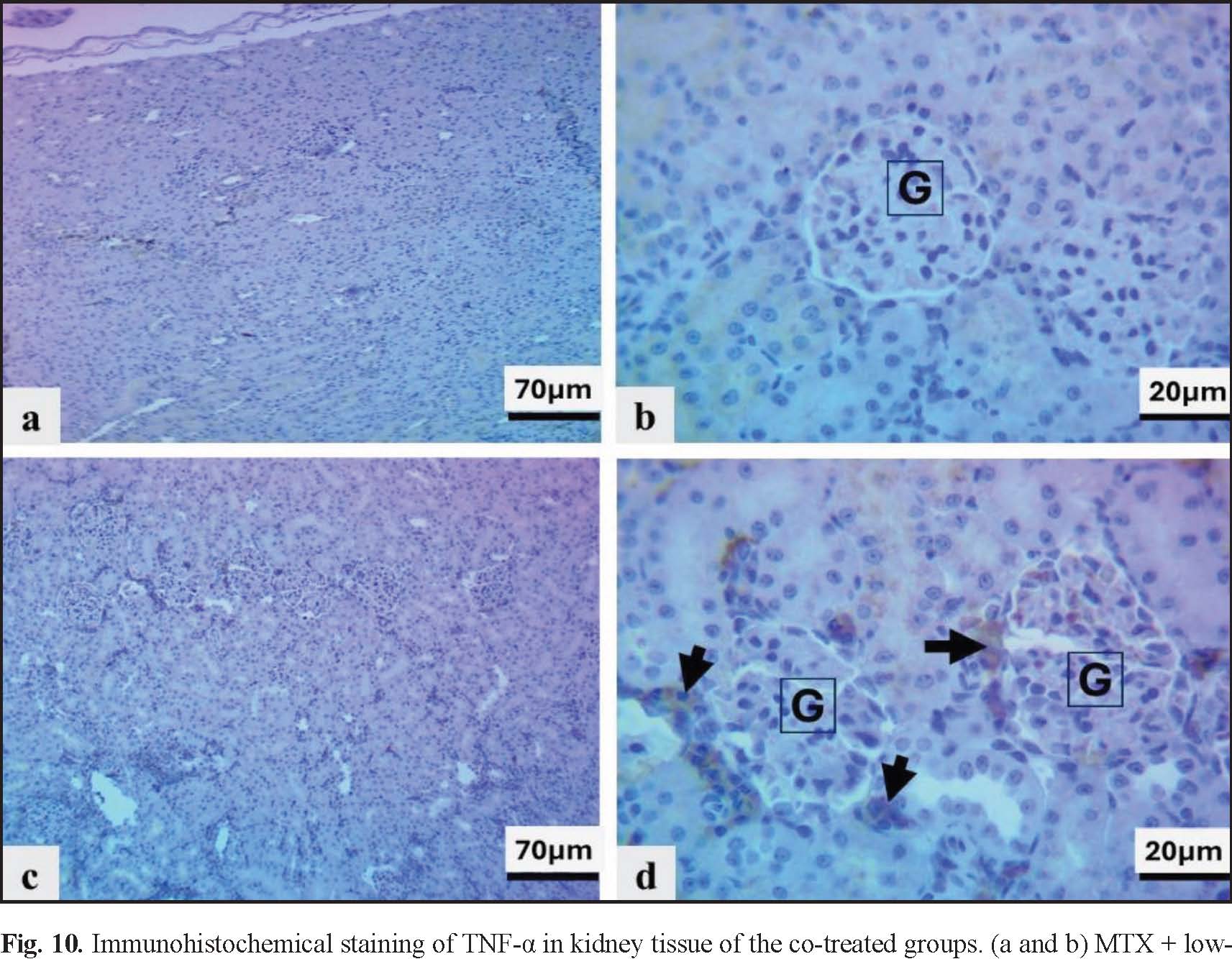
Fig. 9. Immunohistochemical staining of TNF-α in kidney tissue of the rosemary-treated groups. (a and b) Low-dose group and (c and d) high-dose group showing no detectable immunoreactivity (H-score=0). Diffuse moderate to strong immunostaining was induced in the kidney with MTX (H-score=16, 80%) (Fig. 8c–d). Rosmarinus officinalis extract decreased TNF-α expression in a dose-dependent manner, with weak focal staining observed in the MTX + low-dose group (25%, H-score=1) (Fig. 10a–b), and moderate focal staining observed in the MTX + high-dose group (45%, H-score=2) (Fig. 10c–d).
Fig. 10. Immunohistochemical staining of TNF-α in kidney tissue of the co-treated groups. (a and b) MTX + low-dose rosemary showing weak focal immunostaining (H-score=1, 25%). (c and d) MTX + high-dose rosemary showing focal moderate immunostaining (H-score=2, 45%).
Fig. 11. Immunohistochemical staining of TNF-α in liver tissue samples. (a and b) Control group showing no immunoreactivity (H-score=0). (c and d) MTX-treated group showing diffuse moderate to strong staining in hepatocytes, endothelial cells, Kupffer cells, and inflammatory cells (arrows) (H-score=16, 85%).
Fig. 12. Immunohistochemical staining of TNF-α in liver tissue of the rosemary-treated groups. (a and b) Low-dose and (c and d) high-dose groups showing no detectable immunoreactivity (H-score=0). MTX also induced diffuse moderate to strong expression of TNF-α in the liver (H-score=16, 85%) (Fig. 11c–d), which was inhibited after co-treatment with rosemary extract: focal moderate staining in the low-dose co-treatment group (H-score=2,10%) (Fig. 13a–b) and focal moderate to strong staining for the high-dose co-treatment group (H-score=6,35%) (Fig. 13c–d).
Fig. 13. Immunohistochemical staining of TNF-α in the liver tissue of co-treated groups. (a and b) MTX + low-dose rosemary showing focal moderate immunostaining (H-score=2, 10%). (c and d) MTX + high-dose rosemary showing moderate to strong focal immunostaining (H-score=6, 35%). Furthermore, the semi-quantitative H-score analysis confirmed these observations by showing a significant increase in TNF-α expression in the MTX-treated group and a reduction in TNF-α expression in the rosemary-treated group compared with the MTX-treated animals (Fig. 7).
Fig. 7. Semi-quantitative H-score assessment of TNF-α expression in liver and kidney tissues across experimental groups. DiscussionMTX is an established antifolate agent whose toxicity has been linked not only to folate antagonism but also to oxidative stress, mitochondrial dysfunction, and inflammatory pathway activation. In the present study, MTX administration produced a classical model of hepatorenal toxicity, characterized by biochemical disturbances, oxidative imbalance, histopathological alterations, and increased TNF-α levels in the liver and kidneys. These findings are consistent with current evidence, suggesting that interconnected oxidative and inflammatory pathways drive MTX toxicity (Armagan et al., 2015; Bedoui et al., 2019; Ezhilarasan, 2021). Increased serum levels of liver enzymes (ALT, AST, ALP, and GGT) indicate hepatocellular injury and potential cholestatic involvement. These changes are often attributed to membrane damage and mitochondrial impairment induced by oxidative stress, which allows intracellular enzymes to leak into the bloodstream (Mehrzadi et al., 2018; Adikwu and Nnaedozie, 2020; Roghani et al., 2020). The significant elevation of AST further corroborates the mitochondrial involvement, as a proportion of AST is localized within the mitochondria (Schmidt et al., 2022). Moreover, elevated urea and creatinine levels substantiated MTX-induced nephrotoxicity, reflecting impaired renal function. Importantly, the significantly higher creatinine level detected in MTX-treated rats reflects acute impairment of kidney function, likely stemming from MTX-mediated oxidative and inflammatory injury leading to acute tubular damage and diminished glomerular filtration (Severin et al., 2019; Dar et al., 2021; Kutbi, 2024). These results are consistent with previous studies showing that MTX is taken up by renal tubules, where it can mediate oxidative injury, mitochondrial dysfunction, inflammatory activation, tubular degeneration, and reduced glomerular filtration (Elseady et al., 2023; Mishriki et al., 2024). Oxidative stress was a key characteristic of MTX toxicity, as demonstrated by elevated MDA levels and depletion of both enzymatic and non-enzymatic antioxidants, including SOD, CAT, GPx, GSH, ascorbic acid, and vitamin E, reflecting excessive ROS generation. Similar oxidative disruption has been repeatedly observed in different models of MTX toxicity (Montasser et al., 2017; Srinivas, 2021). Concurrently, MTX significantly increased TNF-α levels and immunohistochemical expression within hepatic and renal tissues, indicating inflammatory pathways activation. TNF-α is one of the key mediators regulated by NF-κβ signaling and plays an essential role in exacerbating tissue injury through leukocyte recruitment, endothelial dysfunction, and apoptosis (El-Sheikh et al., 2015; Roghani et al., 2020; Turk et al., 2022). The association between oxidative stress markers and TNF-α expression corroborates the notion of oxidative-inflammatory crosstalk in MTX-mediated toxicity (Roghani et al., 2020). These observations were further supported by histopathological findings, which demonstrated severe hepatic and renal structural damage, including vascular congestion, cellular degeneration, and inflammatory infiltration, in MTX-treated animals. These findings are consistent with the classical descriptions of MTX-associated organ toxicity (Hamed et al., 2022; Kamel et al., 2023). Rosmarinus officinalis extract treatment significantly improved these parameters in a dose-dependent manner. The strong protective effects of rosemary extract (Ahmed and Babakir-Mina, 2020; Ghasemzadeh Rahbardar and Hosseinzadeh, 2025) were evidenced by the normalization of liver enzymes, renal biomarkers, antioxidant status, and TNF-α expression, coupled with improved histological architecture. The protective efficacy of R. officinalis may be attributed to its abundance of bioactive phenolic compounds, particularly rosmarinic acid and carnosic acid. These compounds exert strong antioxidant effects by acting as free radical scavengers and suppressing lipid peroxidation through the induction of endogenous antioxidant defense systems. Moreover, they modulate inflammatory signaling pathways by inhibiting NF-κβ activation, thereby reducing the production of proinflammatory cytokines such as TNF-α (Rašković et al., 2014; Dhouibi et al., 2023; Li Pomi et al., 2023). The dose-dependent protective effect observed in this study further supports the phytochemical pharmacological activity of rosemary. Higher doses brought about more significant restoration of biochemical and histological parameters, suggesting enhanced bioavailability of active compounds (Kosmopoulou et al., 2024; Jalali and Ghasemzadeh Rahbardar, 2025). Despite the restoration of most parameters toward control values, some changes remained significant. Such findings are consistent with previous studies demonstrating that antioxidant treatment achieves only partial recovery following severe oxidative damage (De Oliveira et al., 2019; Nadeem et al., 2019; Goncalves et al., 2022). Despite these encouraging findings, this study has some limitations. Experiments were performed only in Wistar rats; therefore, further research is needed to verify these effects in a clinical setting and other experimental models. Moreover, the exact molecular mechanisms by which rosemary exerts its protective effects require further investigation. In summary, the results of this study indicate that oxidative stress primarily mediates MTX-induced toxicity, resulting in the subsequent activation of inflammatory pathways, including TNF-α- mediated signaling. The protective effects of R. officinalis are mediated through its combined antioxidant and anti-inflammatory properties, with consequent attenuation of oxidative damage, inflammatory responses, and preservation of tissue structure and function (Goncalves et al., 2022; Li Pomi et al., 2023). Only male Wistar rats were included; therefore, potential sex-related differences could not be evaluated. In addition, the relatively small sample size may limit the generalizability of the findings. A limitation of the present study is the absence of a standard reference protective agent for comparison with rosemary extract and the lack of detailed phytochemical characterization of the extract. Future studies, including established hepatoprotective or nephroprotective agents, together with comprehensive phytochemical and molecular analyses, are recommended to further validate the protective efficacy of R. officinalis. ConclusionThe present findings suggest that the crude ethanolic extract of R. officinalis exerts protective effects against MTX-induced hepatorenal toxicity, possibly through antioxidant and anti-inflammatory mechanisms. However, further studies involving detailed phytochemical characterization, molecular investigations, and the inclusion of standard reference drugs are required to confirm these effects and validate the therapeutic potential of this drug. AcknowledgmentsNone. Conflict of interestThe authors declare no conflicts of interest regarding the publication of this study. FundingSelf-supported. Authors’ contributionsConceptualization, N.S.; Methodology, L.A. and N.S.; Validation, L.A. and N.S. Formal Analysis, L.A.; Investigation, L.A. and N.S.; Data Curating, L.A. and N.S.; Writing—Original Draft Preparation, L.A. Writing—Review & Editing, N.S.; Visualization, L.A. and N.S.; Supervision, N.S.; Project Administration, N.S. Data availabilityAll data were provided in the manuscript. ReferencesAdikwu, E. and Nnaedozie, F.E. 2020. Hepatotoxicity and drugs. EXCLI. J. 19, 900. Ahmed, A.S. and Babakir-Mina, S. 2020. Assessment of anti-cancer activity of Mentha spicata, Origanum vulgare and Rosmarinus officinalis plant extracts on human adenocarcinoma. Indian J. Public. Health. Res. Develop. 11(3), 1159–1163. Ali, H.A.A., Abdel-Aziem, M.E., Elshora, M.S., Hassan, M.E. and Nasser, M.M. 2024. Hepatoprotective effect of alpha-lipoic acid and silymarin against methotrexate-induced hepatotoxicity in rats. J. Pharm. Res. Int. 36(11), 4353–4371. Al-Khawalde, H., Khawaja, M., Rahmani, H. and Razavi, M. 2022. The protective effects of vitamin E against MTX-induced hepatotoxicity. Molecules 27(7), 2261. Almalki, Z.S., Albadr, A., Elrasheed, M.M. and Al-Ghamdi, M.A. 2021. Hepatoprotective and antioxidant activities of pomegranate against methotrexate induced hepatotoxicity. J. Pharm. Res. Int. 33(36A), 88–103. Armagan, G., Tunali-Akbay, G., Kuru, Z., Oguz, C., Unver-Saraydin, S., Gümüstekin, Y., Kantarci, E., Uygun, Y. and Kalayci, Ş. 2015. Protective effects of Salvia triloba on methotrexate-induced oxidative liver damage in rats. Pharm. Biol. 53(2), 170–176. Bancroft, J.D. and Gamble, M. 2008. Theory and practice of histological techniques, 6th ed. Philadelphia, PA: Churchill Livingstone Elsevier. Bedoui, S., Guillot, S., Ruat, F., Chevallier, C. and Meraihi, J. 2019. Oxidative mechanisms of methotrexate toxicity. Toxicol. Rep. 6, 179–191. Dar, N.I., Dar, S., Dar, S., Din, A., Dar, S. and Dar, M. 2021. Methotrexate induced acute kidney injury and rhabdomyolysis. Med. J. Armed. Forces. India. 77(S1), S212–S215. De Oliveira, M.R., De Freitas, M., De Souza, C. and Behr, J. 2019. Antioxidant effects of sulfhydryl compounds in experimental models of aging-related neurodegenerative diseases. J. Mol. Neurosci. 67(3), 383–396. Dhouibi, R., Alves, G., Duarte, C., Nunes, L. and Jamoussi, H. 2023. Anti-inflammatory and antioxidant properties of polyphenol-rich essential oils from medicinal plants. Molecules 28(5), 2163. Dogra, V., Gupta, N., Gill, R. and Sindhu, V. 2025. Hepatoprotective role of Rosmarinus officinalis in pharmacologically-induced models. PhytoMed. Int. 8(2), 112–128. El-Dessouki, S.S., Elbaz, K.M., Mohamed, N.H. and Elbakry, T.A. 2025. Role of NF-kappaB signaling in MTX-induced hepatorenal toxicity and the protective effects of plant phenolics. J. Pharmacol. Sci. 24(1), 45–67. Elseady, M.A., Raslan, M., Abd-Elsayed, N. and Ahmed, N. 2023. Protective effects of L-carnitine against methotrexate-induced nephrotoxicity in rats. Med. J. Islamic World Acad. Sci. 31(5), 289–298. El-Sheikh, M.A., Ibrahim, A., Adel, A.A. and El-Fattah, A.A. 2015. Protective effects of resveratrol against methotrexate-induced renal toxicity in rats. Am. J. Pharmacol. Toxicol. 10(1), 23–33. Ezhilarasan, D. 2021. Mechanisms and management of methotrexate toxicity. Int. J. Mol. Sci. 22(18), 9921. Fedchenko, N. and Reifenrath, J. 2014. Sample preparation protocols for immunohistochemical staining of formalin-fixed, paraffin-embedded tissue microarray sections. J. Visualized. Experiments. 86, e51642. Ghasemzadeh Rahbardar, M. and Hosseinzadeh, H. 2025. Protective effects of Rosmarinus officinalis against chemotherapy-induced organ toxicity. Phytotherapy. Res. 39(2), 234–256. Goncalves, A., Martins, M., Ferreira, C., Martins, R. and Lourenço, J. 2022. Antioxidant and anti-inflammatory strategies in managing drug-induced hepatotoxicity. Curr. Pharma. Design 28(15), 1244–1268. Hamam, H., Demir, H., Aydın, M. and Demir, C. 2022. Determination of some antioxidant activities (superoxide dismutase, catalase, reduced glutathione) and oxidative stress level (malondialdehyde acid) in cirrhotic liver patients. Middle Black Sea J. Health Sci. 8(4), 506–514. Hamed, B.S., Dighriri, M., Alakhras, R.A. and Alharbi, A.A. 2022. Methotrexate-induced organ toxicity: molecular mechanisms and protective strategies. Curr. Pharm. Biotechnol. 23(6), 751–768. Irato, P. and Santovito, G. 2021. Enzymatic and non-enzymatic molecules with antioxidant function. MDPI 10, 579. Jafaripour, S., Ramezani, M., Ansari, M. and Karimi, A.A. 2021. Hepatoprotective activity of Rosmarinus officinalis extract against drug-induced liver injury. Phytomedicine Plus 1(1), 100003. Jalali, A. and Ghasemzadeh Rahbardar, M. 2025. Bioavailability and pharmacokinetics of rosemary active compounds. J. Herbal Med. 40(2), 101–115. Kamel, E. M., Eman, A., Hassan, M.E., Wasfy, W.Y. and Salih, N.A. 2023. Protective effects of curcumin against methotrexate-induced hepatorenal injury: biochemical and histopathological evaluation. Toxicol. Mech. Methods. 33(4), 312–324. Kosmopoulou, V., Antifakos, N., Tsartsaflis, D., Karavasilis, G. and Loukopoulos, E. 2024. Enhancement of antioxidant properties of plant extracts through extraction methods. Molecules 29(3), 789. Kutbi, A. 2024. Nephrotoxicity of methotrexate and protective strategies: a review. Int. J. Environ. Res. Public. Health. 21(1), 78. Lesnik, P., Hršak, I., Kramer, M. and Prgomet, A. 2021. Anti-inflammatory and antioxidant properties of Rosmarinus officinalis essential oils. Antioxidants 10(8), 1273. Li Pomi, F., Seminerio, M., Frasca, M., Di Rosa, M. and Tibullo, A. 2023. Role of rosemary polyphenols in attenuating oxidative and inflammatory stress. Phytomedicine 103, 154863. Mehrzadi, S., Kohansal, M., Hajiasgharzadeh, S. and Sadeghnia, R. 2018. Protective effects of silymarin against methotrexate-induced hepatotoxicity: the role of oxidative stress and inflammatory markers. J. Evidence-Based Complement. Alternative Med. 2018, 2957315. Mishriki, H.S., Shamseddin, A., Mansour, E. and Elhadad, M. 2024. Methotrexate-induced renal dysfunction: molecular pathways and therapeutic interventions. Biomolecules 13(2), 195. Montasser, A.F., Souilem, R., Bouajila, N.S., Fares, F., Labaied, E., Karray, H. and Elleuch, S. 2017. Protective effect of phenolic-rich extracts against methotrexate-induced hepatotoxicity. Saudi. Pharm. J. 25(7), 1031–1038. Nadeem, A., Al-Hariri, H., Ansari, M. and Janssen-Megens, A. 2019. Recovery mechanisms following methotrexate-induced oxidative stress. Oxid. Med. Cell. Longev. 2019, 8734745. Ohkawa, H., Ohishi, N. and Yagi, K. 1979. Assay for lipid peroxides in animal tissues by thiobarbituric acid reaction. Anal. Biochem. 95(2), 351–358. Rašković, A., Milanović, I., Pavlović, N., Ćebović, T., Vukmirović, S. and Mikov, M. 2014. Antioxidant activity of rosemary (Rosmarinus officinalis L.) essential oil and its hepatoprotective potential. BMC Complementary Alternative Med. 14(1), 225. Roghani, M., Sadeghnia, H., Ghaffari, M.A., Salahshoor, R. and Karamali, A. 2020. Ameliorative effects of rosmarinic acid on methotrexate-induced hepatorenal toxicity. Iranian. J. Vet. Res. 21(4), 245–255. Schmidt, P.L., Schrader, B., Schmid-Burgk, C., Mack, T. and Lang, H. 2022. Mitochondrial aspartate aminotransferase in hepatocellular integrity. Cell Tissue Res. 388(2), 205–218. Severin, J.S., Schaafsma, M., Miedema, A., Verschuuren, E. and van Oost, W. 2019. Acute kidney injury following methotrexate administration. Nephrology 24(5), 495–502. Srinivas, G. 2021. Oxidative stress biomarkers in methotrexate-induced organ toxicity. Biomarkers. Med. 15(4), 267–284. Turk, J., Alev, B., Akmal, M. and Ozel, V. 2022. TNF-alpha signaling pathways in chemotherapy-induced hepatotoxicity. Int. J. Mol. Med. 50(5), 159. | ||
| How to Cite this Article |
| Pubmed Style Muhammed LJ, Salih NA. Protective effects of Rosmarinus officinalis against methotrexate-induced hepatorenal toxicity in Wistar rats: Biochemical, histopathological, and immunohistochemical evaluation. Open Vet. J.. 2026; 16(7): 4526-4539. doi:10.5455/OVJ.2026.v16.i7.36 Web Style Muhammed LJ, Salih NA. Protective effects of Rosmarinus officinalis against methotrexate-induced hepatorenal toxicity in Wistar rats: Biochemical, histopathological, and immunohistochemical evaluation. https://www.openveterinaryjournal.com/?mno=316821 [Access: July 15, 2026]. doi:10.5455/OVJ.2026.v16.i7.36 AMA (American Medical Association) Style Muhammed LJ, Salih NA. Protective effects of Rosmarinus officinalis against methotrexate-induced hepatorenal toxicity in Wistar rats: Biochemical, histopathological, and immunohistochemical evaluation. Open Vet. J.. 2026; 16(7): 4526-4539. doi:10.5455/OVJ.2026.v16.i7.36 Vancouver/ICMJE Style Muhammed LJ, Salih NA. Protective effects of Rosmarinus officinalis against methotrexate-induced hepatorenal toxicity in Wistar rats: Biochemical, histopathological, and immunohistochemical evaluation. Open Vet. J.. (2026), [cited July 15, 2026]; 16(7): 4526-4539. doi:10.5455/OVJ.2026.v16.i7.36 Harvard Style Muhammed, L. J. & Salih, . N. A. (2026) Protective effects of Rosmarinus officinalis against methotrexate-induced hepatorenal toxicity in Wistar rats: Biochemical, histopathological, and immunohistochemical evaluation. Open Vet. J., 16 (7), 4526-4539. doi:10.5455/OVJ.2026.v16.i7.36 Turabian Style Muhammed, Lana J., and Nadia A. Salih. 2026. Protective effects of Rosmarinus officinalis against methotrexate-induced hepatorenal toxicity in Wistar rats: Biochemical, histopathological, and immunohistochemical evaluation. Open Veterinary Journal, 16 (7), 4526-4539. doi:10.5455/OVJ.2026.v16.i7.36 Chicago Style Muhammed, Lana J., and Nadia A. Salih. "Protective effects of Rosmarinus officinalis against methotrexate-induced hepatorenal toxicity in Wistar rats: Biochemical, histopathological, and immunohistochemical evaluation." Open Veterinary Journal 16 (2026), 4526-4539. doi:10.5455/OVJ.2026.v16.i7.36 MLA (The Modern Language Association) Style Muhammed, Lana J., and Nadia A. Salih. "Protective effects of Rosmarinus officinalis against methotrexate-induced hepatorenal toxicity in Wistar rats: Biochemical, histopathological, and immunohistochemical evaluation." Open Veterinary Journal 16.7 (2026), 4526-4539. Print. doi:10.5455/OVJ.2026.v16.i7.36 APA (American Psychological Association) Style Muhammed, L. J. & Salih, . N. A. (2026) Protective effects of Rosmarinus officinalis against methotrexate-induced hepatorenal toxicity in Wistar rats: Biochemical, histopathological, and immunohistochemical evaluation. Open Veterinary Journal, 16 (7), 4526-4539. doi:10.5455/OVJ.2026.v16.i7.36 |


