





| Case Report | ||
Open Vet. J.. 2026; 16(7): 4288-4294 Open Veterinary Journal, (2026), Vol. 16(7): 4288-4294 Case Report Portal vein hypoplasia in a Dalmatian dogLetícia Lopes Carrijo1, Vitor Eduardo Arantes de Barros2, Giulia de Castro Silva1, Mariana Moreira Lopes1, Alexandra Moura Fraga1, Luísa Pinheiro Moreira1, Iago Martins Oliveira3*, Marco Augusto Machado Silva4, Aline Maria Vasconcelos Lima4 and Veridiana Maria Brianezi Dignani de Moura41Programa de Residência Multiprofissional em Saúde, Clínica e Cirurgia de Pequenos Animais, Escola de Veterinária e Zootecnia, Universidade Federal de Goiás, Rodovia Goiânia - Nova Veneza, Goiânia, Brazil 2Programa de Pós-graduação em Ciência Animal, Escola de Veterinária e Zootecnia, Universidade Federal de Goiás, Rodovia Goiânia - Nova Veneza, Goiânia, Brazil 3Escola de Ciências Médicas e da Vida, Pontifícia Universidade Católica de Goiás, Goiânia, Brazil 4Departamento de Medicina Veterinária, Escola de Veterinária e Zootecnia, Universidade Federal de Goiás, Rodovia Goiânia - Nova Veneza, Goiânia, Brazil *Corresponding Author: Iago Martins Oliveira. Escola de Ciências Médicas e da Vida, Pontifícia Universidade Católica de Goiás, Goiânia, Goiás, Brazil. Email: iago.vetufg [at] gmail.com Submitted: 27/04/2026 Revised: 25/05/2026 Accepted: 30/05/2026 Published: 02/07/2026 © 2026 Open Veterinary Journal
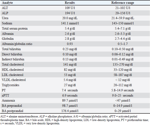
ABSTRACTBackground: Portal vein hypoplasia (PVH) is a congenital vascular anomaly described in dogs. It is characterized by abnormally small intrahepatic or extrahepatic portal veins, resulting in hepatic hypoperfusion and the potential development of portal hypertension (PH), which may progress to the formation of portosystemic shunts. Case Description: A 3-year-old female Dalmatian dog presented with ascites and anemia with a history of 7 months. Diagnostic investigation included laboratory tests and contrast-enhanced computed tomography, which revealed chronic hepatopathy associated with multiple acquired extrahepatic portosystemic shunts, consistent with PH. Histopathological evaluation of liver biopsy samples confirmed PVH as the underlying cause of PH. Clinical management consisted of a hepatic diet, hepatoprotective agents, and associated complications treatment, resulting in significant clinical improvement. Conclusion: PVH should be considered among the main differential diagnoses in young dogs presenting with PH, particularly when vascular manifestations, such as ascites, congestion, and portosystemic shunts, are observed. Definitive diagnosis requires correlation between clinical, imaging, and histopathological findings, and appropriate medical management may lead to effective control of clinical signs. Keywords: Canine, Chronic hepatopathy, Hepatic vascular anomalies, Portosystemic shunt. IntroductionPortal vein hypoplasia (PVH) is a congenital vascular anomaly described in dogs. It is characterized by abnormally small intrahepatic or extrahepatic portal veins, resulting in hepatic hypoperfusion and the potential development of portal hypertension (PH). Clinical manifestations such as organ congestion, gastrointestinal bleeding, ascites, and acquired portosystemic shunts may be observed as portal vein pressure increases (Akiyoshi et al., 2017). PVH typically affects young dogs aged between 5 years and is commonly reported in both sexes of purebred animals, with a higher prevalence in large-breed dogs. Clinical manifestations arise from the combination of PH and PVH severity, with ascites being a prominent clinical finding. In addition, gastrointestinal signs, polydipsia, and weight loss may be observed, whereas hepatic encephalopathy is less common (Center et al., 2022). Definitive diagnosis is established through histopathological evaluation of liver biopsy samples, in combination with ultrasonographic findings or computed tomography angiography (Akiyoshi et al., 2017; Webster et al., 2019; Center et al., 2022). The management of PVH includes the use of hepatoprotective and antioxidant agents, dietary sodium restriction and protein modulation, and diuretics and paracentesis for managing ascites. Lactulose is indicated for the treatment of hepatic encephalopathy, and in cases of portal hypertensive gastropathy, gastroprotective drugs may be used (Akiyoshi et al., 2017; Webster et al., 2019; Calleja et al., 2023). In this context, this study aimed to describe a case of PVH associated with PH and acquired portosystemic shunts in a Dalmatian dog, with emphasis on the diagnostic approach. Case DetailsA 3-year-old spayed female Dalmatian dog weighing 16.8 kg was presented for evaluation due to persistent ascites and anemia for 7 months. According to the clinical history, the patient exhibited normal appetite, water intake, urination, and defecation, with no episodes of vomiting. On physical examination, the patient was alert, with pale oral and ocular mucous membranes, a capillary refill time of 2 seconds, adequate hydration status, enlarged mandibular lymph nodes, a rectal temperature of 38.7°C, a body condition score of 3/9 (Chun et al., 2019), and mild abdominal distension. Cardiac auscultation revealed a grade 3/6 murmur at the mitral and tricuspid areas, with a heart rate of 130 beats per minute. Pulmonary auscultation indicated a respiratory rate of 24 breaths per minute. Complementary diagnostic tests were requested based on the clinical presentation, including complete blood count, reticulocyte count, biochemical profile, lipid panel, pre- and postprandial bile acids, sodium measurement, urinalysis, urine protein-to-creatinine ratio, blood glucose, analysis of cavitary fluid, ammonia levels, coagulation profile, echocardiography, and contrast-enhanced computed tomography of the thorax and abdomen. Hematological evaluation revealed microcytic normochromic anemia (hematocrit 27%; reference: 37%–55%), reticulocytosis (absolute reticulocyte count: 152,820/mm³, consistent with moderate regeneration), eosinophilia (1,572/mm³; reference: 100–1,250/mm³), lymphopenia (655/mm³; reference: 1,000–4,800/mm³), and thrombocytopenia (160 ×10³/µl; reference: 200–500 ×10³/µl). The biochemical profile showed a mild decrease in serum urea (20.0 mg/dl; reference: 21.4–59.9 mg/dl), hypoproteinemia (5.6 g/dl; reference: 6.0–8.0 g/dl), and mild increases in alanine aminotransferase (109 U/l; reference: 21–102 U/l) and alkaline phosphatase (ALP) (194 U/l; reference: 20–156 U/l), as well as hyponatremia (141.1 mmol/l; reference: 143–150 mmol/l). The total protein, albumin, globulins, glucose, and bilirubin levels were within normal limits. Bile acids were markedly increased in both pre- and postprandial measurements (preprandial: 98.7 µmol/l; reference: 0–14.9 µmol/l; postprandial: 176.4 µmol/l; reference: 0–29.9 µmol/l). The detailed biochemical results are presented in Table 1. Table 1. Biochemical profile of a female dog with portal hypertension.
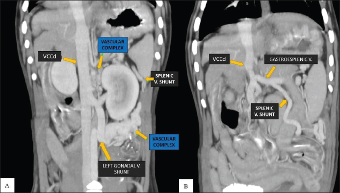
Urinalysis revealed turbid urine, acidic pH of 5.0, and the presence of ammonium urate crystals (++++) with a normal urine protein-to-creatinine ratio. The cavitary fluid analysis was consistent with a simple transudate, characterized by low cellularity and protein content. Serum ammonia concentration (89.7 µmol/l; reference: <47 µmol/l) indicated hyperammonemia. Coagulation tests, including PT and aPTT, were within normal ranges. Echocardiography demonstrated preserved systolic and diastolic function, with normal cardiac chamber morphology and dimensions. Cardiac murmur was considered secondary to anemia. Contrast-enhanced abdominal computed tomography revealed two anomalous and tortuous vascular complexes located between the kidneys and in the caudal left abdominal region. These complexes communicated between the portal system and systemic circulation, particularly via the splenic and left gonadal veins, consistent with multiple acquired extrahepatic portosystemic shunts (Fig. 1A and B). Additional findings included microhepatia, moderate splenomegaly, gastric mucosa thickening, mild peritoneal effusion (after ascites drainage), hepatic and jejunal lymphadenomegaly, and urinary bladder microcalculi.
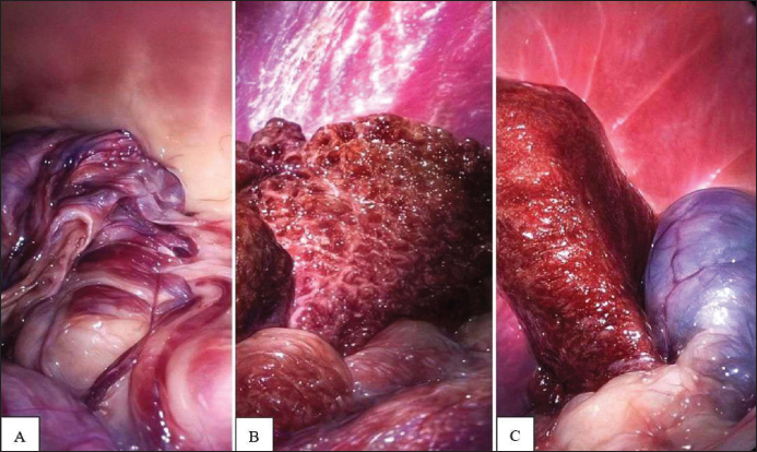
Fig. 1. Post-contrast tomographic image in the soft tissue window in the dorsal plane of a female dog with portal hypertension. A small complex of anomalous vessels is noted between the caudal vena cava and the left kidney, and a second complex of larger-caliber anomalous vessels is noted caudally to the kidney (B). Communication with the left gonadal (A) and splenic (B) veins. Source: CEDIV (Veterinary Diagnostic Imaging Center), Goiânia, Goiás, Brazil. Based on clinical signs and complementary findings, a diagnosis of chronic hepatopathy associated with PH and multiple acquired extrahepatic portosystemic shunts, resulting in ascites and anemia, was established. Supportive treatment for chronic hepatopathy was initiated, including S-adenosylmethionine (20 mg/kg PO SID) for 30 days, vitamin E (10 IU/kg PO SID), ursodeoxycholic acid (15 mg/kg PO SID), silymarin (6 mg/kg PO TID), lactulose (0.3 mL/kg PO TID), and a hepatic diet (Hepatic®, Royal Canin®) for continuous use. In addition, spironolactone (2 mg/kg PO SID), furosemide (1 mg/kg PO SID), and dexamethasone (0.1 mg/kg PO SID) were prescribed for 7 days, along with omeprazole (1 mg/kg PO BID) administered before feeding for 10 days. At the first follow-up visit (7 days later), mild pallor of the mucous membranes persisted, with an increase in hematocrit (31.5%; reference: 37.0%–55.0%) compared to the initial value, persistent lymphopenia (492/mm³), and borderline plasma protein levels (5.9 g/dL). Spironolactone and long-term therapy were continued. At the second follow-up (5 days later), the patient remained free of ascites, alert, and active, with normal mucous membrane coloration. Hematocrit increased to 35%, and cardiac murmur was no longer detected. After clinical stabilization, a liver biopsy was performed via exploratory laparoscopy to determine the cause of PH. Intraoperative findings included multiple portosystemic shunts, predominantly between the splenic and left gonadal veins, microhepatia, and hepatic fibrosis, particularly affecting the left liver lobes (Fig. 2). Six liver fragments were collected, two for each of the right medial, left medial, and left lateral lobes.
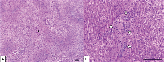
Fig. 2. Macroscopy of the liver of a Dalmatian female dog with PVH during exploratory laparoscopy. (A) Multiple dilated and tortuous vessels in the mesogastrium, corresponding to acquired portosystemic shunts. (B) Right medial hepatic lobe and (C) left lateral hepatic lobe with an irregular surface and areas of fibrosis. Source: Veterinary Hospital EVZ/UFG, 2025. Histochemical staining included hematoxylin and eosin, periodic acid–Schiff (PAS), and rubeanic acid (rhodanine), the latter two aimed at detecting glycogen and copper accumulation, respectively. Histopathological evaluation revealed portal area malformation, characterized by arteriolar proliferation, ductular hyperplasia, and PVH, along with mild portal fibrosis. In addition, a loss of normal hepatic architecture and marked hepatocellular cytoplasmic vacuolization were observed. PAS and rhodamine staining were negative. Based on these findings, a histomorphological diagnosis of PVH was established (Fig. 3), and treatment with hepatoprotective agents, lactulose, and hepatic diet was continued.
Fig. 3. Photomicrographs of the liver of a Dalmatian female dog. (A) Loss of hepatic architecture, diffuse and marked vacuolar degeneration, and portal fibrosis (asterisk). (B) Periportal space. Arteriole proliferation (arrowheads), ductal hyperplasia (arrows), and absence of the portal vein HE. Source: Animal Pathology Service, EVZ/UFG, 2025. DiscussionThe patient was diagnosed with PVH and its associated consequences, including PH, acquired portosystemic shunts, and chronic hepatopathy, based on the results of the complementary diagnostic tests (Bunch et al., 2001; Buob et al., 2011; Akiyoshi et al., 2017; Calleja et al., 2023). In this context, it is important to highlight that the clinical manifestations in patients with PVH are related to the severity of the hypoplasia and the clinical signs associated with PH (Cullen et al., 2006; Rothuizen, 2009). Abdominal distension secondary to ascites was the main clinical finding in this patient and occurs in up to 92% of dogs with PH (Calleja et al., 2023). Ascites is primarily associated with increased hydrostatic pressure and lymphatic overload in dogs with PH (García Leiva et al., 2007; Buob et al., 2011; Center et al., 2022). The patient exhibited mild hypoalbuminemia, and although decreased albumin levels may contribute to ascites formation in dogs with liver disease, increased hydrostatic pressure is the primary determinant of fluid accumulation in PH (James et al., 2008; Hou and Sanyal, 2009; Buob et al., 2011). Anemia and thrombocytopenia are commonly reported in dogs with PH and may result from blood loss and consumption, including gastrointestinal bleeding due to portal hypertensive gastropathy, coagulopathies, or anemia of chronic disease (Webster et al., 2019). Although overt gastrointestinal bleeding was not observed in the present case, chronic occult blood loss associated with portal hypertensive gastropathy cannot be excluded as a contributing factor to regenerative anemia. Biochemical analysis revealed mild increases in ALT and moderate increases in ALP. The mild increases in ALT and ALP levels observed in this patient were compatible with chronic hepatocellular injury and hepatic fibrosis associated with PVH (Webster et al., 2019). Urinalysis also provided important diagnostic information, revealing acidic urine with ammonium urate crystals, which can be explained by acquired portosystemic shunts (Lulich et al., 2016). In addition, the Dalmatian breed is genetically predisposed to defects in uric acid metabolism, resulting in increased urinary excretion of urate and a higher risk of crystal and urolith formation (James et al., 2008; Lulich et al., 2016). Hyponatremia observed in this patient may be attributed to PH-associated ascites, which can lead to dilutional hyponatremia (Hou and Sanyal, 2009; Buob et al., 2011). Similarly, increased pre- and postprandial bile acids, reduced urea levels, and hyperammonemia, which are commonly reported in dogs with PH, were observed. Reduced urea concentration may also reflect impaired hepatic conversion of ammonia into urea due to decreased functional hepatic perfusion. Increased bile acids have been described in up to 100% of cases, and hyperammonemia in approximately 81.8%, making these among the most sensitive tests for detecting portosystemic shunts (Buob et al., 2011; Adam et al., 2012; Calleja et al., 2023). Despite hyperammonemia, the patient did not exhibit neurological signs, which are less commonly observed in dogs with acquired portosystemic shunts than in those with congenital shunts (Adam et al., 2012; Gow, 2017; Konstantinidis et al., 2023). Imaging findings revealed microhepatia, irregular liver contours, and a tendency for regenerative nodule formation, along with multiple portosystemic shunts, supporting the diagnosis of PVH-induced chronic hepatopathy secondary to hepatic hypoperfusion. Although abdominal ultrasonography was not available in the present case, contrast-enhanced computed tomography associated with histopathological evaluation provided sufficient information for the characterization of PH and the confirmation of PVH (Tobias et al., 2018). The development of shunts is considered a compensatory response to PH (Berent and Tobias, 2009; Bertolini, 2019; Calleja et al., 2023). In addition, pancreatic edema, splenomegaly, and gastric wall thickening were likely secondary to congestion caused by PH (Buob et al., 2011). The histopathological findings reflected the effects of reduced portal perfusion, characterized by decreased or absent portal veins. Consequently, to increase hepatic blood flow, the hepatic arterioles become hypertrophic, undergo hyperplasia, and sometimes assume tortuous shapes (Devriendt et al., 2014; Akiyoshi et al., 2017; Argenta et al., 2017). Also, centrilobular degeneration and necrosis, portal fibrosis, and ductular proliferation occur in response to chronic hypoperfusion and hypoxia (Cullen et al., 2006). Despite these significant and diffuse histomorphological alterations, the patient’s baseline hepatic function remained relatively preserved based on biochemical parameters, except for increased bile acids due to acquired portosystemic shunting. The large functional reserve of hepatic parenchyma, which can maintain basal metabolism even when up to 70% of functional tissue is compromised, supports this preservation (Washabau and Day, 2020; Center et al., 2022; Yang et al., 2023). The clinical improvement observed in this patient likely reflects the therapeutic effects of antioxidant and hepatoprotective agents (S-adenosylmethionine, silymarin, ursodeoxycholic acid, and vitamin E), which contribute to improved hepatic metabolism and reduced nitrogenous waste accumulation (Webster et al., 2019). Although these therapies do not directly improve hepatic perfusion, metabolic stabilization may reduce hepatocellular degeneration and indirectly contribute to improved hepatic perfusion and function. The management of complications associated with PVH, such as ascites and hepatic encephalopathy, is also essential. In this case, ascites was managed through paracentesis and diuretic therapy, combined with lactulose administration to reduce serum ammonia levels and prevent neurological signs associated with HE (Buob et al., 2011; Devriendt et al., 2014; Webster et al., 2019). The patient was followed up for 538 days and remains clinically stable, with no evidence of ascites or other complications, indicating a favorable outcome up to the last follow-up. However, PVH is not a curable condition, and its prognosis depends on the severity of hepatic lesions and the patient’s response to long-term therapy (Devriendt et al., 2014; Harris et al., 2017). ConclusionPVH should be considered among the main differential diagnoses in young dogs with chronic hepatopathy and clinical manifestations of vascular disorders secondary to PH. In this context, the measurement of bile acids and ammonia, contrast-enhanced computed tomography, and hepatic histopathological evaluation are essential for diagnosing the disease and identifying signs of PH, including ascites, splenic, gastric, and pancreatic congestion, as well as acquired portosystemic shunts. AcknowledgmentsTo the Veterinary Hospital of the School of Veterinary Medicine and Animal Science, Federal University of Goiás. FundingThis study received no specific funding. Authors' contributionsConceptualization: V.E.A.B., L.L.C., and G.C.; Methodology: V.E.A.B. and I.M.O.; Formal analysis: L.L.C. and G.C.; Data curation: L.L.C., V.E.A.B., I.M.O., and L.P.M.; Clinical case management: L.L.C., V.E.A.B., and I.M.O.; Laparoscopic procedure: M.A.M.S., M.M.L., A.M.F., and L.P.M.; Writing – original draft: L.L.C. and V.E.A.B.; Supervision and case guidance: V.M.B.D.M. and A.M.V.L.; Writing – review and editing: V.E.A.B., I.M.O., and L.L.C. All authors read and approved the final version of the manuscript. Conflict of interestThe authors have no conflicts of interest to declare. Data availabilityAll data supporting this study’s conclusions are available within the manuscript. ReferencesAdam, F.H., German, A.J., Mcconnell, J.F., Trehy, M.R., Whitley, N., Collings, A., Watson, P.J. and Burrow, R.D. 2012. Clinical and clinicopathologic abnormalities in young dogs with acquired and congenital portosystemic shunts: 93 cases (2003–2008). J. Am. Vet. Med. Assoc. 241, 760–767; doi:10.2460/javma.241.6.760 Akiyoshi, M., Hisasue, M. and Akiyoshi, M. 2017. Clinicopathological findings and prognosis in canine cases diagnosed as primary hypoplasia of the portal vein. Front. Vet. Sci. 4, 224; doi:10.3389/fvets.2017.00224 Argenta, F.F., Hobbhahn, S., Witz, M.I., Fallavena, L.C.B. and Esmeraldino, A.T. 2017. Hepatite lobular dissecante em dois caninos Golden Retriever não relacionados geneticamente. Acta Sci. Vet. 45(Suppl. 1), 20; doi: 10.22456/1679-9216.86080 Berent, A.C. and Tobias, K.M. 2009. Portosystemic vascular anomalies. Vet. Clin. North Am. Small Anim. Pract. 39, 513–541; doi:10.1016/j.cvsm.2009.02.004 Bertolini, G. 2019. Anomalies of the portal venous system in dogs and cats as seen on multidetector-row computed tomography: an overview and systematization proposal. Vet. Sci. 6, 10; doi:10.3390/vetsci6010010 Bunch, S.E., Johnson, S.E. and Cullen, J.M. 2001. Idiopathic noncirrhotic portal hypertension in dogs: 33 cases (1982–1998). J. Am. Vet. Med. Assoc. 218, 392–399; doi:10.2460/javma.2001.218.392 Buob, S., Johnston, A.N. and Webster, C.R.L. 2011. Portal hypertension: pathophysiology, diagnosis, and treatment. J. Vet. Internal Med. 25, 169–186; doi:10.1111/j.1939-1676.2011.00691.x Calleja, S., Shiel, R., Bree, L., Silvestrini, P., Trehy, M. and McMahon, L. 2023. Prognostic indicators in dogs with intra-hepatic portal hypertension. Can. Vet. J. 64, 174–180. Center, S.A., Nelson, R.W. and Couto, C.G. 2022. Pathophysiology of liver disease.In Medicina interna de pequenos animais. Nelson, R.W. and Couto, C.G 6th, Rio de Janeiro, Brazil: Guanabara Koogan, pp: 605–18. Chun, J.L., Bang, H.T., Ji, S.Y., Jeong, J.Y., Kim, M., Kim, B., Lee, S.D., Lee, Y.K., Reddy, K.E. and Kim, K.H. 2019. A simple method to evaluate body condition score to maintain the optimal body weight in dogs. J. Anim. Sci. Technol. 61, 366–370; doi:10.5187/jast.2019.61.6.366 Cullen, J.M., Van Den Ingh, T.S.G.A.M., Bunch, S.E., Rothuizen, J., Washabau, R.J. and Desmet, V.J. 2006. Morphological classification of circulatory disorders of the canine and feline liver.In WSAVA Standards for Clinical and Histological Diagnosis of Canine and Feline Liver Disease. Eds., Rothuizen J., Bunch S.E., Charles J.A., Cullen J.M., Desmet V.J., Szatmári V., Twedt D.C. and Van den Ingh T.S.G.A.M. Philadelphia, PA: Saunders Elsevier, pp: 41–56. Devriendt, N., Or, M., Paepe, D., Vandermeulen, E., Hesta, M., De Cock, H.E.V. and De Rooster, H. 2014. Portal vein hypoplasia in dogs. Vlaams. Diergeneeskd. Tijdschr. 83, 234–242; doi:10.21825/vdt.v83i5.16635 García Leiva, J., Martínez Salgado, J., Estradas, J., Torre, A. and Uribe, M. 2007. Pathophysiology of ascites and dilutional hyponatremia: contemporary use of aquaretic agents. Ann. Hepatol. 6, 214–221; doi:10.1016/S1665-2681(19)31901-5 Gow, A.G. 2017. Hepatic encephalopathy. Vet. Clin. North Am. Small Anim. Pract. 47, 585–599; doi:10.1016/j.cvsm.2016.11.008 Harris, L., Reems, M. and Dial, S. 2017. An unusual case of portal hypertension secondary to primary hypoplasia of the portal vein. J. Am. Animopathic. Hospatism. Assoc. 53, 331–337; doi:10.5326/JAAHA-MS-6428 Hou, W. and Sanyal, A.J. 2009. Ascites: diagnosis and management. Med. Clin. Nursing. 93, 801–817; doi:10.1016/j.mcna.2009.03.007 James, F., Knowles, G., Mansfield, C. and Robertson, I. 2008. Ascites due to pre-sinusoidal portal hypertension in dogs: a retrospective analysis of 17 cases. Austral. Vet. J. 86, 180–186; doi:10.1111/j.1751-0813.2008.00284.x Konstantinidis, A.O., Patsikas, M.N., Papazoglou, L.G. and Adamama-Moraitou, K.K. 2023. Congenital portosystemic shunts in dogs and cats: classification, pathophysiology, clinical presentation and diagnosis. Vet. Sci. 10, 160; doi:10.3390/vetsci10020160 Lulich, J.P., Berent, A.C., Adams, L.G., Westropp, J.L., Bartges, J.W. and Osborne, C.A. 2016. ACVIM consensus recommendations on the treatment and prevention of uroliths in dogs and cats. J. Vet. Internal Med. 30, 1564–1574; doi:10.1111/jvim.14559 Qiu T Yang., Peng, J., Xiang, C., Huang, H., Ling, L. and Luo, Y. 2023. Diagnosis of liver fibrosis and liver function reserve through non-invasive imaging methods. J. Clin. Imag. 10, 145–158; doi:10.1016/j.pacs.2023.100562 Rothuizen, J. 2009. Important clinical syndromes associated with liver disease. Vet. Clin. North. Am. Small. Anim. Pract. 39, 419–437; doi:10.1016/j.cvsm.2009.02.007 Tobias, K., Johnston, A.N. and Tobias, K.M. 2018. Liver and biliary system.In Veterinary Surgery: small Animal. Johnston, A.N. and Tobias, K.M 2nd, St. Louis, MO: Elsevier pp: 1564–600. Washabau, R.J. and Day, M.J. 2020. Liver: physiology and clinical implications of functional reserve. Liver Int. 40, 9–21. Webster, C.R.L., Center, S.A., Cullen, J.M., Penninck, D.G., Richter, K.P., Twedt, D.C., Watson, P.J., Rothuizen, J., Lidbury, J.A., Gerding, P.A., Vasanjee, S.C. and Callan, M.B. 2019. ACVIM consensus statement on the diagnosis and management of chronic hepatitis in dogs. J. Vet. Intern. Med. 33, 1173–1200; doi:10.1111/jvim.15467 | ||
| How to Cite this Article |
| Pubmed Style Carrijo LL, Barros VEAD, Silva GDC, Lopes MM, Fraga AM, Moreira LP, Oliveira IM, Silva MAM, Lima AMV, . Portal vein hypoplasia in a Dalmatian dog. doi:10.5455/OVJ.2026.v16.i7.16 Web Style Carrijo LL, Barros VEAD, Silva GDC, Lopes MM, Fraga AM, Moreira LP, Oliveira IM, Silva MAM, Lima AMV, . Portal vein hypoplasia in a Dalmatian dog. https://www.openveterinaryjournal.com/?mno=318774 [Access: June 30, 2026]. doi:10.5455/OVJ.2026.v16.i7.16 AMA (American Medical Association) Style Carrijo LL, Barros VEAD, Silva GDC, Lopes MM, Fraga AM, Moreira LP, Oliveira IM, Silva MAM, Lima AMV, . Portal vein hypoplasia in a Dalmatian dog. doi:10.5455/OVJ.2026.v16.i7.16 Vancouver/ICMJE Style Carrijo LL, Barros VEAD, Silva GDC, Lopes MM, Fraga AM, Moreira LP, Oliveira IM, Silva MAM, Lima AMV, . Portal vein hypoplasia in a Dalmatian dog. doi:10.5455/OVJ.2026.v16.i7.16 Harvard Style Carrijo, L. L., Barros, . V. E. A. D., Silva, . G. D. C., Lopes, . M. M., Fraga, . A. M., Moreira, . L. P., Oliveira, . I. M., Silva, . M. A. M., Lima, . A. M. V. & (2026) Portal vein hypoplasia in a Dalmatian dog. doi:10.5455/OVJ.2026.v16.i7.16 Turabian Style Carrijo, Letícia Lopes, Vitor Eduardo Arantes De Barros, Giulia De Castro Silva, Mariana Moreira Lopes, Alexandra Moura Fraga, Luísa Pinheiro Moreira, Iago Martins Oliveira, Marco Augusto Machado Silva, Aline Maria Vasconcelos Lima, and Veridiana Maria Brianezi Dignani De Moura. 2026. Portal vein hypoplasia in a Dalmatian dog. doi:10.5455/OVJ.2026.v16.i7.16 Chicago Style Carrijo, Letícia Lopes, Vitor Eduardo Arantes De Barros, Giulia De Castro Silva, Mariana Moreira Lopes, Alexandra Moura Fraga, Luísa Pinheiro Moreira, Iago Martins Oliveira, Marco Augusto Machado Silva, Aline Maria Vasconcelos Lima, and Veridiana Maria Brianezi Dignani De Moura. "Portal vein hypoplasia in a Dalmatian dog." doi:10.5455/OVJ.2026.v16.i7.16 MLA (The Modern Language Association) Style Carrijo, Letícia Lopes, Vitor Eduardo Arantes De Barros, Giulia De Castro Silva, Mariana Moreira Lopes, Alexandra Moura Fraga, Luísa Pinheiro Moreira, Iago Martins Oliveira, Marco Augusto Machado Silva, Aline Maria Vasconcelos Lima, and Veridiana Maria Brianezi Dignani De Moura. "Portal vein hypoplasia in a Dalmatian dog." doi:10.5455/OVJ.2026.v16.i7.16 APA (American Psychological Association) Style Carrijo, L. L., Barros, . V. E. A. D., Silva, . G. D. C., Lopes, . M. M., Fraga, . A. M., Moreira, . L. P., Oliveira, . I. M., Silva, . M. A. M., Lima, . A. M. V. & (2026) Portal vein hypoplasia in a Dalmatian dog. doi:10.5455/OVJ.2026.v16.i7.16 |


