





| Research Article | ||
Open Vet. J.. 2026; 16(7): 4503-4509
Open Veterinary Journal, (2026), Vol. 16(7): 4503-4509 Research Article Relationship between gastric pH, lactic acid bacteria, and gastric lesions in pigsZuzana Krepelková1, Jaroslav Novotný1*, Vanda Hajdučková2, Katarína Bárdová1 and František Zigo31Clinic for Swine, University Veterinary Hospital, University of Veterinary Medicine and Pharmacy in Košice, Slovakia 2Department of Microbiology and Immunology, University of Veterinary Medicine and Pharmacy in Košice, Slovakia 3Department of Animal Nutrition and Husbandry, University of Veterinary Medicine and Pharmacy, Košice, Slovakia *Corresponding Author: Jaroslav Novotný. Clinic for Swine, University Veterinary Hospital, University of Veterinary Medicine and Pharmacy in KošiceKošice, Slovakia. Email: jaroslav.novotny [at] uvlf.sk Submitted: 15/05/2026 Revised: XX/XX/XX Accepted: 30/06/2026 Published: XX/XX/XX © 2025 Open Veterinary Journal
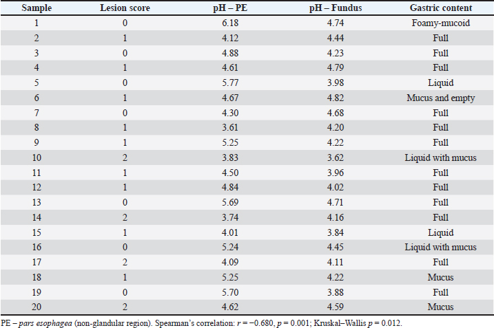
AbstractBackground: Lesions of the pars esophagea in pigs are a common multifactorial, predominantly non-infectious gastrointestinal disorder associated with physicochemical, nutritional, and management-related factors. Disruption of the physiological pH gradient between glandular and non-glandular gastric regions is considered a key mechanism in lesion development, while Lactobacillus spp. represent a stable component of the porcine gastric microbiota. Aim: This study aimed to analyse the relationship between the severity of gastric mucosal damage, gastric pH values, and the presence of lactic acid bacteria, predominantly morphologically consistent with Lactobacillus spp., using culture and cytological methods. Methods: A total of 20 porcine stomachs were analysed. The pH values were measured in the pars esophagea and in the glandular region of the stomach. Microbiological examination included cultivation on selective media and impression smear cytology. Results: The results demonstrated a decrease in pH in the pars esophagea associated with increasing lesion severity, from 5.39 ± 0.64 in score 0 lesions to 4.07 ± 0.40 in score 2 lesions (p=0.012), whereas pH values in the glandular region remained relatively stable (mean 4.42). Lactic acid bacteria, predominantly morphologically consistent with Lactobacillus spp., were detected in all samples regardless of lesion severity. Conclusion: The findings suggest that local acidification of the pars esophagea is a key factor in the development of gastric lesions, whereas lactic acid bacteria, predominantly morphologically consistent with Lactobacillus spp., appear to represent an acid-tolerant but likely secondary component of the gastric microbiota. Keywords: Gastric ulceration, Lactic acid bacteria, Microbiota, Pars esophagea, Pigs. IntroductionLesions of the pars esophagea of the porcine stomach constitute a significant pathological problem in intensive pig production systems, and their development results from a complex interaction of physicochemical, nutritional, and management-related factors. Their high prevalence in slaughterhouses indicates substantial economic losses and a negative impact on animal welfare (Guise et al., 1997; Amory et al., 2006). Recent large-scale field studies in finishing pigs have confirmed the high prevalence of gastric ulceration and highlighted the importance of herd management factors in its occurrence (Cybulski et al., 2024). The pathogenesis of gastric ulceration in pigs is multifactorial and primarily non-infectious, with significant influence of management and production-related factors (Cybulski et al., 2024; Taillieu et al., 2024). In addition to physicochemical factors, infectious agents have also been proposed to contribute to gastric ulceration through modulation of gastric acid secretion and alteration of the gastric microbiota (De Witte et al., 2018). Dietary organic acids have also been studied for their effects on the gastrointestinal environment and microbial balance in pigs (Suiryanrayna and Ramana, 2015). Physical properties of feed play a key role by influencing the structure and stratification of gastric contents. Finely ground or pelleted feed disrupts the natural stratification of stomach contents and increases the exposure of the sensitive pars esophagea mucosa to the acidic gastric environment (Mikkelsen et al., 2004). The non-glandular part of the stomach lacks protective mucus and bicarbonate mechanisms and is therefore highly susceptible to the effects of low pH. A decrease in pH results in epithelial damage, disruption of cellular integrity, and the development of erosions and ulcerations (Straw et al., 2006). An important risk factor is also herd management and housing conditions, which affect feed intake, stress load, and the overall physiology of digestion. These factors have repeatedly been associated with an increased occurrence of gastric lesions in finishing pigs (Amory et al., 2006). From a microbiological perspective, the gastric and intestinal microbiota of pigs develop after birth and is characterized by relative stability of the major bacterial groups. The gastrointestinal microbiota plays an important role in digestion, immune function, and maintenance of intestinal homeostasis in pigs (Kiernan et al., 2023; Szabó et al., 2023; Wang et al., 2025). Lactobacillus spp. are among the major bacteria adapted to the acidic environment of the gastrointestinal tract and constitute a common component of the porcine microbiota (Leser et al., 2002; Konstantinov et al., 2006). The porcine stomach is colonized by microbiota in which Lactobacillus spp. are frequently present. Their occurrence appears relatively consistent regardless of the condition of the gastric mucosa, suggesting high acid resistance and the ability to survive passage through the gastric environment (Dowarah et al., 2017; Wang et al., 2017). Current evidence suggests that Lactobacillus spp. are an integral component of the host microbial ecosystem, while their functional significance is influenced by local physicochemical conditions (Walter, 2008). At the same time, disruption of the pH gradient between the glandular and non-glandular regions of the stomach has been identified as a key pathogenetic mechanism. Under physiological conditions, this gradient acts as a protective mechanism, and its loss leads to increased susceptibility of the pars esophagea (Straw et al., 2006; Peralvo-Vidal et al., 2021). This study aimed to analyse the relationship between the severity of gastric mucosal damage, pH values in different regions of the stomach, and the presence of lactic acid bacteria, predominantly morphologically consistent with Lactobacillus spp., with particular emphasis on their potential association with gastric lesions. Materials and MethodsA total of 20 stomachs from finishing pigs obtained post mortem at a slaughterhouse during the spring season of 2026 were analysed in this study. Samples were collected without any intervention in live animals. The degree of macroscopic gastric mucosal damage was evaluated using a semiquantitative scoring system ranging from 0 to 3, where score 0 represented physiologically normal, intact mucosa, score 1 parakeratosis, score 2 the presence of erosions, and score 3 the occurrence of gastric ulcers. Gastric pH values were measured within 1 hour post mortem in two anatomical regions, specifically in the pars esophagea (PE) and in the glandular region of the stomach (fundus), using a glass pH electrode (GE 135 pH electrode, Greisinger, Germany). The pH meter (portable pH meter G 1501, Greisinger, Germany) was calibrated before each measurement according to the manufacturer’s recommendations. Statistical analysis was performed using Spearman’s rank correlation coefficient to assess the relationship between lesion severity and pH values in the pars esophagea. Differences among lesion scores were evaluated using the Kruskal–Wallis test. Statistical significance was set at p < 0.05. Cultivation examination was performed using selective de Man, Rogosa, and Sharpe (MRS) agar (de Man et al., 1960), which supports the growth of Lactobacillus spp. and other lactic acid bacteria. Inoculated samples were incubated at 37°C for 48 hours under anaerobic conditions. After incubation, the grown colonies were subcultured in order to obtain pure cultures. Subsequently, the isolated strains were evaluated macroscopically and subjected to microscopic examination following Gram staining. Impression smear cytology was also included in the examination. Mucosal samples were fixed and stained with Giemsa stain (Merck, Germany). Morphological characteristics of bacterial forms present on the mucosal surface were evaluated microscopically at 1000 × magnification. Photographs were taken using a PrimoStar light microscope (Zeiss, Germany) connected to a digital camera (PROMICAM PRO3-CP), and all images were documented using QuickPhoto Industrial software. Samples were collected post mortem at a commercial slaughterhouse, and no experimental procedures were performed on live animals. ResultsIn the analysis of 20 porcine stomachs, the degree of mucosal damage, pH values in different regions of the stomach, and the presence of lactic acid bacteria, with a predominance of morphologically consistent Lactobacillus spp., were evaluated. The distribution of mucosal lesion severity showed that no macroscopic changes (score 0) were observed in seven samples (35%), whereas parakeratosis (score 1) was present in nine samples (45%). Erosive lesions (score 2) were recorded in four samples (20%), while ulcerations (score 3) were not observed in any case. Analysis of pH values demonstrated a significant negative association between pH in the pars esophagea and lesion severity. In samples without mucosal damage (score 0), the mean pH value was 5.39 ± 0.64, whereas in parakeratotic lesions (score 1), it decreased to 4.54 ± 0.55. The lowest pH values were recorded in erosive lesions (score 2), where the mean pH reached 4.07 ± 0.40. Differences among lesion scores were significant (Kruskal–Wallis test, p=0.012). Spearman’s rank correlation confirmed a significant negative association between lesion severity and pH values in the pars esophagea (rs=−0.680, p=0.001). In contrast, pH values in the glandular region of the stomach (fundus) remained relatively stable, with a mean value of 4.42 ± 0.37 (range 3.62–4.82), and no apparent trend related to lesion severity in the pars esophagea was observed (Table 1). Table 1. Gastric pH values and gastric content characteristics in pigs.
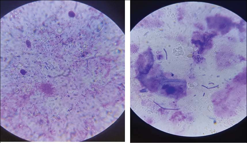
Cytological examination using the impression smear method revealed the presence of rod-shaped bacterial forms, often arranged in long chains (streptobacillary forms), morphologically compatible with lactic acid bacteria, predominantly Lactobacillus spp. (Fig. 1) These bacteria were also observed at low pH values (below 4.0), indicating their ability to survive in the acidic gastric environment and potentially colonise the gastric mucosa.
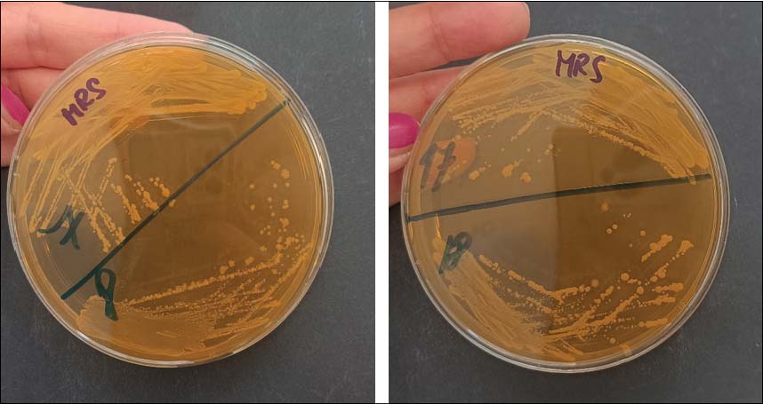
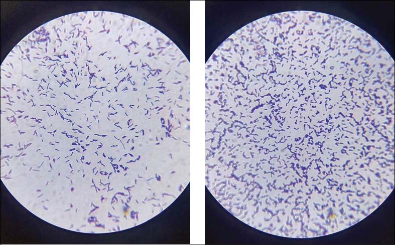
Fig. 1. Impression smear cytology showing gram-positive, rod-shaped bacteria morphologically consistent with Lactobacillus spp. on the gastric mucosa. Magn. ×1000.. No other bacterial morphotypes, particularly spiral-shaped forms compatible with the genus Helicobacter, were observed in the analysed samples. Microbiological examination confirmed the presence of lactic acid bacteria, predominantly morphologically consistent with Lactobacillus spp. (100% of samples), regardless of pH or lesion severity (Figs. 2 and 3).
Fig. 2. Growth of lactic acid bacteria, predominantly morphologically consistent with Lactobacillus spp., on MRS agar.
Fig. 3. Gram-stained gastric microflora with predominance of Gram-positive, rod-shaped bacteria morphologically consistent with Lactobacillus spp. Magn. ×1000. DiscussionThe results of this study suggest that lactic acid bacteria, predominantly morphologically consistent with Lactobacillus spp., constitute a stable component of the gastric microbiota in pigs and remain present even in cases of marked pathological changes in the pars esophagea. This finding is consistent with previous studies suggesting that the development of gastric lesions is not primarily associated with major alterations in the composition of the commensal microbiota, but rather with disturbances in the physicochemical conditions of the gastric environment, particularly pH and the characteristics of gastric contents (Mikkelsen et al., 2004; Straw et al., 2006). A key finding of this study was the relationship between decreasing pH in the pars esophagea and increasing lesion severity. This association was statistically significant, confirming that lower pH values in the pars esophagea were associated with more severe gastric lesions. This observation supports the hypothesis that the main pathogenetic mechanism is not overall gastric acidification, but rather local disruption of the physiological pH gradient between the glandular and non-glandular regions of the stomach. Under physiological conditions, this gradient represents an important protective mechanism, and its disruption leads to increased exposure of the sensitive pars esophagea mucosa to acidic gastric contents (Straw et al., 2006; Peralvo-Vidal et al., 2021). This mechanism is closely associated with the physical properties of feed and the subsequent stratification of gastric contents. Finely ground or pelleted diets disrupt the natural layering of digesta and facilitate the movement of acidic contents into the proximal regions of the stomach, thereby increasing acid exposure of the non-glandular mucosa (Mikkelsen et al., 2004). Similar conclusions were reported by Peralvo-Vidal et al. (2021), who emphasized the role of increased fluidity of gastric contents as a factor promoting gastric ulceration. In the present study, pH values in the glandular region of the stomach remained relatively stable regardless of lesion severity, supporting the concept that pathological changes occur primarily in the pars esophagea and are associated with local environmental alterations rather than overall gastric acidity. From a microbiological perspective, lactic acid bacteria, predominantly morphologically consistent with Lactobacillus spp., were detected in all analysed samples regardless of pH values or lesion severity. This finding is consistent with recent studies reporting that Lactobacillus spp. are a stable component of the porcine gastric microbiota (Taillieu et al., 2024). Consistently, higher counts of Lactobacillus have been reported in healthy gastric mucosa compared with ulcerated stomachs in pigs, suggesting a potential protective role of these bacteria (Almeida et al., 2018). Recent studies investigating the gastric microbiota of pigs have indicated that changes in the abundance of Lactobacillus spp. may accompany the development of gastric lesions, although a direct causal relationship has not been established (Almeida et al., 2018; Taillieu et al., 2024). This finding demonstrates their marked acid tolerance and ability to survive within the gastric environment. Mechanisms of acid resistance in lactic acid bacteria include regulation of intracellular pH, protection of cellular structures, and activation of stress adaptation responses, enabling survival even at low pH values (Wang et al., 2017). Impression smear cytology additionally suggested the ability of Lactobacillus spp. to adhere to the gastric mucosa. Rod-shaped bacterial cells arranged in chains, together with successful cultivation on selective media, confirmed their consistent presence in the analysed samples. Despite their constant occurrence, no direct association was observed between the presence of lactic acid bacteria, predominantly morphologically consistent with Lactobacillus spp., and the severity of gastric mucosal lesions. The role of Lactobacillus spp. in gastric ulceration remains controversial, with evidence suggesting their abundance may reflect rather than drive gastric environmental changes (Almeida et al., 2018; Taillieu et al., 2024). This finding supports the hypothesis that these bacteria represent an adapted and likely secondary component of the gastric microbiota rather than a direct etiological factor in lesion development. Similar conclusions were reported by Walter (2008) and Dowarah et al. (2017), who suggested that the functional significance of lactobacilli is strongly influenced by local environmental conditions and nutrient availability. Recent reviews have further emphasized the importance of microbial stability and host–microbiota interactions for maintaining gastrointestinal health in pigs (Upadhaya and Kim, 2022). No spiral-shaped bacterial forms morphologically compatible with the genus Helicobacter were observed in the cytological preparations, suggesting that Helicobacter suis infection was unlikely to be a significant etiological factor in the analysed animals. This observation further supports the multifactorial and predominantly non-infectious nature of gastric ulceration in pigs (Pluske et al., 1995; Friendship, 2004). From a pathogenetic perspective, gastric lesions appear to result from a complex interaction of several factors, including disruption of the pH gradient, physical characteristics of feed, fluidity of gastric contents, and individual host susceptibility. These findings are consistent with both experimental and field studies describing gastric ulceration as a multifactorial process without a dominant infectious agent (Pluske et al., 1995; Friendship, 2004; Peralvo-Vidal et al., 2021). Furthermore, recent field studies have emphasized the role of herd management factors in the occurrence of gastric ulceration in finishing pigs (Cybulski et al., 2024). From a practical standpoint, the results suggest that modulation of lactic acid bacteria alone is unlikely to play a major role in the prevention of gastric lesions. Greater importance should instead be placed on the optimisation of feed physical properties and maintenance of physiological gastric stratification and pH balance. A limitation of this study is the relatively low number of analysed samples, which may affect the strength of the obtained conclusions. Another limitation is the absence of quantitative microbiota analysis, which would allow a more precise evaluation of bacterial population dynamics in relation to the degree of mucosal damage. Furthermore, due to the culture-based and cytological methods used, the presence of other genera of lactic acid bacteria, such as Bifidobacterium spp., cannot be completely excluded. Precise taxonomic identification would require the use of molecular methods, such as biochemical tests (API), MALDI-TOF analysis, or PCR and 16S rRNA sequencing. Future studies applying these quantitative and molecular approaches may contribute to a deeper understanding of the role of microbiota in the pathogenesis of gastric lesions in pigs. Recent advances in molecular and culture-independent approaches have substantially improved the understanding of the composition, function, and dynamics of the porcine gastrointestinal microbiota and may provide more comprehensive insights into host–microbiota interactions associated with gastrointestinal health and disease (Tang et al., 2025). ConclusionThis study suggests that lactic acid bacteria, predominantly morphologically consistent with Lactobacillus spp., constitute a stable component of the gastric microbiota in pigs without a direct association with the severity of gastric lesions. In contrast, an association was observed between decreasing pH in the pars esophagea and the degree of mucosal damage. The findings support the hypothesis that local acidification of the non-glandular region of the stomach, resulting from disruption of the physiological stratification of gastric contents, represents a key pathogenetic factor in the development of gastric lesions. From a practical perspective, preventive measures should primarily focus on optimisation of the physical properties of feed and stabilisation of the gastric environment rather than modulation of the microbiota. AcknowledgmentsThe authors thank all personnel involved in sample collection and laboratory processing. Conflicts of interestThe authors declare no conflict of interest. FundingThis work was supported by the VEGA grant 1/0040/24: Research on the prevalence, etiological, and predisposing factors of gastric ulcerative lesions in pigs. Authors’ contributionsZK – Investigation, Data curation, Writing – original draft. JN – Conceptualization, Supervision, Funding acquisition, Sample transport, Writing – review and editing, Translation. VH – Methodology, Investigation. KB – Methodology, Investigation, Sample processing. FZ – Supervision, Expert consultation. Ethical approvalNot needed for this study. Data availability All data generated or analysed during this study are included in this published article. ReferencesAlmeida, L.R., Costa, P.S., Nascimento, A.M.A., Reis, M.D.P., Barros, K.O., Alvim, L.B., Nunes, A.C., Queiroz, D.M.M., Rocha, G.A., Nicoli, J.R. and De Moura, S.B. 2018. Porcine stomachs with and without gastric ulcer differ in Lactobacillus load and strain characteristics. Can. J. Microbiol. 64, 493–499; doi: 10.1139/cjm-2017-0758 Amory, J.R., Mackenzie, A.M. and Pearce, G.P. 2006. Factors in the housing environment of finisher pigs associated with the development of gastric ulcers. Vet. Rec. 158, 260–264; doi: 10.1136/vr.158.8.260 Cybulski, P., Woźniak, A., Larska, M., Jabłoński, A. and Stadejek, T. 2024. Gastric ulcers in finishing pigs: the evaluation of selected non-dietary risk factors and impact on production performance. Porcine Health Manage. 10, 11; doi: 10.1186/s40813-024-00362-0 De Man, J.C., Rogosa, M. and Sharpe, M.E. 1960. A medium for the cultivation of lactobacilli. J. Appl. Bacteriol. 23, 130–135. De Witte, C., Ducatelle, R. and Haesebrouck, F. 2018. The role of infectious agents in the development of porcine gastric ulceration. Vet. J. 236, 56–61; doi: 10.1016/j.tvjl.2018.04.015 Dowarah, R., Verma, A.K. and Agarwal, N. 2017. The use of Lactobacillus as an alternative of antibiotic growth promoters in pigs: a review. Anim. Nutr. 3(1), 1–6; doi : 10.1016/j.aninu.2016.11.002 Friendship, R.M. 2004. Gastric ulceration in swine. J. Swine Health Prod. 12, 34–35. Guise, H.J., Carlyle, W.W.H., Penny, R.H.C., Abbott, T.A., Riches, H.L. and Hunter, E.J. 1997. Gastric ulcers in finishing pigs: their prevalence and failure to influence growth rate. Vet. Rec. 141, 563–566; doi: 10.1136/vr.141.22.563 Kiernan, D.P., O’Doherty, J.V. and Sweeney, T. 2023. The effect of prebiotic supplements on the gastrointestinal microbiota and associated health parameters in pigs. Animals 13, 3012; doi: 10.3390/ani13193012 Konstantinov, S.R., Awati, A.A., Williams, B.A., Miller, B.G., Jones, P., Stokes, C.R., Akkermans, A.D.L., Smidt, H. and De Vos, W.M. 2006. Post-natal development of the porcine microbiota composition and activities. Environ. Microbiol. 8, 1191–1199; doi: 10.1111/j.1462-2920.2006.01009.x Leser, T.D., Amenuvor, J.Z., Jensen, T.K., Lindecrona, R.H., Boye, M. and Møller, K. 2002. Culture-independent analysis of gut bacteria: the pig gastrointestinal tract microbiota revisited. Appl. Environ. Microbiol. 68(2), 673–690; doi: 10.1128/AEM.68.2.673-690.2002 Mikkelsen, L.L., Naughton, P.J., Hedemann, M.S. and Jensen, B.B. 2004. Effects of physical properties of feed on microbial ecology and survival of Salmonella enterica serovar Typhimurium in the pig gastrointestinal tract. Appl. Environ. Microbiol. 70, 3485–3492; doi: 10.1128/AEM.70.6.3485-3492.2004 Peralvo-Vidal, J.M., Weber, N.R., Nielsen, J.P., Bache, J.K., Haugegaard, S. and Pedersen, A.Ø. 2021. Risk factors for gastric ulceration in nursery pigs. Prev. Vet. Med. 189, 105298; doi: 10.1016/j.prevetmed.2021.105298 Pluske, J.R., Williams, I.H. and Aherne, F.X. 1996. Maintenance of villous height and crypt depth in piglets by providing continuous nutrition after weaning. Anim. Sci. 62, 131–144; doi: 10.1017/S1357729800014417 Straw, B.E., Zimmerman, J.J., D’Allaire, S. and Taylor, D.J. 2006. Diseases of swine. Suiryanrayna, M.V.A.N. and Ramana, J.V. 2015. A review of the effects of dietary organic acids fed to swine. J. Anim. Sci. Biotechnol. 6, 45; doi: 10.1186/s40104-015-0042-z Szabó , C., Kachungwa Lugata, J. and Ortega, A.D.S.V. 2023. Gut health and influencing factors in pigs. Animals 13, 1350; doi: 10.3390/ani13081350 Taillieu, E., Taelman, S., De Bruyckere, S., Goossens, E., Chantziaras, I., Van Steenkiste, C., Yde, P., Hanssens, S., De Meyer, D., Van Criekinge, W., Stock, M., Maes, D., Chiers, K. and Haesebrouck, F. 2024. The role of Helicobacter suis, Fusobacterium gastrosuis, and the pars oesophageal microbiota in gastric ulceration in slaughter pigs receiving meal or pelleted feed. Vet. Res. 55, 32; doi: 10.1186/s13567-024-01274-1 Tang, Q., Yin, X., Wen, G., Luo, Z., Zhang, L. and Tan, S. 2025. Unraveling the composition and function of pig gut microbiome from metagenomics. Anim. Microbiome. 7, 60; doi: 10.1186/s42523-025-00419-7 Upadhaya, S.D. and Kim, I.H. 2022. Maintenance of gut microbiome stability for optimum intestinal health in pigs – a review. J. Anim. Sci. Biotechnol. 13, 140; doi: 10.1186/s40104-022-00790-4 Walter, J. 2008. Ecological role of lactobacilli in the gastrointestinal tract: implications for fundamental and biomedical research. Appl. Environ. Microbiol. 74, 4985–4996; doi: 10.1128/AEM.00753-08 Wang, C., Cui, Y. and Qu, X. 2018. Mechanisms and improvement of acid resistance in lactic acid bacteria. Arch. Microbiol. 200, 195–201; doi:10.1007/s00203-017-1446-2 Wang, J., Tong, T., Yu, C. and Wu, Q. 2025. The research progress on the impact of pig gut microbiota on health and production performance. Front. Vet. Sci. 12, 1564519; doi:10.3389/fvets.2025.1564519 | ||
| How to Cite this Article |
| Pubmed Style Krepelková Z, Novotni J, Hajdučková V, Bárdová K, Zigo F. Relationship between gastric pH, lactic acid bacteria, and gastric lesions in pigs. Open Vet. J.. 2026; 16(7): 4503-4509. doi:10.5455/OVJ.2026.v16.i7.34 Web Style Krepelková Z, Novotni J, Hajdučková V, Bárdová K, Zigo F. Relationship between gastric pH, lactic acid bacteria, and gastric lesions in pigs. https://www.openveterinaryjournal.com/?mno=320997 [Access: July 15, 2026]. doi:10.5455/OVJ.2026.v16.i7.34 AMA (American Medical Association) Style Krepelková Z, Novotni J, Hajdučková V, Bárdová K, Zigo F. Relationship between gastric pH, lactic acid bacteria, and gastric lesions in pigs. Open Vet. J.. 2026; 16(7): 4503-4509. doi:10.5455/OVJ.2026.v16.i7.34 Vancouver/ICMJE Style Krepelková Z, Novotni J, Hajdučková V, Bárdová K, Zigo F. Relationship between gastric pH, lactic acid bacteria, and gastric lesions in pigs. Open Vet. J.. (2026), [cited July 15, 2026]; 16(7): 4503-4509. doi:10.5455/OVJ.2026.v16.i7.34 Harvard Style Krepelková, Z., Novotni, . J., Hajdučková, . V., Bárdová, . K. & Zigo, . F. (2026) Relationship between gastric pH, lactic acid bacteria, and gastric lesions in pigs. Open Vet. J., 16 (7), 4503-4509. doi:10.5455/OVJ.2026.v16.i7.34 Turabian Style Krepelková, Zuzana, Jaroslav Novotni, Vanda Hajdučková, Katarína Bárdová, and František Zigo. 2026. Relationship between gastric pH, lactic acid bacteria, and gastric lesions in pigs. Open Veterinary Journal, 16 (7), 4503-4509. doi:10.5455/OVJ.2026.v16.i7.34 Chicago Style Krepelková, Zuzana, Jaroslav Novotni, Vanda Hajdučková, Katarína Bárdová, and František Zigo. "Relationship between gastric pH, lactic acid bacteria, and gastric lesions in pigs." Open Veterinary Journal 16 (2026), 4503-4509. doi:10.5455/OVJ.2026.v16.i7.34 MLA (The Modern Language Association) Style Krepelková, Zuzana, Jaroslav Novotni, Vanda Hajdučková, Katarína Bárdová, and František Zigo. "Relationship between gastric pH, lactic acid bacteria, and gastric lesions in pigs." Open Veterinary Journal 16.7 (2026), 4503-4509. Print. doi:10.5455/OVJ.2026.v16.i7.34 APA (American Psychological Association) Style Krepelková, Z., Novotni, . J., Hajdučková, . V., Bárdová, . K. & Zigo, . F. (2026) Relationship between gastric pH, lactic acid bacteria, and gastric lesions in pigs. Open Veterinary Journal, 16 (7), 4503-4509. doi:10.5455/OVJ.2026.v16.i7.34 |


