| Original Article | ||
Open Vet. J.. 2022; 12(4): 469-480 Open Veterinary Journal, (2022), Vol. 12(4): 469–480 Original Research Pre- and postcapillary pulmonary hypertension in dogs: Circulating biomarkersDmitrij Arkadievich Oleynikov1*, and Ma Yi21Veterinary clinic “Belij Klyk”, Moscow, Russia 2Laboratory of Transfusion and Efferent Therapy, Nacional’nyj Medicinskij Issledovatel’skij Centr Imeni V A Almazova: Sankt Peterburg, Saint Petersburg, Russia *Corresponding Author: Dmitrij Arkadievich Oleynikov. Veterinary clinic “Belij Klyk”, Moscow, Russia.Email: wolfberg.guard [at] gmail.com Submitted: 23/02/2022 Accepted: 19/06/2022 Published: 14/07/2022 © 2022 Open Veterinary Journal
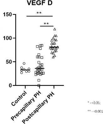
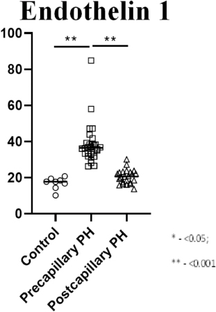
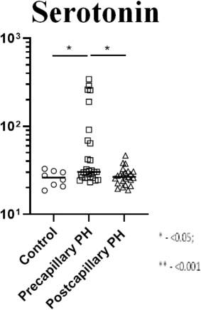
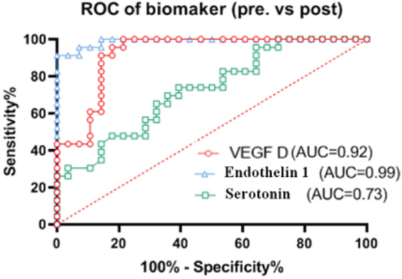
AbstractBackground: Pulmonary hypertension (PH) in dogs is a syndrome that could be primary or secondary due to pulmonary disease, pulmonary thromboembolism, heartworm disease, and heart failure. Due to the inability of right heart catheterization in veterinary patients, there is a lack of differential criteria between PH forms. In some acute cases, it is impossible to provide a full EchoCG or catheterization study. In this situation, circulating markers may be useful to discover the possible mechanism of PH form and provide specific therapy. Aim: Following all previous data in human and veterinary studies, we assumed that plasm concentration of serotonin, endothelin-1 (ET-1), and vascular endothelial growth factor D (VEGF-D) would show a predominance in affected part of pulmonary circulation. Methods: We studied 59 small-breed dogs of different sexes and ages. Groups were formed according to a primary pathology: healthy dogs (n=8); dogs with myxomatous mitral valve disease (MMVD) and postcapillary PH (PostPH, n=23); dogs with MMVD and precapillary PH (PrePH, n=28). Animals in the study were diagnosed with the primary disease by standard echocardiographic methods and algorithms. Blood samples were collected at the moment of presentation and frozen in a −80°C fridge. For biochemistry analysis, we used species-specific ELISA kits, provided by Cloud-Clone Corp. (USA). The tests were provided by the means of Almazov National Medical Research Center, IEM laboratory. Results: Dogs with EchoCG-proved PostPH had a higher concentration of VEGF-D in comparison to control and PrePH (р <0.001, for both). There was no difference between the control and PrePH groups (р >0.05). ET-1 was higher in PrePH in comparison to PostPH and control dogs (р <0.001, for both). In addition, there was no difference between the control and PostPH groups (р >0.05). Serotonin concertation did not have a difference between controls and PostPH. However, it was higher in PrePH than in control (р <0.033) and PostPH group (р <0.006). Receiver operating curve analysis showed that plasma concentrations of ET-1 (0.99) and VEGF-D (0.92) had high effectiveness in the differentiation of PostPH and PrePH. Conclusion: This study showed a correlation between circulating biomarkers (serotonin, ET-1, and VEGF-D). We found a connection between ET-1 and right-sided heart failure as well as VEGF-D and left heart failure in the PH context. Keywords: Biomarkers, Endothelin-1, Pulmonary hypertension, Serotonin, VEGF-D. IntroductionPulmonary hypertension (PH) in dogs is a syndrome that could be primary or secondary due to pulmonary disease, pulmonary thromboembolism, heartworm disease, and heart failure (Reinero et al., 2020). There is a growing body of data for echocardiography assessment in PH diagnostics. But there is still a lack of “gold standard” in right heart catheterization studies, therefore some of the EchoCG-based parameters may be inaccurate (Johnson et al., 1999; Kellihan and Stepien, 2010; Visser, 2017; Jaffey et al., 2019; Menciotti et al., 2021). Even so, in acute cases, sometimes it is impossible to provide full EchoCG to identify PH and its possible cause. In dogs, acute respiratory distress could be associated with PH-associated states: progression of left-heart disease, pulmonary edema (a reactive form of PH), lung disease, dirofilariasis, and thromboembolism of the pulmonary artery (Reinero et al., 2020). In this acute context, circulating markers that show the prevalence of the damaged part of lung circulation is in demand. Human studies show defying circulating markers: ANP, brain natriuretic peptide (BNP), NTproBNP, ADMA, PDGF, vascular endothelial growth factor (VEGF), PIGF, etc. (Hewes et al ., 2020). Nowadays, the most valuable circulating biomarkers in human medicine are natriuretic peptides. In human research, BNP was found to be the most reliable factor of right chambers overload, due to its production predominantly by overstretched right ventricle muscle (Säleby et al., 2019). In studies, BNP showed prognostic value in chronic pulmonary artery thromboembolism (PAT) but was not suspected as an early marker of PH (Hewes et al., 2020). In veterinary medicine, there are several studies of natriuretic peptides, most of them associated with NT-proBNP clinic relevance to pulmonary edema or left-sided heart failure (LSHF) (Nagaya et al., 1998; Heishima et al., 2018; Hezzell et al., 2018). Moreover, there are studies on endothelin-1 (ET-1) concentration in PH patients and its correlation with pulmonary artery pressure (Nootens et al., 1995; Dupuis and Hoeper, 2008; Warwick et al., 2008). Also, there is a lot of information about VEGF’s role in PH, but the data is not homogenous, possibly because of different VEGF ligands (A, B, C, D) and variable effects with different receptors (Kanazawa et al., 2007; Voelkel and Gomez-Arroyo, 2014; Ahmed et al., 2020). By contrast, in veterinary medicine, several studies are elucidating the role of serotonin in lung remodeling and LSHF due to myxomatous mitral valve disease (MMVD) (Mangklabruks and Surachetpong, 2014; Höglund et al., 2018; Sakarin et al., 2020). Following all previous data in human and veterinary studies, we supposed that plasm concentration of serotonin, ET-1, and VEGF-D would show a predominance of affected part of pulmonary circulation. Materials and MethodsIn this pilot study, we included dogs of different sexes, predominantly of age above 10 years old, with a history of pre-existent MMVD at stage C of ACVIM classification. The dogs were treated with standard therapy (Pimobendan, Furosemide, Spironolactone +/−ACEi). All the dogs were divided into three groups: a control group with MMVD but without signs of PH (n=8); dogs with MMVD and signs of precapillary PH (PrePH) (n=28); dogs with signs of postcapillary PH (PostPH) and MMVD (n=23). All the included dogs were taken to the intensive care unit with signs of acute respiratory distress while being on standard MMVD stage C medication. At the moment of examination, all of them had endocardial murmurs, harsh lung sounds, and crackles. The control group included dogs with diagnoses of MMVD at stage C. They were treated with standard protocol and did not have signs of respiratory insufficiency, previous pulmonary disease, and PH. In addition, most of them underwent a routine oral cavity sanitation. In parallel, echocardiography and radiology (radiology study of the thoracic cavity in two projections) studies were performed to exclude subclinical forms of diseases. The dogs that appeared in the intensive care unit had a radiology examination to characterize the insensitivity of pulmonary edema. Then, they were placed in the oxygen camera and were treated in the standard way until clinical stabilization. After that, they underwent blood sampling for biomarkers study and an echocardiographic study to estimate their heat performance. To differentiate PrePH and PostPH, we used phenotypic markers. We assumed that all presented dogs previously had the phenotype of LSHF: left chambers dilation, pulmonary veins distention, preserved systolic function, diastolic dysfunction above class 2, absence of right chamber’s dilatation, and low velocity of tricuspid regurgitation. The cases, with preserved phenotypic markers of LSHF and addition of new-onset PH features (tricuspid regurgitation above 2.7 m/s, presence of pulmonary artery regurgitation, decreased right pulmonary artery distensibility index, right ventricle wall hypertrophy, and right chambers dilation), were assumed as patients with PostPH according to ACVIM consensus in PH. Meanwhile, dogs with signs of PH onset (as mentioned above), but with loss of LSHF signs were marked as PrePH. This subjective method is mentioned to find out some features closely connected with different forms of PH from the blank. We excluded dogs with pre-existing PH and its therapy, congenital defects, arrhythmia, lung disease, and systemic diseases affecting blood flow (arterial hypertension, cushing’s disease, PAT, dirofilariasis, sepsis, electrolyte abnormalities, etc.) and dogs without previous history of MMVD. We performed echocardiography in standard methods, with an estimation of chamber diameter, and systolic and diastolic function of the left ventricle. Left atria and right atria diameter were studied in long parasternal 4 chambers-view axis. Right ventricle diameter and right ventricle free wall thickness were estimated in the long parasternal 4 chambers-view axis. Additional criteria included subjective characterization of dilated/non-dilated chambers and right wall hypertrophy presence. The longitudinal systolic function of the right heart was studied by TPASE. Systolic and diastolic diameter of the left ventricle, wall thickness, and shortening fraction was studied in M-mode by the standard methods. The velocity measurements were performed in standard methods with PW-and CW-Doppler. The pulmonary flow was studied on the right short axis: we analyzed anterograde flow, its phenotype, the ratio between acceleration time and effusion time, and regurgitation velocity. In this projection, the ratio between PA and aorta was subjectively estimated. In the context of associated changes of the right ventricle, the velocity of tricuspid regurgitation above 2.7 m/s was assumed as a PH marker. Velocity less than 2.2 m/s was declared as non-significant. The transmitral flow was studied in the context of mitral regurgitation, a diastolic function of the left ventricle (velocity and ratio between E-wave and A-wave calculation). Additionally, tissue Doppler measurements were performed: S’-wave on the tricuspid valve, e’-wave on the interventricular septum, and its ratio with transmitral E-wave. In parallel, we performed subjective characterization of right pulmonary artery distensibility. The PH diagnosis was verified by echocardiography means, using conventional criteria (echocardiography data could be found: Oleynikov and Yi, 2022) Blood samples were collected at the moment of presentation and frozen in a −80°C fridge. For biochemistry analysis, we used species-specific ELISA kits, provided by Cloud-Clone Corp. (USA) The tests were provided by the means of Almazov National Medical Research Center, IEM laboratory. Statistical analysis was performed by SPSS version 23.0 software (IBM Corporation, Armonk, NY) and GraphPad Prism 8.00 (GraphPad Software Inc., La Jolla, CA). The comparison of group characteristics, and echocardiographic indices was performed by the nonparametric Mann–Whitney test (Appendix Table 1) significance of the differences in categorical variables was calculated by Fisher’s exact test. All p values were 2-sided, and p < 0.05 was considered to be statistically significant. The correlation between variables was evaluated by Spearman’s rank correlation coefficient analysis. Ethical approvalThe study was approved at the internal clinical conference. ResultsThis study included 59 dogs of a senior (9–11 years old) age, different sexes, and breeds, predominantly of toy and small breeds. Statistically, a significant weight difference was not observed (р > 0.05, for all groups). Average heart rhythm was more rapid in the group with PostPH, but did not statistically different from other groups (р > 0.05, for all groups, Table 1). The PH diagnosis was verified by echocardiography means, using conventional criteria (echocardiography data could be found: Oleynikov and Yi, 2022). In the PostPH (Table 2, Fig. 1) group plasma, VEGF-D concentration was significantly elevated in comparison to the control dogs and PrePH group (р < 0.001, for both). There was no difference between the control and PrePH groups (р > 0.05). The plasma concentration of ET-1 in the PrePH (Table 2, Fig. 2) group was statistically higher than in the control and PostPH groups. There was no difference between control and PostPH groups (р > 0.05). There was no significant difference between serotonin (Table 2, Fig. 3) concentration in control and PostPH groups. Albeit, plasma serotonin was increased in the PrePH group in comparison to the control dogs (р=0.033) and the PostPH group (р=0.006). Receiver operating curve (ROC) analysis of the VEGF D, ET-1, serotonin showed that VEGF D AUC is 0,92 (95%; CI=0.85–1.0; p < 0.001); for the ET-1 is 0.99 (95%; CI=0.97–1.0; p < 0.001); for the serotonin is 0.73 (95% CI=0.59–0.86; p=0.0057) (Fig. 4). However, AUC showed predictive value: ET-1> VEGF D> serotonin, which means that ET-1 and VEGF-D has high diagnostic value for differentiation between Pre- and PostPH in dogs. Table 1. Weight and heart rate analyze.
Table 2. Serum biomarkers.
Fig. 1. Graphic representation of the VEGF-D plasma concentration in PrePH, PostPH, and control groups. Plasma markers showed correlation to EchoCG findings, analyzed by Spearman Heatmap. We observed reverse correlation between VEGF-D and LVFW for PrePH dogs (r=−0.53, p=0.004), ET-1 and PA effusion time (r=−0.50, p=0.006); positive correlation for serotonin and presence of PA regurgitation (r=0.60, p=0.035). In Post PH group, we found reverse correlation of these serotonin (r=−0.59, p=0.003) and VEGF-D (r=−0.62, p=0.002) to the IVS width; reverse correlation of ET-1 and MR (r=−0.51, p=0.012); positive correlation of the serotonin concentration and PA acceleration time (r=0.58, p=0.004). DiscussionIn this study, we tried to find a correlation between EchoCG parameters and serum biomarkers in PH forms differentiation. We suspect that our findings in biomarkers could help with PH diagnosis in cases where EchoCG should be avoided or is unavailable. There is a tendency in human medicine to replace or find alternative circulating biomarkers to avoid invasive methods. In the context of this study, which includes dogs with previously diagnosed LSHF, we could not use natriuretic peptides due to possible difficulties in the interpretation.
Fig. 2. Graphic representation of the ET-1 plasma concentration in PrePH, PostPH, and control groups. The pathogenies of chronic pulmonary congestion predispose to compensatory increased volume drainage, which is done by the lymphatic system. Physiological dog studies discovered that lymph drainage could be enhanced above 300% in case of chronic volume overload (Uhley et al., 1962). VEGF-D is a predominantly lymphatic vessels growth factor, taking part in lymphatic pool remodeling both in chronic interstitial lung diseases and LSHF (Achen et al., 1998). Human clinical studies found that patients with dyspnea and later diagnosed heart failure had elevated plasma VEGF-D concentration. Authors suspected that this condition/symptom developed as a consequence of pulmonary circulation congestion and compensatory lymphatic system remodeling. This could also be associated with PostPH development due to pulmonary arterial wedge pressure (PAWP) increase and alteration in the microcirculatory drainage (Borné et al., 2018). This statement was validated by the study which showed a correlation between PAWP and VEGF-D (Houston et al., 2019). Additionally, VEGF-D rise was admitted in cases of chronic PAT, which was interpreted as a remodeling due to local congestion in the context of the redistributive edema (Säleby et al., 2019). In both cases, VEGF-D was associated with increased pulmonary vascular resistance, vessels remodeling due to chronic congestion. Moreover, VEGF-D is a mortality factor for patients with PostPH (Miller et al., 2013). One more interesting study in terminal heart failure and cardiac transplantation showed lowering of VEGF-D levels due to a decrease in signs of pulmonary congestion and pulmonary vascular resistance (Ahmed et al., 2020).
Fig. 3. Graphic representation of the serotonin plasma concentration in PrePH, PostPH, and control groups. Another rather specific biomarker is ET-1. This factor is more connected with the rise of pulmonary vascular resistance. ET-1 could be increased both by hyperproduction and by altered clearance (Cody et al., 1992; Staniloae et al., 2004). Human clinical studies showed that ET-1 clearance decreased in patients with PrePH and there was a correlation between ET-1 plasma level and pulmonary vascular resistance determined by right heart catheterization (Meoli et al., 2018). In pathophysiological dog studies, ET-1 rise was found in experimental chronic PAT and decreased ET-1 clearance, showing utilization alteration in pulmonary circulation (Kim et al., 2000). Additionally, we found data about successful bosentan therapy in PrePH and increased circulating ET-1 level (Kim et al., 2000). There is an enhancing factor for ET-1 associated PH—serotonin, which could be a synergist for ET-1 sensitivity in the context of its decreased clearance (Channick et al., 2001; Rubin et al., 2002; Eickelberg et al., 2003; Campbell, 2007). Additionally, serotonin is a proliferating factor for pulmonary vessels media and muscles (Eddahibi et al., 2000; Marcos et al., 2004). In veterinary studies, dogs with myxomatous valvular disease showed a significant role of serotonin pathways in PrePH and PostPH development (Hashimoto et al., 2000; Oyama and Levy, 2010; Ljungvall et al., 2013; Roels et al., 2015). In a recent study, serotonin pathway proteins were studied in dogs with PostPH (Sakarin et al., 2020). Increased serotonin-associated proteins were found in pulmonary vessels, which was interpreted as a consequence of chronic hypoxia and vascular muscular cells remodeling (Eddahibi et al., 2000; Marcos et al., 2004). However, the study did not show a correlation between plasma concentration and pulmonary tissue findings. In our data, we did not find a clear difference in serotonin plasma between groups. This data should be assumed in the context of pulmonary circulation changes associated with two paths. The first one is associated with enhancing effect on ET-1-induced vasoconstriction, as mentioned above, in this variant serotonin plasma level could differ in an unrecognizable limit, but its action on the receptors and increasing to ET-1 sensibility would be enough to provoke severe pulmonary vessel remodeling. The second path is associated with increased serotonin pulmonary circulation and an increased window of acting; this could be sustained by VEGF and serotonin interactions and prolonged pulmonary transition time, observed in dogs with myxomatous valvular disease (Lord et al., 2003).
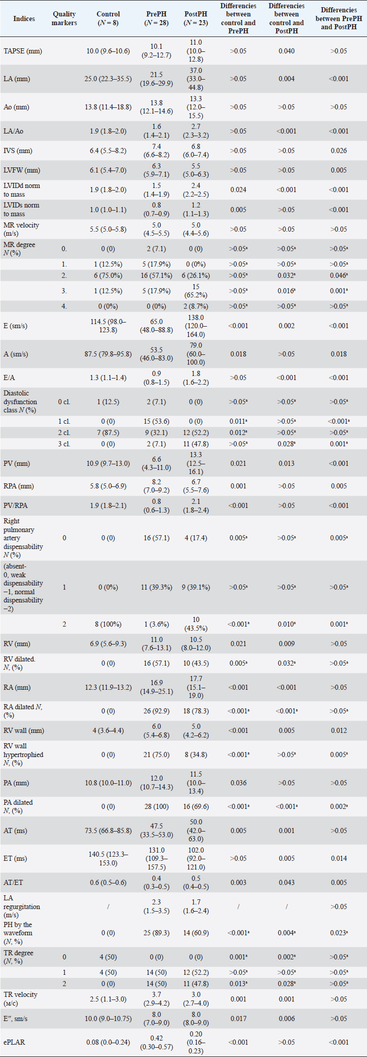
Fig. 4. Grafical ROC with diagnostic ability of the blood markers. Our VEGF-D observations in this study showed that in dogs with EchoCG diagnosed PostPH, VEGF-D concentration almost doubled. This result is similar to a prospective study in human medicine, which elucidates the correlation between PAWP and VEGF-D (Houston et al., 2019). Based on the physiological features of lymphatic drainage in cases of chronic volume overload described in early physiological studies, we suspected these changes are mostly attributed to lymphatics proliferation to prevent pulmonary congestion in dogs with myxomatous valvular disease (Uhley et al., 1962). Logically we can predict a correlation between LA pressure, PAWP and blood VEGF-D concentration. As for ET-1, it was significantly higher in dogs with PrePH, than in the PostPH and control groups. This factor could be explained by microvascular bed remodeling both in the late stages of LSHF and secondary pulmonary arterial remodeling due to lung interstitial disease or pneumonitis. In this context, we can suspect a correlation between ET-1 and right-sided heart failure, because in human studies we can find data about the dependence between ET-1 level and right atrial pressure, and pulmonary artery oxygen saturation (Nootens et al., 1995). Moreover, a recent veterinary study found that dogs with PrePH have a median lifespan of about 276 days, while PostPH dogs—576 days, which shows us possible benefits in diagnosis, monitoring and treatment of the found PH (Borgarelli et al., 2015; Jaffey et al., 2019). In conclusion, we can say, that the most valuable circulating marker in this study is ET-1. It rose in both Post- and PrePH, which reflects its own diagnostic power of the PH diffentiation. But in combination with VEGF-D, it could show the difference between the forms. Subjectively we can recommend these markers as diagnostic criteria of PH in absence of echocardiography, and prediction of the right heart or the left heart failure phenotype. In this study, several limitations are present. We lack the opportunity to differentiate some specific diseases such as hemangiomatosis or veno-occlusive disease, which could be presented with similar symptoms, observed in this study’s dogs, and could show EcoCG signs of both Pre- and PostPH or shifting between phenotypes (Reinero et al., 2019). But our findings are pointing at the leading wing of the pulmonary microcirculation bed damage. Additionally, we studied acute manifestations, predisposing to speculations in chronic states interpretations or comorbidities. Of course, this study should be powered by more wide prospective study, including more dogs and re-analyzing time and treatment interactions. And we have to find out the specific circulatory and respiratory systems threshold. AcknowledgmentSpecial thanks to Toropova J.V., Zelinskaya I.V. for providing an opportunity to analyze studied substrates. Author’s contributionOleynikov D.: collected data, provided diagnostics and treatment, coordinated the data-analysis and contributed to the writing of the manuscript. Ma Yi: coordinated the data analysis and contributed to the writing of the manuscript. ReferencesAchen, M., Jeltsch, M., Kukk, E., Mäkinen, T., Vitali, A., Wilks, A., Alitalo, K. and Stacker, S. 1998. Vascular endothelial growth factor D (VEGF-D) is a ligand for the tyrosine kinases VEGF receptor 2 (Flk1) and VEGF receptor 3 (Flt4). Proc. Natl. Acad. Sci. USA. 95(2), 548–553. Ahmed, S., Ahmed, A., Säleby, J., Bouzina, H., Lundgren, J. and Rådegran, G. 2020. Elevated plasma tyrosine kinases VEGF-D and HER4 in heart failure patients decrease after heart transplantation in association with improved haemodynamics. Heart Vessels 35(6), 786–799. Borgarelli, M., Abbott, J., Braz-Ruivo, L., Chiavegato, D., Crosara, S., Lamb, K., Ljungvall, I., Poggi, M., Santilli, R. and Haggstrom, J. 2015. Prevalence and prognostic importance of pulmonary hypertension in dogs with myxomatous mitral valve disease. J. Vet. Intern. Med. 29, 569–574. Borné, Y., Gränsbo, K., Nilsson, J., Melander, O., Orho-Melander, M., Smith, J. and Engström, G. 2018. Vascular endothelial growth factor D, pulmonary congestion, and incidence of heart failure. J. Am. Coll. Cardiol. 71, 580–582. Campbell, F. 2007. Cardiac effects of pulmonary disease. Vet. Clin. North Am. Small Anim. Pract. 37(5), 949–962. Channick, R., Simonneau, G. and Sitbon, O. 2001. Effects of the dual endothelin-receptor antagonist bosentan in patients with pulmonary hypertension: a randomised placebo-controlled study. Lancet 358, 1119–1123. Cody, R., Haas, G., Binkley, P., Capers, Q. and Kelley, R. 1992. Plasma endothelin correlates with the extent of pulmonary hypertension in patients with chronic congestive heart failure. Circ. 85, 504–509. Dupuis, J. and Hoeper, M. 2008. Endothelin receptor antagonists in pulmonary arterial hypertension. Eur. Respir. J. 31, 407–415. Eddahibi, S., Hanoun, N., Lanfumey, L., Lesch, K., Raffestin, B. and Hamon, M. 2000. Attenuated hypoxic pulmonary hypertension in mice lacking the 5-hydroxytryptamine transporter gene. J. Clin. Invest. 105, 1555–1562. Eickelberg, O., Yeager, M. and Grimminger, F. 2003.The tantalizing triplet of pulmonary hypertension BMP receptors, serotonin receptors and angiopoietins. Circ .Res. 60, 465–467. Hashimoto, Y., Hirota, K., Yoshioka, H., Hashiba, E., Kudo, T. and Ishihara, H. 2000. Spasmolytic effects of prostaglandin E1 on serotonin-induced bronchoconstriction and pulmonary hypertension in dogs. Br. J. Anaesth. 85, 460–462. Heishima, Y., Hori, Y., Nakamura, K., Yamashita, Y., Isayama, N., Kanno, N., Katagi, M., Onodera, H., Yamano, S. and Aramaki, Y. 2018. Diagnostic accuracy of plasma atrial natriuretic peptide concentrations in cats with and without cardiomyopathies. J. Vet. Cardiol. 20(4), 234–243. Hewes, J., Lee, J., Fagan, K. and Bauer, N. 2020. The changing face of pulmonary hypertension diagnosis: a historical perspective on the influence of diagnostics and biomarkers. Pulm. Circ. 10(1), 2045894019892801. Hezzell, M., Block, C., Laughlin, D. and Oyama, M. 2018. Effect of prespecified therapy escalation on plasma NT-proBNP concentrations in dogs with stable congestive heart failure due to myxomatous mitral valve disease. J. Vet. Intern. Med. 32(5), 1509–1516. Höglund, K., Häggström, J., Hanås, S., Merveille, A., Gouni, V., Wiberg, M., Lundgren Willesen, J., Entee, K., Mejer Sørensen, L., Tiret, L., Seppälä, E., Lohi, H., Chetboul, V., Fredholm, M., Lequarré, A. and Ljungvall, I. 2018. Interbreed variation in serum serotonin (5-hydroxytryptamine) concentration in healthy dogs. J. Vet. Cardiol. 20(4), 244–253. Houston, B., Tedford, R., Baxley, R., Sykes, B., Powers, E., Nielsen, C., Steinberg, D., Maran, A., Fernandes, V., Todoran, T., Jones, J. and Zile, M. 2019. Relation of lymphangiogenic factor vascular endothelial growth factor-D to elevated pulmonary artery wedge pressure. Am. J. Cardiol. 124(5), 756–762. Jaffey, J., Wiggen, K., Leach, S., Masseau, I., Girens, R. and Reinero, C. 2019. Pulmonary hypertension secondary to respiratory disease and/or hypoxia in dogs: clinical features, diagnostic testing and survival. Vet. J. 251, 105347. Johnson, L., Boon, J. and Orton, E. 1999. Clinical characteristics of 53 dogs with Doppler derived evidence of pulmonary hypertension: 1992–1996. J. Vet. Intern. Med. 13(5), 440–447. Kanazawa, H., Asai, K. and Nomura, S. 2007. Vascular endothelial growth factor as a non-invasive marker of pulmonary vascular remodeling in patients with bronchitis-type of COPD. Respir. Res. 8(1), 22. Kellihan, H. and Stepien, R. 2010. Pulmonary hypertension in dogs: diagnosis and therapy. Vet. Clin. North Am. Small Anim. Pract. 40(4), 623–641. Kim, H., Yung, G., Marsh, J., Konopka, R., Pedersen, C., Chiles, P., Morris, T. and Channick, R. 2000. Endothelin mediates pulmonary vascular remodelling in a canine model of chronic embolic pulmonary hypertension. Eur. Respir. J. 15(4), 640–648. Ljungvall, I., Hoglund, K., Lilliehook, I., Oyama, M.A., Tidholm, A. and Tvedten, H. 2013. Serum serotonin concentration is associated with severity of myxomatous mitral valve disease in dogs. J. Vet. Intern. Med. 27, 1105–1112. Lord, P., Eriksson, A., Häggström, J., Järvinen, A., Kvart, C., Hansson, K., Maripuu, E. and Mäkelä, O. 2003. Increased pulmonary transit times in asymptomatic dogs with mitral regurgitation. J. Vet. Intern. Med. 17(6), 824–829. Mangklabruks, T. and Surachetpong, S. 2014. Plasma and platelet serotonin concentrations in healthy dogs and dogs with myxomatous mitral valve disease. J. Vet. Cardiol. 16(3), 155–162. Marcos, E., Fadel, E., Sanchez, O., Humbert, M., Dartevelle, P. and Simonneau, G. 2004. Serotonin-induced smooth muscle hyperplasia in various forms of human pulmonary hypertension. Circ. Res. 94, 1263–1270. Menciotti, G., Abbott, J., Aherne, M., Lahmers, S. and Borgarelli, M. 2021. Accuracy of echocardiographically estimated pulmonary artery pressure in dogs with myxomatous mitral valve disease. J. Vet. Cardiol. 35, 90–100. Meoli, D., Su, Y., Brittain, E., Robbins, I., Hemnes, A. and Monahan, K. 2018. The transpulmonary ratio of endothelin 1 is elevated in patients with preserved left ventricular ejection fraction and combined pre- and post-capillary pulmonary hypertension. Pulm. Circ. 8(1), 2045893217745019. Miller, W., Grill, D. and Borlaug, B. 2013. Clinical features, hemodynamics, and outcomes of pulmonary hypertension due to chronic heart failure with reduced ejection fraction: pulmonary hypertension and heart failure. JACC Heart Fail. 1, 290–299. Nagaya, N., Nishikimi, T. and Okano, Y. 1998. Plasma brain natriuretic peptide levels increase in proportion to the extent of right ventricular dysfunction in pulmonary hypertension. J. Am. Coll. Cardiol. 31, 202–208. Nootens, M., Kaufmann, E. and Rector, T. 1995. Neurohormonal activation in patients with right ventricular failure from pulmonary hypertension: relation to hemodynamic variables and endothelin levels. J. Am. Coll. Cardiol. 26, 1581–1585. Oleynikov, D. and Yi, M. 2022. Echocardiographic pulmonary to left atrial ratio in dogs (ePLAR): a differential marker of pre- and postcapillary pulmonary hypertension. Am. J. Anim. Vet. Sci. 17(1), 42–52. Oyama, M. and Levy, R.J. 2010. Insights into serotonin signaling mechanisms associated with canine degenerative mitral valve disease. J. Vet. Intern. Med. 24, 27–36. Reinero, C., Jutkowitz, L., Nelson, N., Masseau, I., Jennings, S. and Williams, K. 2019. Clinical features of canine pulmonary veno-occlusive disease and pulmonary capillary hemangiomatosis. J. Vet. Intern. Med. 33(1), 114–123. Reinero, C., Visser, L.C., Kellihan, H.B., Masseau, I., Rozanski, E., Clercx, C., Williams, K., Abbott, J., Borgarelli, M. and Scansen, B.A. 2020. ACVIM consensus statement guidelines for the diagnosis, classification, treatment, and monitoring of pulmonary hypertension in dogs. J. Vet. Intern. Med. 34(2), 549–573. Roels, E., Krafft, E., Antoine, N., Farnir, F., Laurila, H. and Holopainen, S. 2015. Evaluation of chemokines CXCL8 and CCL2, serotonin, and vascular endothelial growth factor serum concentrations in healthy dogs from seven breeds with variable predisposition for canine idiopathic pulmonary fibrosis. Res. Vet. Sci. 101, 57–62. Rubin, L., Badesch, D. and Barst, R. 2002. Bosentan therapy for pulmonary arterial hypertension. N. Eng. J. Med. 346, 896–903. Sakarin, S., Surachetpong, S. and Rungsipipat, A. 2020. The expression of proteins related to serotonin pathway in pulmonary arteries of dogs affected with pulmonary hypertension secondary to degenerative mitral valve disease. Front. Vet. Sci. 7, 612130. Säleby, J., Bouzina, H., Ahmed, S., Lundgren, J. and Rådegran, G. 2019. Plasma receptor tyrosine kinase RET in pulmonary arterial hypertension diagnosis and differentiation. ERJ Open Res. 5, 00037. Staniloae, C., Dupuis, J., White, M., Gosselin, G., Dyrda, I., Bois, M., Crepeau, J., Bonan, R., Caron, A. and Lavoie, J. 2004. Reduced pulmonary clearance of endothelin in congestive heart failure: a marker of secondary pulmonary hypertension. J. Card. Fail. 10, 427–432. Uhley, H., Leeds, S., Sampson, J. and Friedman, M. 1962. Role of pulmonary lymphatics in chronic pulmonary edema. Circ. Res. 11, 966–970. Visser, L. 2017. Right ventricular function: imaging techniques. Vet. Clin. North Am. Small Anim. Pract. 47, 989–1003. Voelkel, N. and Gomez-Arroyo, J. 2014. The role of vascular endothelial growth factor in pulmonary arterial hypertension. The angiogenesis paradox. Am. J. Respir. Cell Mol. Biol. 51(4), 474–484. Warwick, G., Thomas, P. and Yates, D. 2008. Biomarkers in pulmonary hypertension. Eur. Respir. J. 32(2), 503–512. Appendix Table 1. Echocardiographic-derived measurements and indices. Complex analyze and statistics.
| ||
| How to Cite this Article |
| Pubmed Style Dmitrij Oleynikov|. Pre- and postcapillary pulmonary hypertension in dogs. Circulating biomarkers. Open Vet. J.. 2022; 12(4): 469-480. doi:10.5455/OVJ.2022.v12.i4.8 Web Style Dmitrij Oleynikov|. Pre- and postcapillary pulmonary hypertension in dogs. Circulating biomarkers. https://www.openveterinaryjournal.com/?mno=89784 [Access: December 02, 2025]. doi:10.5455/OVJ.2022.v12.i4.8 AMA (American Medical Association) Style Dmitrij Oleynikov|. Pre- and postcapillary pulmonary hypertension in dogs. Circulating biomarkers. Open Vet. J.. 2022; 12(4): 469-480. doi:10.5455/OVJ.2022.v12.i4.8 Vancouver/ICMJE Style Dmitrij Oleynikov|. Pre- and postcapillary pulmonary hypertension in dogs. Circulating biomarkers. Open Vet. J.. (2022), [cited December 02, 2025]; 12(4): 469-480. doi:10.5455/OVJ.2022.v12.i4.8 Harvard Style Dmitrij Oleynikov| (2022) Pre- and postcapillary pulmonary hypertension in dogs. Circulating biomarkers. Open Vet. J., 12 (4), 469-480. doi:10.5455/OVJ.2022.v12.i4.8 Turabian Style Dmitrij Oleynikov|. 2022. Pre- and postcapillary pulmonary hypertension in dogs. Circulating biomarkers. Open Veterinary Journal, 12 (4), 469-480. doi:10.5455/OVJ.2022.v12.i4.8 Chicago Style Dmitrij Oleynikov|. "Pre- and postcapillary pulmonary hypertension in dogs. Circulating biomarkers." Open Veterinary Journal 12 (2022), 469-480. doi:10.5455/OVJ.2022.v12.i4.8 MLA (The Modern Language Association) Style Dmitrij Oleynikov|. "Pre- and postcapillary pulmonary hypertension in dogs. Circulating biomarkers." Open Veterinary Journal 12.4 (2022), 469-480. Print. doi:10.5455/OVJ.2022.v12.i4.8 APA (American Psychological Association) Style Dmitrij Oleynikov| (2022) Pre- and postcapillary pulmonary hypertension in dogs. Circulating biomarkers. Open Veterinary Journal, 12 (4), 469-480. doi:10.5455/OVJ.2022.v12.i4.8 |


